
8. Eukaryotic Translation
Slides in this set (53)
-
- understand how mRNAs are translated into proteins
- know what the translators of genetic code are
- understand the underlying mechanisms of translation of mRNA to proteins: steps and components involved
- understand how translation is regulated at many different levels
- know some of the methods used in studying translation
-
- Synthesis of polypeptide from mRNA template
- Each amino acid (aa) is encoded by a 3 base “codon”
- The “translators” of the genetic code are tRNAs & aminoacyl-tRNA synthetases
- Recognition by base-pairing between tRNA anticodon & mRNA codon
- Important to translate in correct reading frame- if not forms entirely different polypeptide
-
- Triplet code is degenerate- 64 codons in total for 20 amino acids
- 3 stop codons, and AUG start codon which codes for Met
- Only 51 tRNAs in humans
- Some tRNAs read more than one codon by wobble base pairing eg G:U
- Flexibility in how tRNAs read and pair the 3rd base in a codon on mRNA
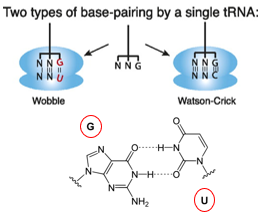
-
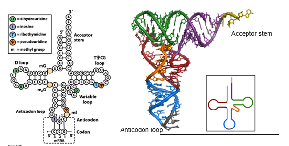
- tRNA is small (~76nts)
- Anticodon loop= where codon is recognised on mRNA
- Amino acid attached to acceptor stem and forms charged tRNA
- Specialised bases and modifications stabilise tRNA and facilitate wobble base pairing
- Eg inosine which is the deaminated form of adenine, and can pair with C/U or A
-
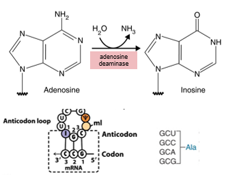
- Generated in tRNA from adenosine by RNA editing, post-transcriptionally (deamination)
- Inosine in anticodon can pair with C, U or A in codon
- Maximises the potential of wobble base pairing
- Eg 3 of the 4 codons for alanine can be read by a tRNA with IGC anticodon (codons vary by 3rd base)
-
- Amino acids are attached to tRNA by esterification by aminoacyl-tRNA synthetases (aaRS):
- Esterify aa to tRNAs- one aaRS per aa, but usually can aminoacylate >1 tRNA
- 2 step process that requires ATP:
- Enzyme has sites that specifically bind particular aa and particular tRNA
- 1. Covalently links O of tRNA to C of specific aa and loses water, using ATP= linked tRNA to specific aa
- 2. Anticodon of tRNA is selected by its codon on mRNA
- Mutations in aaRS genes cause Charcot-Marie-Tooth disease: Inherited neurological disorder, incurable, progressive loss of muscle + touch sensation
-
- Fidelity= accuracy of the process
- Accuracy of DNA, RNA and protein synthesis relies of base complementarity (DNA:DNA, DNA:RNA, or mRNA:tRNA)
- Protein synthesis has largest error rate
- Matching tRNAs to aa by aaRS is base-pairing independent
- tRNA needs to interact with correct aa, aa's that have similar side chains make this difficult
- Identity of aa is not checked on ribosome, so matching correct aa to tRNA is essential
-
- Fidelity is achieved by editing by Aminoacyl-tRNA Synthases:
- Challenging for aaRS to distinguish aa's- tRNAs are larger with more distinguishing features, aa's are small
- aaRS's have editing sites to cope with the problem: editing sites hydrolyse aa's from misacylated tRNAs
- An editing defective aaRS causes neurodegeneration in mice
- Mouse mutation causing neurodegeneration was traced to a substiturtion in the editing domain of aaRS for ala
- Mischarged tRNA (point mutation- wrong aa incorporated) caused misfolded protein accumulation in neurons
-
Sources of Error in Protein Synthesis
- tRNA Mischarging
- Coupling wrong aa to a tRNA
- Mischarging generally involves similar aa's which reduces the impact of error
- tRNA Mismatching
- Matching mRNA codon to the wrong tRNA anticodon
- Wobble base pairing increases mismatching frequency
-
Protein Synthesis Takes Place in Ribosomes
- Ribosomes made up of RNA and protein, and act as ribozymes where rRNA is the catalytic subunit (40S and 60S= 80S)
- Human ribosomes are 4.3 MDa - 4 rRNAs & 80 proteins
- Many copies of tandemly repeated rDNA (rRNA genes)
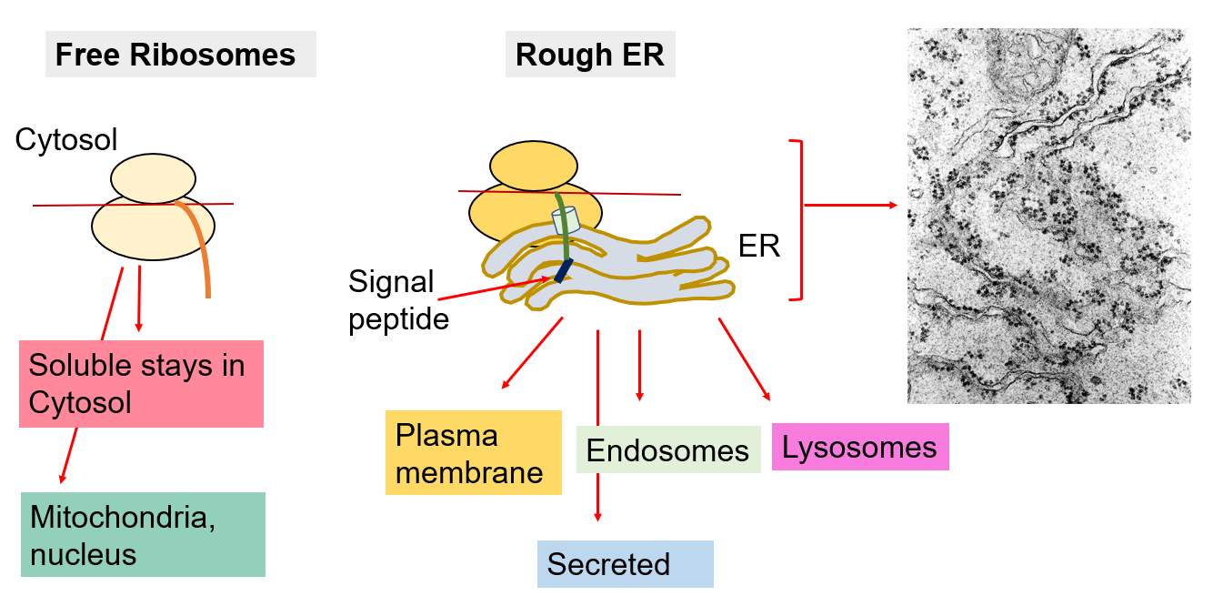
- Found in cytosol and attached to ER
- Proteins synthesised by ribosome attached to ER are destined for secretion, association with membrane or delivery to organelles
- Proteins synthesised by ribosomes free in cytosol are generally soluble proteins
-
Protein Synthesis in the Ribosome
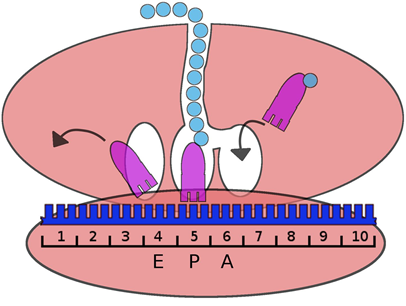
- There are 3 sites that can accomodate tRNA within the ribosome:
- A= aminoacyl-tRNA enters the A site
- P=peptidyl-tRNA occupies P site
- E= exit site
- 2 tRNAs occupy adjacent codons on the mRNA within the ribosome:
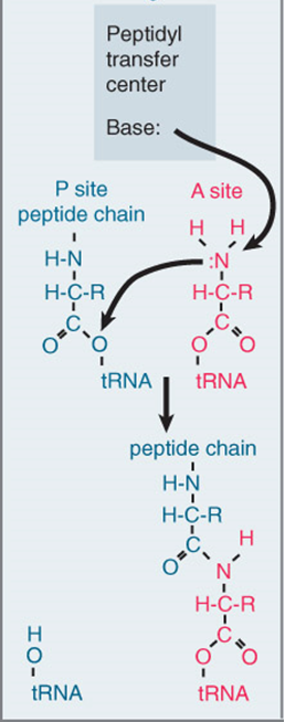
- When new tRNA arrives on A site, the growing pp transfers onto the tRNA in the A site as a peptide bond forms
- Leaves tRNA in the P site with no aa attached to it and moves to E site
- Translocation of ribosome moves the peptidyl-tRNA into P site and ejects the previous tRNA from the E site
- PP extends through tunnel in larger ribosomal subunit
-
-
- Peptidyl transferase centre within ribosome (28S rRNA) catalyses peptide bond formation,
- Amino group of aminoacyl-tRNA attacks ester linkage between pp chain and tRNA in P site and forms peptide bond
- Movement of ribosome causes uncharged tRNA (previously in P site after pp has been transferred to A site tRNA) is released from E
- A site now free to receive aminoacyl-tRNA
-
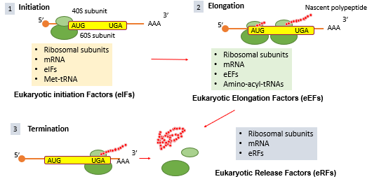
- Multiple translation factors are involved in protein synthesis in eukaryotes:
- Initiation requires: ribsomal subunits, mRNA, eIFs (eukaryotic Initiation Factors), Met-tRNA (AUG codon on mRNA signals for recruitment of Met-tRNA anticodon)
- Elongation requires: ribosomal subunits, mRNA, eEFs, aminoacyl-tRNAs (charged)
- Termination requires: ribosomal subunits, mRNA, eRFs (release- hydrolyse pp chain and release it)
- Highly translated mRNAs have multiple ribosomes attached to them simultaneously= polysomal
-
-
- Complex and multistep process, requires positioning of 40S and 60S ribosomal subunits with initiator met-tRNA at mRNA start codon, AUG
- Assembly of entire ribosome and bring to right AUG start site, and bring right tRNA
- Most highly regulated phase of translation- rate limiting
- Involves ~13 eIFs (eukaryotic initiation factors)
-
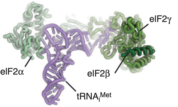
- eIF2= delivers initiator tRNA (met) to start site at 40S ribosomal RNA
- Ternary complex= eIF2 bound to met-tRNA and GTP
-
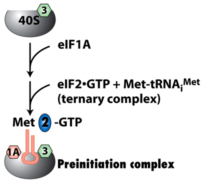
- Ternary complex joins eIF1A and 40S subunit of ribosome to form preinitiation complex (PIC)
- eIF1A:
- Stabilises binding by ternary complex at A site
- Blocks access of met-tRNA to ribosomal P site
-
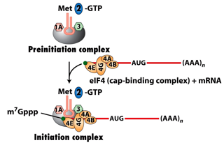
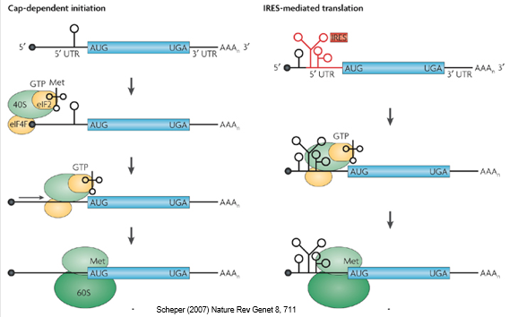
- mRNA is recruited to PIC by eIF4 cap binding complex
- Cap is an inverted 7-methylguanosine group at 5' end of mRNAs
- Cap binds eIF4E to aid recruitment to the PIC
-
Initiation Step 2: eIF4 Complex
- eIF4E= mRNA cap recognition protein, highly regulated, not very abundant, deregulated in cancer
- eIF4G= serves as scaffold, recognises 4E and recruits 4A and 4B, mutations in Parkinsons
- eIF4A= helicase, unwinds secondary structures in 5' region of mRNA to allow access by PIC
- eIF4B= interacts with PIC to bring complexes together
-
- Polycistronic messages in prokaryotes= may have several start sites
- Monocistronic in eukaryotes= ribosome scanning for one start site
- mRNA scanning in eukaryotes:
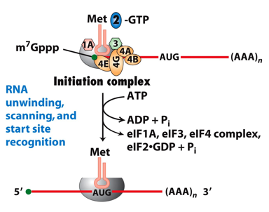
- Assembly of PIC happens at cap end of mRNA, once bound to cap PIC scans along mRNA until it finds AUG codon
- Strong start codons have optimal bases flanking AUG- consensus is called kozak sequence that distinguishes start AUG from other AUGs
- PIC scanning through other suboptimal AUGs that dont contain kozak= leaky scanning
-
Internal Ribosome Entry Sites (IRES)
- IRES allow cap-independent translation (alternative pathway to recruit mRNA to PIC without cap)
- IRES= region of complex secondary structures in 5' UTR of some mRNAs recognised by proteins in PIC and recruit ribosome independent of cap
- Discovered in picornaviruses:
- All translation of these viruses done by IRES and not cap
- Viruses recognise IRES and degrade cap complex of host - degrade eIF4E
- IRES dependent expression of proteins (mainly viral) as most host genes are cap dependent
-
2 Alternative Pathways to Initiation
-
- Once start codon recognised:
- eIF2 hydrolyses GTP into GDP and induces conformational change
- eIF1A, eIF2.GDP and eIF4 dissociate- PIC dissociation
- Met-tRNA now occupies P site - at correct site of start AUG
-
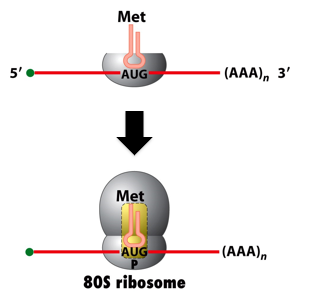
- 60S ribosomal subunit recruited = fully formed ribosome that can begin translation
-
- Need elongation factors
- eEF1a-GTP brings aa-tRNA and enters A site
- If tRNA anticodon doesnt match mRNA codon, complex dissociates
- If anticodon matches, eEF1a hydrolyses GTP (similar to eIF2)
- Ribosome judges match by assessing codon/anticodon geometry & ability to H-bond with rRNA
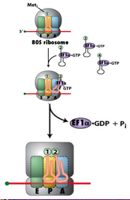
-
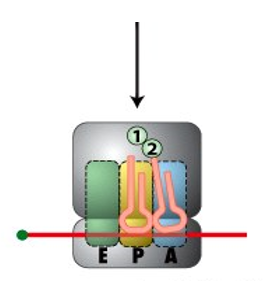
- GTP hydrolysis causes conformational change that allows peptide bond formation
- Peptide bond forming in peptidyl transferase centre between Met in P and aa in A site
-
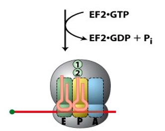
- Ribosome moves along mRNA driven by eEF2 mediated GTP hydrolysis
-
Rare Codons for Rare Amino Acids
- Rare codons are translated more slowly than common codons
- Reflects differences in abundance of cognate tRNAs- tRNAs that decode common codons are more abundant than tRNAs that decode rare codons
- Changes in relative levels of tRNAs may influence which mRNAs are translated most efficiently
- Cancers overexpress particular tRNAs- may promote expression of proteins required for tumorigenesis
-
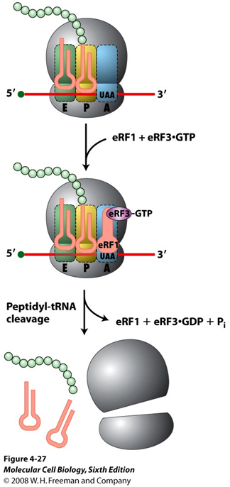
- Stop codons recognised by release factor eRF1 in A site
- eRF1 promotes hydrolysis (using water) of the bond linking pp to tRNA at P site- releases PP
- Hydrolysis catalysed by peptidyl transferase centre of ribosome
- Release facotr eRF3 stimulates process with GTP hydrolysis
-
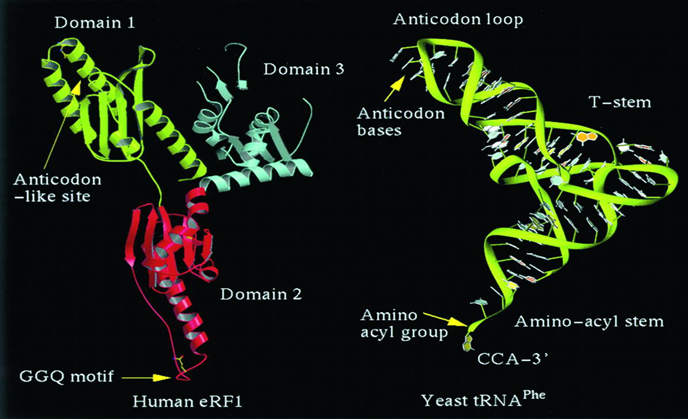
Termination Factor eRF1 Mimics a tRNA
- eRF1 brought into A site instead of charged tRNA
- Has similar size and shape to a tRNA
- Domain 1= recognises stop codon on mRNA (like anticodon loop recognises codon on mRNA)
- Tip of domain 2 promotes cleavage by ribosome peptidyl transferase
-
Regulating Rate Limiting Step: eIF2
- Initiation is rate limiting step, regulated through eIF2 and eIF4
- eIF2 has 3 subunits (alpha, beta, gamma) and GTPase activity
- eIF2 delivers met-tRNA to ribosome as ternary complex with GTP
- Once eIF2 joins with ribosome via 40S, cap binding protein eIF4 brings mRNA to form PIC and moves along mRNA to find AUG
- Once start codon recognised eIF2 hydrolyses GTP-> eIF2.GDP released
- Left with tRNA and 40S located at start codon
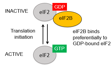
- For eIF2 to be active again= needs to replace GDP for GTP
-
Regulating Rate Limiting Step: eIF2
- Recycling of eIF2 is catalysed by Guanine nucleotide Exchange Factor (GEF) = eIF2B
- Regulation of eIF2 by eIF2B- changes GDP for GTP
- If eIF2B inhibited, eIF2 locked in GDP-inactive form
-
Regulating Rate Limiting Step: eIF2
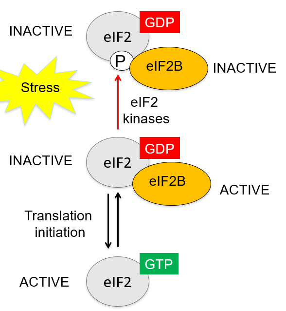
- Stress conditions prevents recycling of eIF2= suppresses translation
- Phosphorylation of eIF2 by eIF2 kinases sequesters eIF2B in inactive complex
- 4 different eIF2 kinases activated by different stress including:
- Hypoxia, viral infection, aa starvation, accumulation of unfolded proteins
- Chronic stress (long-term), kinases constantly activated and stop eIF2= less protein synthesis
-
Neurodegenerative Disease: eIF2-P
- Alzheimer's, Parkinson's and Prion diseases all involve accumulation of misfolded protein aggregates in neurons
- Neurons become chronically enable to continue protein synthesis in a normal way
- These diseases have elevated eIF2-P as part of protective mechanism to stop protein synthesis - part of Unfolded Protein Response
- Neurodegeneration of mice with Prion disease can be improved by overexpressing eIF2 phosphatase (remove phosphate of eIF2)
- In brains of affected mice, overexpressing phosphatases restored translation and synaptic function= increased survival
-
Mutation in eIF2B= Vanishing White Matter
- Vanishing white matter disease in humans is a recessive neurological disease caused by mutations in genes for eIF2B subunits
- Involves loss of cerebral white matter
- Because eIF2B mutations alter/inhibit function
- Degeneration often precipitated by physiological stress or trauma eg head bang
-
Regulating Rate Limiting Step: eIF4
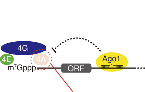
- miRNAs can inhibit translation by inactivating eIF4
- miRNAs bound to 3' UTR of mRNA can cause dissociation of eIF4A and thus inhibit translation
- miRNA at 3' end of mRNA affects the cap-binding complex at 5'
- eg. Ago1 dsiplaces eIF4A at 5'
-
Regulating Rate Limiting Step: eIF4
- How can something thousands bp away affect this? Circularisation
- Eukaryotic mRNA are circularised by Poly-A bidning protein (PABP) and eIF4G
- PABP at 3' end of mRNA can interact with eIF4G at 5' end to bring ends together
- Circularisation of mRNA facilitated miRNA (at 3') inhibiting eIF4 (at 5')
- Circularisation of mRNA also facilitates ribosome recycling:
- Once ribosome dissociates from mRNA at 3', it isnt too far from 5' to reassembly and do another round of translation- some genes need to be translated again and again
-
Regulating Rate Limiting Step: eIF4
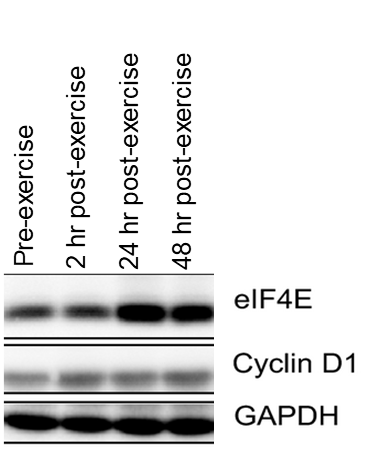
- The least abundant so most limiting translation initiation factor is eIF4E, so changes in eIF4E levels strongly influence protein translation
- Experiment: tissue biopsy of healthy individuals doing resistance training
- Resistance training raises eIF4E levels i skeletal muscle
- Levels of cyclin D1 are elevated when cells undergo division
- But during resistance training levels of cyclin D1 remain constant so exercising is not increasing number of cells but making them bigger= protein synthesis requireD
- GADPH= loading control
-
Regulating Rate Limiting Step: eIF4
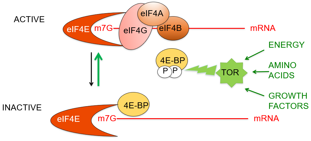
- Starvation inhibits Cap-binding complex
- Starvation- eIF4E bound by 4E-BP (binding protein), inhibits eIF4E binding eIF4G at 5' cap and stops translation
- TOR kinase controls formation of cap-binding complex by phsophorylating 4E-BP:
- Phosphorylated 4E-BP cannot bind eIF4E
- Cap-binding complex assembles and translation proceed
- TOR suppresses the suppressor of translation
- TOR kinase activity responds to growth factors eg insulin, aa, energy availability- Ensures translation is active under favourable conditions
-
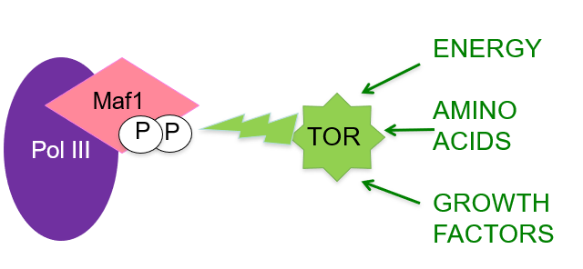
- TOR kinase controls tRNA synthesis:
- Another target of TOR is Maf1- repressor of RNA pol III
- Pol III responsible for transcription of tRNA and small rRNA
- By switching off Mar1, TOR increases tRNA synthesis when conditions favour protein synthesis and growth
-
- Rapamycin is specific inhibitor of TOR kinase and can extend lifespan
- As well as extending lifespan also prolongs health
- Through calorie restriction - eat less= less response to growth factors, less TOR, less protein synthesis
-
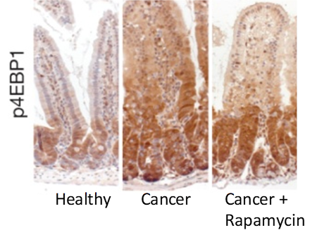
- TOR is hyperactive in cancer
- Stained colonic villi for phsophorylated 4E-BP (4E-BP phosphorylated by TOR)
- In healthy villi- lots of cell division at base to replenish cells, phosphorylated 4E-BP restricted to base to elevate translation
- In cancer- phosphorylated 4E-BP is everywhere-protein synthesis everywhere
- Cancer + rapamycin= P-4E-BP restricted to base of villi again
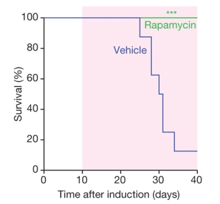
- Survival plot of mice with colon cancer, treated with vehicle or rapamycin
- Start with 100%= all alive at start
- Mice infected with vehicle (no rapamycin) = reduced survival
- Rapamycin= improves survival in mouse model for colon cancer
- TOR inhibutors used in cancer therapy- rapamycin derivatives= rapalogs, however in some cancers, rapalogs trigger other signalling pathways through feedback loops that bypass it
-
-
eIF4E Strongly Implicated in Cancer
- Elevated eIF4E levels in many cancer types:
- Correlates with decreased survival
- Overexpression of eIF4E can induce tumorigenesis in mice by elevating translation
- Phosphoresistant 4E-BP mutant can supress tumorigenesis by eIF4E
- Replace serine that gets phosphorylated for alanine
-
Elevated Protein Synthesis in Cancer
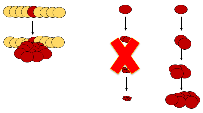
- Cancers express elevated levels of tRNA and rRNA- tRNA needed for protein synthesis
- Why protein synthesis is important for cancer
- Inappropriate growth needed as well as inappropriate cell division to make tumour
- Protein= 80-90% of cells dry mass- protein synthesis needed to grow cell size
-
Elevated Protein Synthesis in Cancer
- Experiment: Effect of overexpression of initiator tRNA on growth of fruitfly
- Growth of fruitfly larvae can be enhanced by overexpressing initiator tRNA (met-tRNA)
- Overexpression= no longer limiting factor
- Larvae grows bigger and faster
- Other tRNAs dont have this effect
- Suggests that translation initiation is rate limiting in protein synthesis (especially met-tRNA levels)
-
Elevated Protein Synthesis in Cancer
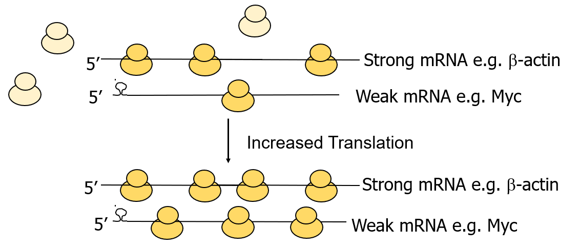
- Elevated protein synthesis has qualitative effect
- Weak mRNAs (eg Myc) aren't translated efficiently due to 5' secondary structure, prevents ribosome binding to form PIC
- When you upregulate translation, weak mRNAs are disproportionately advantaged= especially responsive to increased translation
- Upregulation of translation of strong mRNAs is enhanced a little but not significantly
- Oncogene proteins often encoded by weak mRNA- not usually expressed much but when something goes wrong are selected and favoured disproportionately
-
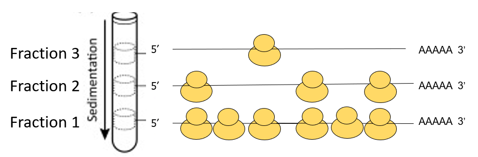
- Polysome Profiles: Measure how well mRNA translated
- Produced by centrifugation of cytoplasmic extracts through sucrose gradients
- Polysome complexes are bigger if a lot of ribosomes are bound to mRNA
- Smaller components at top and larger components sediment to bottom
- Use Northern blot or RT-PCR to determine which fractions contain a particular mRNA (strong or weak)
- Strong mRNAs that are well translated will have many ribosomes bound, sediment faster than mRNA with fewer ribosomes
-
Myc: Production of Translational Machinery
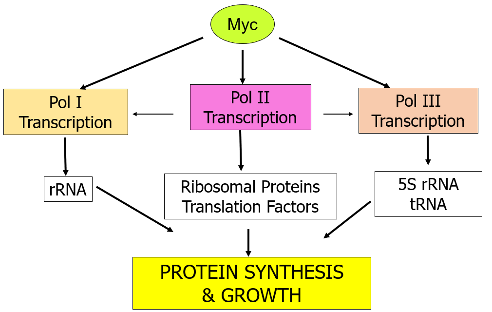
- Myc stimulates production of most components of protein synthetic machinery by directly stimulating transcription of Pol I, II, and III
- Different Pols all transcribe important components of translation
- Upregulated myc enhances transcription of all components of translation
-
Cancers Can Supress Translation: Starved
- Hypoxic (O deprived) regions of advanced breast cancers increase 4E-BP levels to suppress translation and conserve resources
- In large tumours, the cells in the middle are deprived of nutrients and O as they are far away from blood vessels
- Cancer cells resolve this by inhibiting cap-dependent translation and favour IRES-dependent translation
- Several pro-angiogenic factors are encoded by mRNAs with IRES sites that are insensitive to 4E-BP
- Eg mRNA for VEGF (vascular endothelial GF) which promoters blood vessel formation (angiogenesis) in hypoxic regions
-
Initiation Not Always Rate Limiting Step
- Elongation may be rate limiting for mRNAs with many rare codons= slow to find matching tRNA as they are less abundant
-
Information
- Description
- MBBB L15-16
- Created by
- Chloe Cavarretta · May 2019
- Updated
- May 20, 2019
- Views
- 4
- Rating