
Notes
7.3 - Translation
Content of the notes (4)
Page 1
tRNA activating enzymes
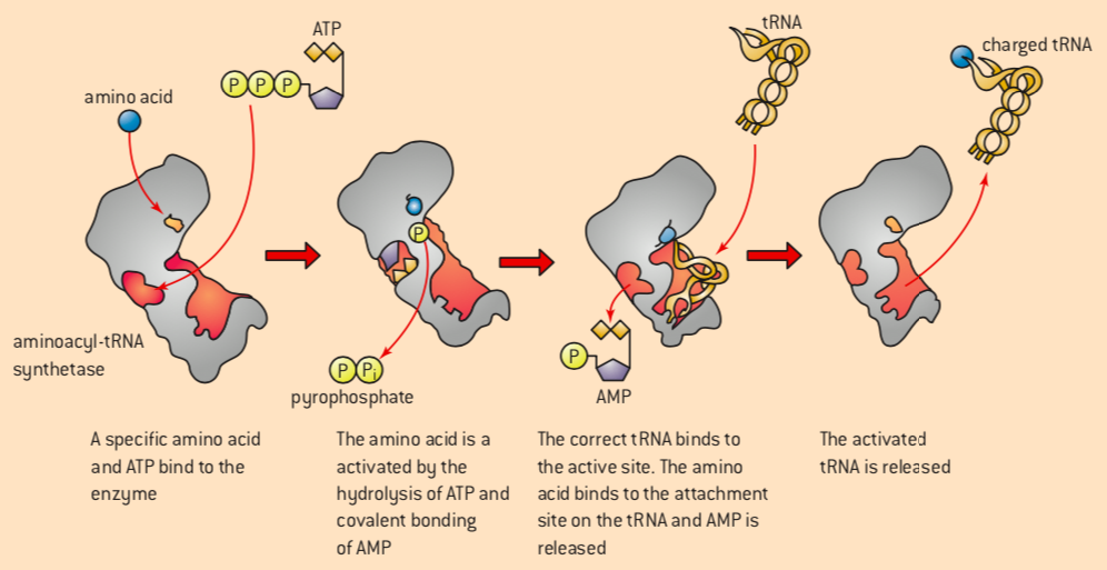
each tRNA molecule is recognized by a tRNA-activating enzyme that attaches a specific amino acid to the tRNA, using ATP for energy
activation involves attaching an amino acid to the 3' terminal to the tRNA
there are 20 d/t tRNA-activating enzymes for each of the 20 amino acids
energy from ATP is needed for the attachment of amino acids
once the amino acid is attached, the amino acid is activated by the formation of a bond between the enzyme and adenosine monophosphate (AMP)
the activated amino acid is then covalently attached to the tRNA
energy from this bond is later used to link the amino acid to the growing polypeptide chain during translation
Page 2
Initiation, Elongation, Termination
Initiation of translation
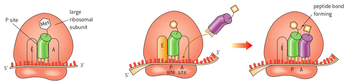
to start, the mRNA molecule binds to the small ribosomal subunit at an mRNA binding site
an initiator tRNA molecule carrying methionine binds at the start codon "AUG"
the large ribosomal subunit then binds to the small one
then,
the initiator tRNA is in the P site
the next codon signals another tRNA to bind
it occupies the A site
a peptide bond is formed between the amino acids in the P and A site
Elongation of translation
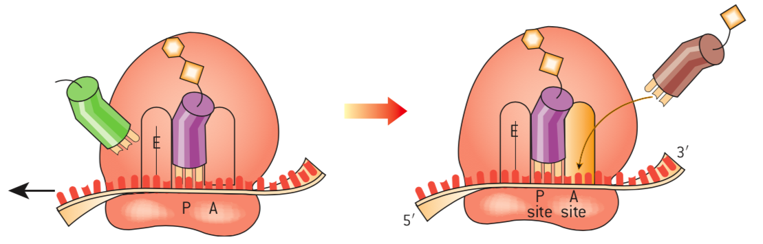
the ribosome moves the tRNA in the P site to the E site, freeing it
this allows a tRNA with the appropriate anticodon to bind to the next codon in the vacant A site
btw, peptide bonds are forming between the amino acids
there are only 2 tRNA molecules on the ribosome at a time, on the P and A
Termination of translation
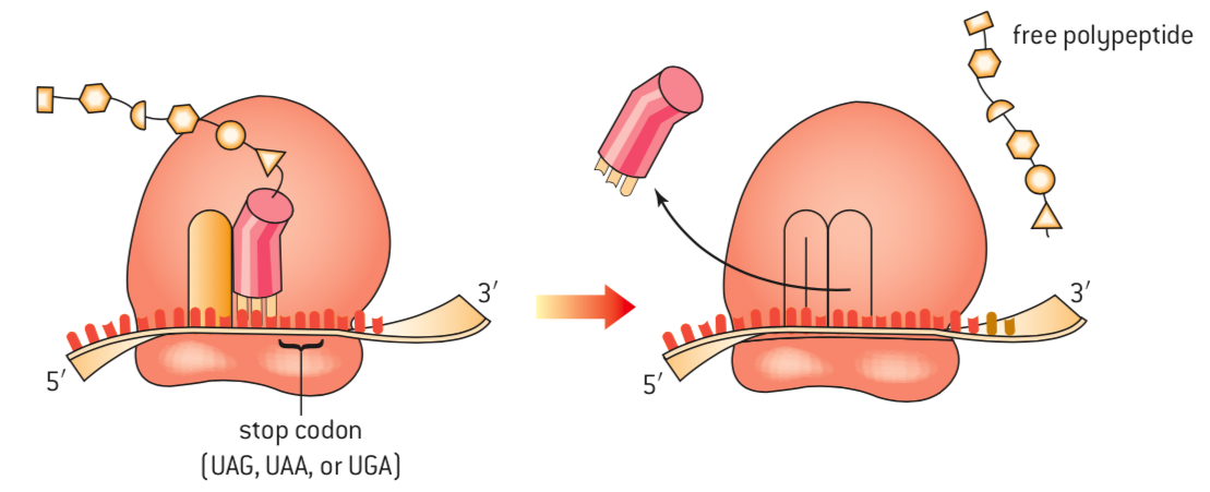
the process continues until a stop codon is reached
the free polypeptide is released
btw, the direction of movement along the mRNA is 5' end to the 3' end
Page 3
Where in the cell is the protein made?
this is determined by the final destination of the protein
it can either be synthesized in the cytoplasm or the endoplasmic reticulum
whether the ribosome free in the cytosol or bound to the ER:
depends on the presence of a signal sequence on the polypeptide being translated
as the signal sequence is created, it becomes bound to a signal recognition protein that stops the translation until it can bind to a receptor on the surface of the ER
once this happens, translation begins again with the polypeptide moving into the lumen of the ER as it is created
Free ribosomes
translation occurs more commonly in the cytosol
synthesized by free ribosomes are:
proteins destined to use in the cytoplasm, mitochondria, and chloroplasts
Bound ribosomes
synthesizes by ribosomes bound to the ER are:
proteins destined for use in the ER, the Golgi apparatus, lysosomes, the plasma membrane or outside the cell
The coupling of transcription and translation in prokaryotes
in eukaryotes:
once transcription is complete, the transcript is modified before leaving the nucleus
there is a delay between transcription and translation due to compartmentalization
in prokaryotes:
as soon as the mRNA is transcribed, translation begins
Polysomes
structures visible in an electron microscope
they represent multiple ribosomes attached to a single mRNA molecule
because transcription and translation occur in the same compartment in prokaryotes, multiple polysomes are visible associated with one gene
in eukaryotes, polysomes occur in both the cytoplasm and next to the ER
Page 4
Protein Structures
the biological activity of a protein is related to its primary, secondary, tertiary and quaternary structure
exposure to high temperatures or changes in pH can lead to alterations in the structure, disrupting its biological activity
this is also known as denaturation
(permanent loss of structure) - more about this in topic 2.5
Primary structure
the sequence of amino acids in a polypeptide is its primary structure
Secondary structure
defined by the pattern of hydrogen bonds between the amino hydrogen and carboxyl oxygen atoms in the peptide backbone
Tertiary structure
the overall three-dimensional shape of the protein
different types of interactions,
positively charged R-groups will interact with negatively charged R-groups
avoid contact with water, because of hydrophobic amino acids
polar R-groups will form H bonds with other polar R-groups
Quaternary structure
when polypeptides fit together when there's more than one chain
it also refers to the addition of non-polypeptide components
Information
- Description
- User has deleted their subject information Note on 7.3 - Translation, created by Deleted user on 20/02/2020.
- Created by
- Deleted user · Feb 2020
- Updated
- Feb 20, 2020
- Views
- 14
- History
-
- Created by Blen Abate · Feb 17, 2020
- Copied by Blen Abate · Feb 20, 2020
- Rating