
Quiz
Study Guide: Appendicular Muscles
This quiz has question types that open in the usual player.
Questions in this quiz (31)
1All of the following are rotator cuff muscles except
- Adeltoid
- Binfraspinatus
- Csupraspinatus
- Dteres minor
2The muscle that shrugs the shoulders is the
- Asternocleidomastoid
- Bdeltoid
- Ctrapezius
- Dpectoralis minor
3The posterior arm muscle that extends the forearm is the
- Atriceps brachii
- Btriceps surae
- Cbrachialis
- Dbiceps brachii
4The olecranon of the ulna is a site of insertion for the
- Abiceps brachii
- Bbrachialis
- Cbrachioradialis
- Dtriceps brachii
5Intrinsic muscles of the hand originate on the forearm and insert on the metacarpals.
- ATrue
- BFalse
6Carpal tunnel syndrome affects the median nerve.
- ATrue
- BFalse
7The deltoid is a good example of a multifunctional muscle.
- ATrue
- BFalse
8The pectoralis major flexes the upper arm
- ATrue
- BFalse
9The biceps brachii is an extensor muscle.
- ATrue
- BFalse
10Label the following illustrations, using the terms provided: Muscles acting on the shoulder girdle
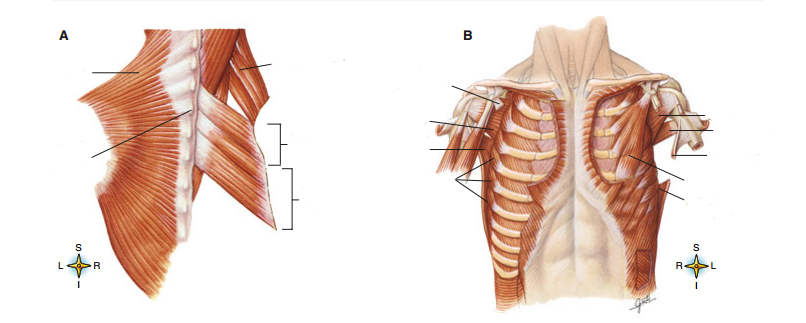
- ATrapezius
- BSeventh vertical vertibra
- CPectorals minor (cut)
- DSubscapularis (anatomical right)
- ELatissimus dorsi (anatomical right)
- FSerratus anterior
- GLatissimus dorsi (anatomical left)
- HPectoralis minor
- ITeres major
- JTeres minor
- KSubscapularis (anatomical left)
- LLevator scapulae
- MRhomboid minor
- NRhomboid major
11Label the following illustrations, using the terms provided: Rotator cuff muscles
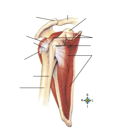
- AClavicle
- BAcromion process
- CCoracoid process
- DInfraspinatus
- EGreater tubercle
- FTeres minor
- GIntertubercula (bicipital) groove
- HHumerus
- ISubscapularis
- JLesser tubercle
- KSupra spinatus
12Label the following illustrations: Muscles that move the upper arm
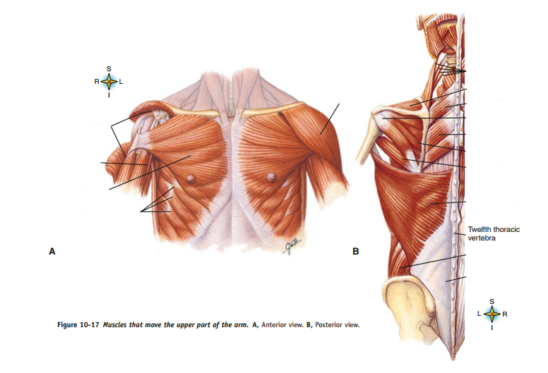
- ADeltoid (cut)
- BCoracobrachiallis
- CPectoralis major
- DSerratus anterior
- EDeltoid
- FLevator scapulae
- GSupraspinatus
- HRhomboideus minor
- ITeres minor
- JRhomboideus major
- KInfraspinatus
- LTeres major
- MLatissimus dorsi
- NExternal abdominal oblique
- OThoracolumbular fascia
13Label the following illustrations: Muscles of the upper arm
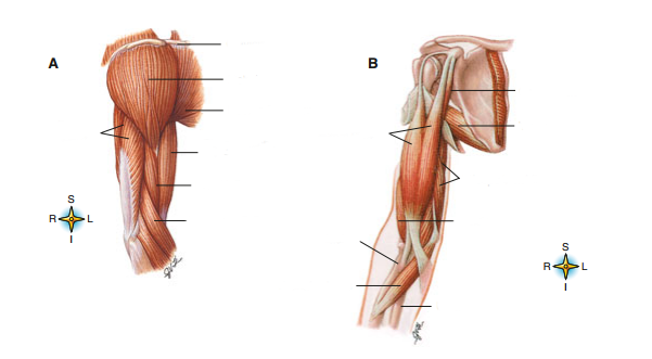
- AClavicle
- BDeltoid
- CPectoralis major
- DTriceps brachii (side view)
- EBiceps brachii (long head)
- FBrachialis
- GBrachioradialis
- HCoracobrachiallis
- ITeres major
- JBiceps brachii
- KTriceps brachii (anterior view)
- LBrachiallis
- MRadius
- NUlna
- OPronator teres
14Label the following illustrations: Muscles that act on the forearm
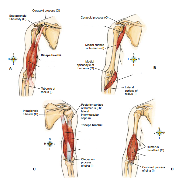
- Ashort head
- Blong head
- Ctriceps - long head
- Dlateral (short head)
- Emedial head
- FCoracobrachialis
- GPronator teres
- HBrachialis
15Label the following illustrations: Muscles of the forearm
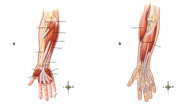
- AMedial epicondyle of humerus
- BPronator ters
- CFlexor carpi radialis
- DPalmaris longus
- EFlexor carpi ulnaris
- FRadius
- GUlna
- HFlexor pollicis brevis (superficial)
- IOpponens pollicis (deep)
- JAbductor digiti minimi
- KFlexor digiti minimi
- LBrachoradialis
- MFlexor digitorum superficialis
16The muscles of the quadriceps femoris include all of the following except:
- Avastus intermedius
- Bvastus medialis
- Cvastus lateralis
- Dvastus femoris
17The anterior superior iliac spine is the site of origin for the
- Asartorius
- Brectus femoris
- Cgracilis
- Diliacus
18A common site for intramuscular injections is the
- Agluteus maximus
- Bgluteus minimus
- Cgluteus medius
- Dtensor fasciae latae
19Plantar flexion of the foot is achieved by the
- Atibialis anterior
- Btibialis posterior
- Cperoneus brevis
- Dsoleus
20The muscles of the hamstrings include all of the following except the
- Ailiopsoas
- Bsemitendinosus
- Csemimembranosus
- Dbiceps femoris
21The Achilles tendon is common to both the gastrocnemius and soleus
- ATrue
- BFalse
22The iliopsoas is composed solely of the psoas major and the iliacus.
- ATrue
- BFalse
23The vastus intermedius originates on the posterior surface of the femur.
- ATrue
- BFalse
24flexor: [blank_start]gastrocnemius[blank_end], [blank_start]biceps brachii[blank_end], [blank_start]iliopsoas[blank_end], [blank_start]pectoralis major[blank_end], [blank_start]gracilis[blank_end]
extensor: [blank_start]trapezius[blank_end], [blank_start]gluteus maximus[blank_end], [blank_start]triceps brachii[blank_end], [blank_start]fibularis longus[blank_end]
abductor: [blank_start]gluteus medius[blank_end], [blank_start]deltoid[blank_end]
adductor:
rotator: [blank_start]rhomboid major[blank_end]
dorsiflexor or plantar flexor: [blank_start]soleus[blank_end]- Abiceps brachii
- Bgastrocnemius
- Cgracilis
- Diliopsoas
- Epectoralis major
- Ffibularis longus
- Ggluteus maximus
- Htrapezius
- Itriceps brachii
- Jdeltoid
- Kgluteus medius
- Lrhomboid major
- Msoleus
25Muscles that are responsible for such movement as dorsiflexion, plantar flexion, inversion, and eversion of the foot are the [blank_start]extrinsic[blank_end] [blank_start]foot[blank_end] [blank_start]muscles[blank_end].
- Aextrinsic
- Bfoot
- Cmuscles
26Another common name for the calcaneal tendon is the [blank_start]achelles[blank_end] tendon.
- Aachilles
27The rotator cuff muscles are: [blank_start]infraspinatus[blank_end], [blank_start]subscapularis[blank_end], [blank_start]supraspinatus[blank_end], [blank_start]teres minor[blank_end]
- Ainfraspinatus
- Bsubscapularis
- Csupraspinatus
- Dteres minor
28If less than 2mL of medication is to be injected into the muscle, the common muscle that is preferred as the site is the [blank_start]deltoid[blank_end] muscle.
- Adeltoid
29Label the following illustrations: Muscles of the forearm
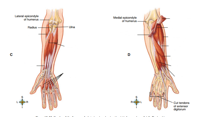
- ASupinator (anatomical)
- BFlexor digitorum profundus
- CPronator quadratus
- DPalmar interosseus
- Eextensor digitorum (cut and reflected)
- FSupinator (deep)
- GExtensor carpi ulnaris (cut)
- HExtensor carpi radialis longus
- IExtensor carpi radialis brevis
- JAbductor pollicis longus
- KExtensor pollicis longus
- LExtensor pollicis brevis
30Label the following illustrations: Muscles of the thigh
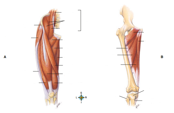
- AIliopsas
- BPsoas minor
- CIliacus
- DPsoas major
- EAnterior superior iliac spine
- FTensor fasciae latae
- GPectineus
- HAdductor longus
- IGracilis
- JSartorius
- KRectus femoris
- LVastus medialis
- MIliotibial tract
- NVastus lateralis
- OPectineus (full view)
- PGracilis (full view)
- QAdductor brevis
- RAdductor longus (full view)
- SFibula
- TTibia
- UInsertion of gracilis in tibia
- VAdductor magnus
31Label the following illustrations: Muscles of the leg
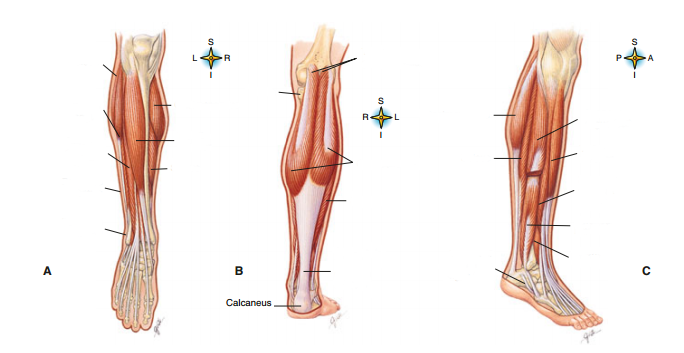
- ASoleus (anterior view, lateral)
- BPeroneus longus
- CExtensor digitorum longus
- DPeroneus brevis
- EPeroneus tertius
- FTibia
- GTwo heads of gastrocnemius
- HGastrocnemius (posterior view)
- ISoleus (posterior)
- JCalcaneal tendon (Achilles tendon)
- KGastrocnemius (side view)
- LSoleus (side view)
- MGastrocnemius (anterior view)
- NTibialis anterior
- OSoleus (anterior view, medial)
- PPeroneus longus (cut)
- QTibialis anterior (side view)
- RExtensor digitorum longus (side view)
- SPeroneus brevis (side view)
- TPeroneus tertius (side view)
- UTendon of peroneus tendon (cut)
Information
- Description
- Study Guide: Appendicular Muscles
- Author
- Alexandra Bozan
- Language
- English
- Views
- 766
- History
- Created on 28 Apr 2017 · updated on 1 May 2017
- Tags
- Rating
- No ratings yet Rate this quiz