7. Apoptosis

Descripción

Cell Biology L15
Chloe Cavarretta
Creado por Chloe Cavarretta
Chloe Cavarretta
Creado por Chloe Cavarretta hace más de 7 años
5
0

Resumen del Recurso

Diapositiva 1

    Learning Outcomes: L15
    Describe the importance of the Bcl-2 family of proteins and the role of mitochondria in the apoptotic pathway Explain the function and activation mechanisms of the caspases, the apoptosis executioners Demonstrate an understanding of the role of trophic factors in cell survival, and how withdrawal of trophic factors induces apoptosis Explain how death receptors, such as Fas, induce apoptosis Give examples of the role of apoptosis in health and disease

Diapositiva 2

Diapositiva 3

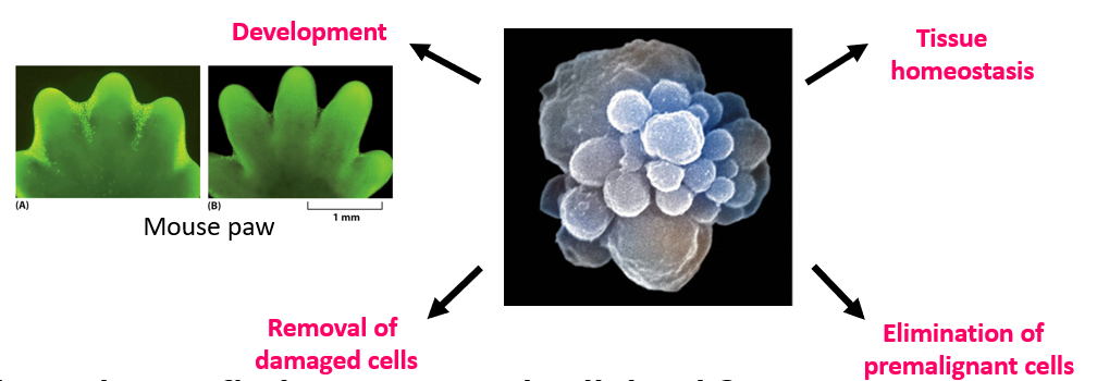
    Where do you find programmed cell death
    Cytotoxic T cells cause killing, NK cells - regulated apoptosis P53 pathway activation (elimination of pre-malignant cells) Tissue homeostasis: small bowel crypts, skin keratinocytes (between digits on hands/feet), thymus (non responsive and self reactive T cells) Outer lens of eye  During development- in particular metamorphosis

Diapositiva 4

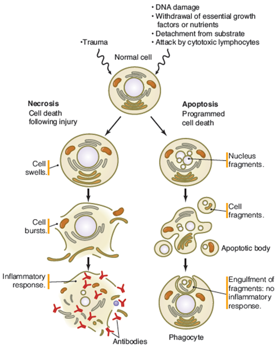
    Apoptosis is Programmed Cell Death
    Necrosis Cell death following injury and results in inflammatory response Cell undergoes injury, contents leak out Apoptosis Controlled non-inflammatory event  No injury, no content leakage Active signal- cell fragmentation and form apoptotic bodies, engulfed by phagosome and taken to lysosome

Diapositiva 5

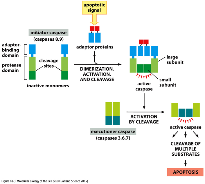
    Intracellular Proteolytic Caspase Cascade
    Initiator caspases (8, 9) dimerize and become active Initiator caspases cleave executioner caspase (3,6,7) subunits-> activate executioners Executioners cleave cellular substrates and cell undergoes apoptosis Once executioners active, only way to stop apoptosis is to digest caspase, happens at end of apoptosis Caspases: Activators/initiators (8,9,19,2) Executioners/effectors (3,6,7) Inflammatory (unrelated to apoptosis) Activated by dimerisation (initatiors) or proteolysis (effectors)- executioners have small and large subunits that need to be cleaved One can activate another (positive feedback)  

Diapositiva 6

Diapositiva 7

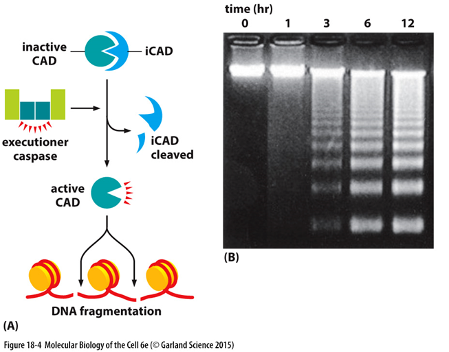
    DNA Fragmentation
    CAD= Caspase Activated DNase In healthy cells- endonuclease CAD is bound to inhibitor - iCAD Apoptosis- Induces iCAD cleavage by executioner caspase, release active DNase which cleaves DNA between nucleosomes  

Diapositiva 8

    Caspase Cascade Amplifies Signal
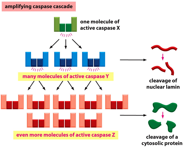
    One caspase activate many other caspases, activate many others and cleave multiple susbtrates Capases cleave nuclear proteins (lamins) and cytoskeletal proteins (actin) Caspases cleave and activate gelsolin which induces actin severing  Result= nuclear fragmentation, disruption of cytoskeleton, membrane blebbing, cell fragmentation  

Diapositiva 9

    Apoptosis Induced in 2 Ways
    Extrinsic: Cell receives explicit signal from extracellular environment to initiate apoptosis- eg death ligand expressed on another cell  Intrinsic: Normally involves release of cytochrome c- induced by stress, damage, developmental cues

Diapositiva 10

    Extrinsic Induced by Death Receptors
    Death receptors= cell surface receptors that initiate apoptosis following ligand binding Receptors are part of TNF family: ​​​​​​​Fas (CD95/APO-1) TNF-R1 (tumour necrosis factor receptor 1) Ligands: ​​​​​​​FasL(CD95L) TNF-a, TRAIL (TNF-related apoptosis inducing factor)

Diapositiva 11

    Extrinsic Induced by Death Receptors
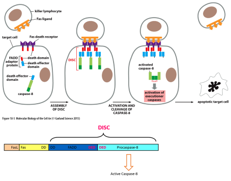
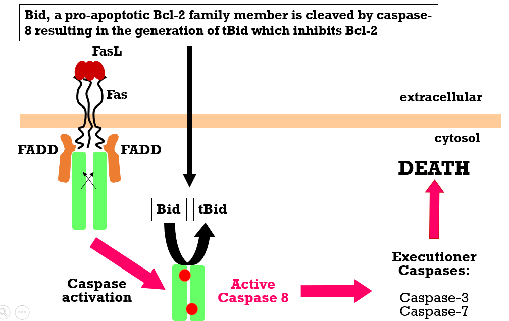
    Death receptors interact with adaptor proteins through mutual death domains (DDs)- Fas and TNRF1 have extracellular DDs FasL binds Fas death receptor  Fas interacts with an adaptor protein FADD (via Fas-associated DD) FADD complexes with procaspase-8 via DED domain (Death Effector Domain)-> activated caspase 8 that can cleave executioner  Forms Death Inducing Signal Complex (DISC)

Diapositiva 12

Diapositiva 13

    Fas in Influenza Defence
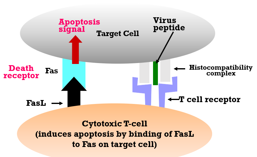
    Infected cell presents a virus peptide on its cell surface with MHC-I MHC-I:peptide interacts with TCR on cytotoxic T cell T cell has Fas ligand that binds Fas receptor on target cell-> sends apoptotic signal

Diapositiva 14

    HIV Infection Results in Loss of CD4 T cells
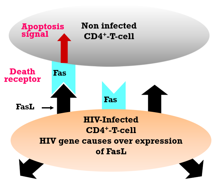
    CD4 T cells express Fas, but HIV-infected cells also express high levels of FasL Induces non-infected CD4 T cell to commit suicide Results in failure of immune response Infected HIV cell survives as high FasL prevents interaction with own Fas

Diapositiva 15

    Intrinsic Apoptosis
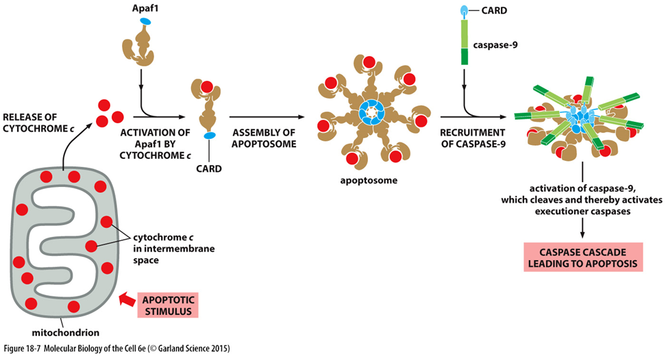
    1. Apoptotic stimulus causes cytochrome c release from mitochondria  2. Cytochrome c binds Apaf1 (Apoptotic protease activating factor) 3. CARD domain of Apaf1 associates with other CARD domains and forms apoptosome (CAspase Recruitment Domains) 4. Caspase 9 (initiator) recruited via its CARD domain and is activated-> cleaves and activates executioner

Diapositiva 16

Diapositiva 17

    Bcl-2 Proteins Regulate Intrinsic Pathway
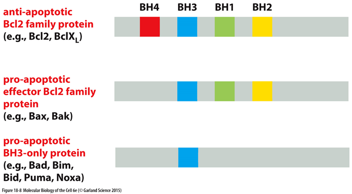
    Bcl-2 family contains both pro- and anti- apoptotic members Bcl-2 is anti-apoptotic Healthy cells maintain balance of pro and anti apoptotic proteins  Relative expression of anti/pro can promote or repress apoptosis All proteins regulated by TFs BH= Bcl-2 homology domain BH3- only proteases that promote apoptosis by inhibiting anti-apoptotic Bcl-2 family proteins

Diapositiva 18

Diapositiva 19

    Bcl-2 Proteins Regulate Intrinsic Pathway
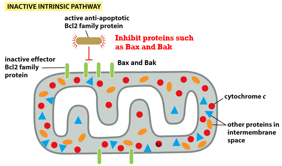
    Pro-apoptotic proteins (Bax/Bak) in mitochondrial membrane Bcl-2 in membrane in dimer with Bcl-xL (antiapoptotic complex) can inhibit Bax and Bak BH3 only BH domain protein that can bind and inhibit Bcl-2 and initiate apoptosis  Activates Bax in membrane and allows release of cytochrome c

Diapositiva 20

Diapositiva 21

    Trophic Factors Inhibit Intrinsic Apoptosis
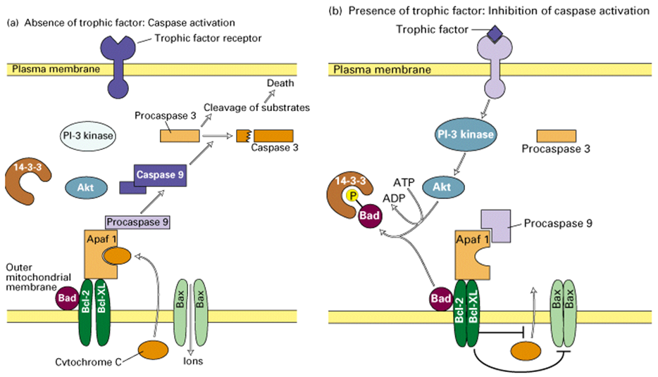
    Trophic factors- inducing differentiation/survival eg NGF (nerve) and EGF (epidermal) Absence of trophic factor:  No signalling via PI3 kinase/Akt pathway Bad (BH3) binds to Bcl2(anti)-> activates Bak(pro), release cytochrome c​​​​​​ Presence and binding to trophic factor: ​​​​​​​Receptor activated, stimulate PI-3 kinase phosphorylates and activate Akt(PKB)-> phsophorylates Bad Phospho-Bad forms complex with 14-3-3 protein (sequesters Bad from mitochondria membrane) Prevents Bad(BH3)from interacting with Bcl-2(anti), no activation of Bax(pro), no cyt c release Survival

Diapositiva 22

Diapositiva 23

    Summary of Bcl-2 Protein
    Bak/Bax= trigger apoptosis (pro-apoptotic) Activator Bim (BH3- pro apoptotic by inhibiting anti apoptotic)activates Bak/Bax Bcl-xL (anti apoptotic) inhibits Bim activator Bad (BH3) de-represses Bim (activated Bim again) by removing Bcl-xL De-repressor depresses the anti-apoptotic, promoting the pro-apoptotic

Diapositiva 24

    Extrinsic Pathway Can Activate Intrinsic
     Caspase 8 (activated in extrinsic pathway) cleaves Bid (pro-apoptotic Bcl2 protein)-> tBid (activated) tBid inhibits Bcl-2 (anti) in mitochondrial membrane Activation of Bax/Bak (pro) and release of cytochrome c  Caspase 9 activation via apoptosome formation Extrinsic= caspase 8 Intrinsic= caspase 9  
Mostrar resumen completo Ocultar resumen completo

Similar

Function and Structure of DNA
Elena Cade
Cells And Cell Techniques - Flashcards (AQA AS-Level Biology)
Henry Kitchen
Cells - Biology AQA B2.1.1
benadyl10
Biology AQA 3.1.3 Cells
evie.daines
GCSE AQA Biology - Unit 2
James Jolliffe
Cells and the Immune System
Eleanor H
Biology Unit 2 - DNA, meiosis, mitosis, cell cycle
DauntlessAlpha
Cell Transport
Elena Cade
Mitosis
Selam H
Unit 1 flashcards
C R
Cells, Tissues and Organs
yusanr98