Fichas
Intraoral Radiography - Anatomy
Fichas del conjunto (24)
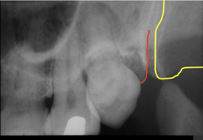
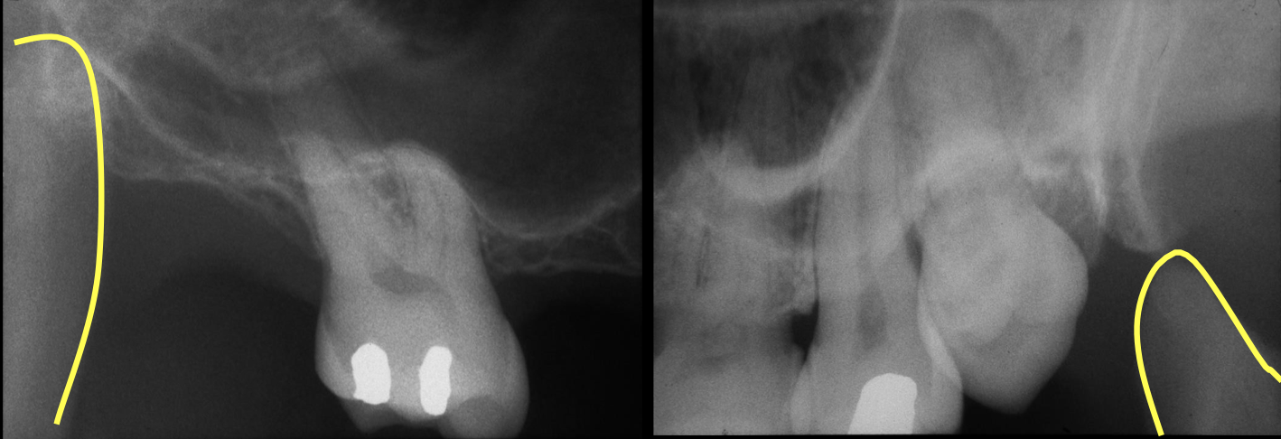
- 1—red - maxillary tuberosity
yellow - pterygoid plate - 2—zygomatic process of maxillary bone = u-shaped radiopacity
zygomatic arch behind it (star)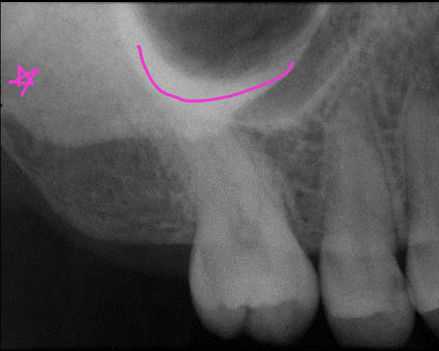
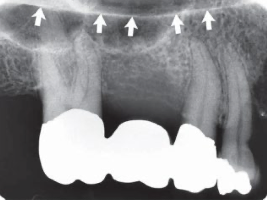
- 3—maxillary sinus - pneumatized bone = oroantral communication
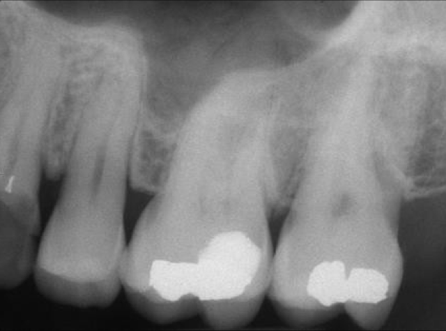
- 4—maxillary sinus - septum
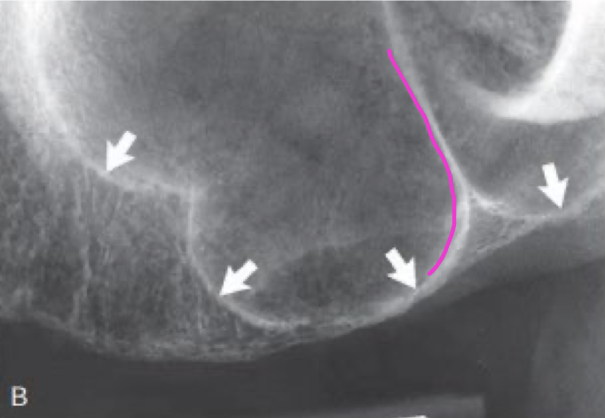
- 5—neurovascular canal (superior alveolar VAN thru the maxillary sinus) = radiolucent line
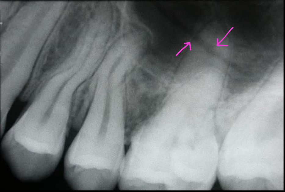
- 6—floor of the nasal cavity = smooth and straight
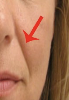
- 7—nasolabial fold
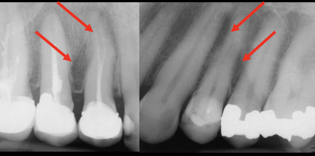
- 8—Y-line: top = nasal floor, vertical = maxillary sinus floor

- 9—soft tissue of nose
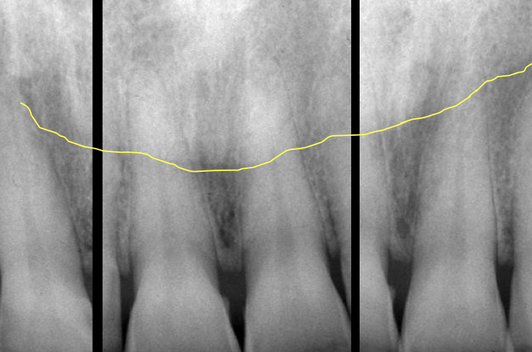
- 10—mylohyoid line = internal oblique line
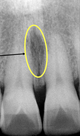
- 11—incisive foramen = radiolucent hole approx 5 mm
- 12—anterior nasal spine = radiopaque structure at midline between central incisors, getting more radiopaque up to the alveolar crest
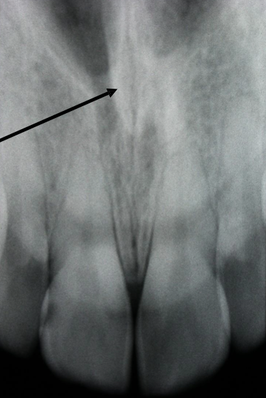
- 13—coronoid process - closer to max post teeth, while condylar head at the TMJ near ear
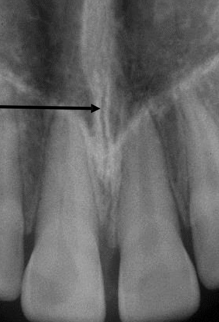
- 14—intermaxillary suture - between palatal plates
- 15—external oblique ridge (of the md)
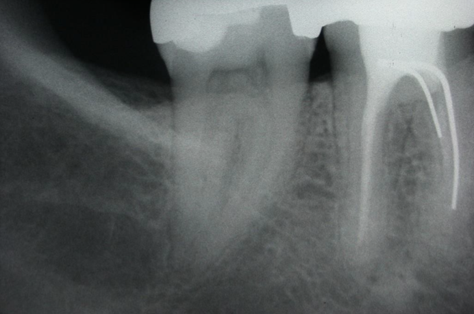
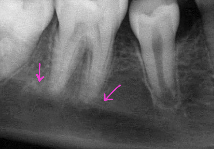
- 16—pink - superior cortex of alveolar canal
green - inferior cortex of alveolar canal
running thru = md canal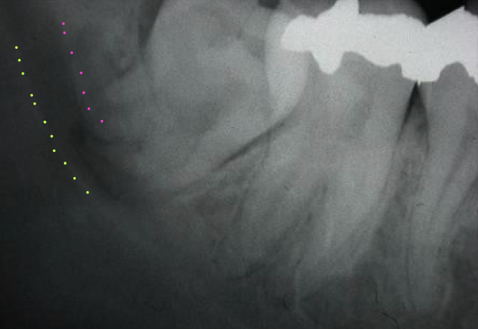
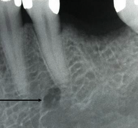
- 17—mental foramen
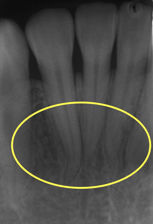
- 18—mental fossa - radiolucent area
- 19—mental ridge
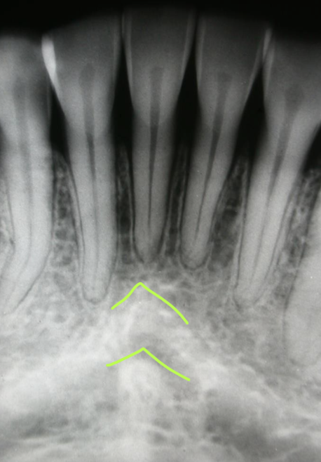
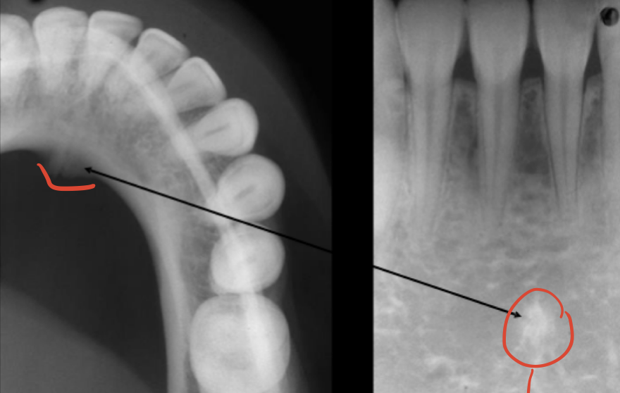
- 20—genial tubercle
- 21—lamina dura - radiopaque line covers whole tooth, continuous with alveolar bone
PDL space - radiolucent line between tooth and lamina dura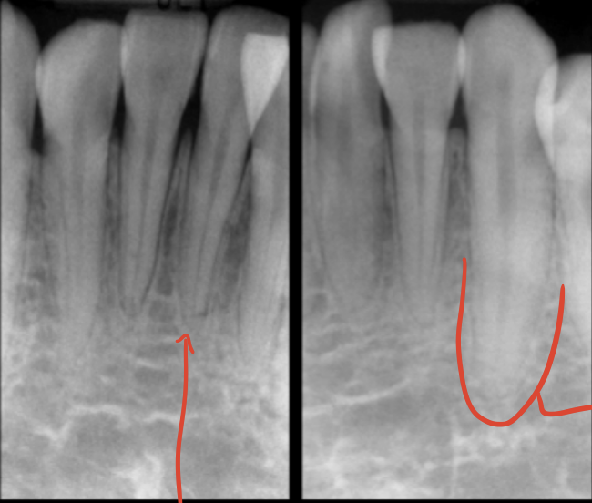
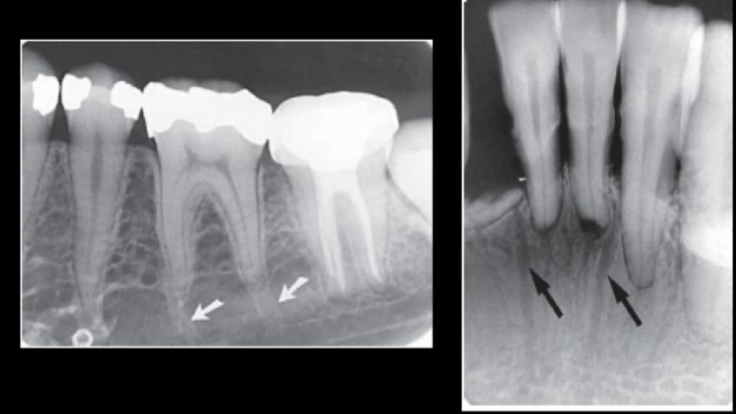
- 22—nutrient canals
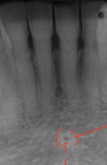
- 23—lingual foramen - between the genial tubercles
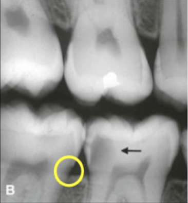
- 24—cary - the enamel shape should b uniform