6873535
Descripción
Fichas por Shauna Ryner, actualizado hace más de 1 año
|
|
Creado por Shauna Ryner
hace más de 7 años
|
|
| Pregunta | Respuesta |
| What side of heart and what chamber fills the apex of the heart? | Left Ventricle |
| Which side of the heart is more muscular and why? | Left. Has to pump blood to the whole body, whereas the right side only pumps blood to the lungs |
| What vessels supply blood to the heart itself? | Coronary vessels - the coronary artery and the coronary vein |
| What are auricles? | No known function, possibly increase surface area. They are at the base of the heart, form the visible portion of the atria. Looks like a floppy ear/cauliflower ear |
| What is the most ventral vessel on the front of the heart? | Pulmonary Artery |
| What is directly behind/caudal to the pulmonary artery? | Aorta |
| Vena cava dumps into: | Right Atrium |
| Contraction of the heart muscle | Systole |
| Relaxation of the heart muscle | Diastole |
| Pericardium | Protective sac-like covering of the heart |
| Pacemaker of the heart | SA Node |
| What blood vessels carry blood from the heart to the lungs? | Pulmonary Artery |
| Chordae Tendineae | Fine, thread-like cords that connect the free edge of the atrioventricular valves to the papillary muscles in the ventricles |
| Semilunar Valve located between the left ventricle and the aorta | Aortic Valve |
| Endocardium | Innermost layer of tissue that lines the chambers of the heart |
| What blood vessels carry blood from the lungs to the heart? | Pulmonary Vein |
| What is the valve between the right atrium and right ventricle of the heart? | Tricuspid Valve |
| Thick muscle layer of the heart wall | Myocardium |
| Mediastinum | The space in the thorax between the lungs that contains the trachea, esophagus, heart, and major blood vessels |
| Renal Artery | Blood vessels that carry blood from the aorta to the kidneys |
| Renal Vein | Blood vessels that carry blood from the kidneys to the caudal vena cava |
| Apex of the heart | When the heart is held in its anatomically correct position, the apex is at the bottom of the heart, and is the point of the heart |
| Base of heart | Wide area of heart where the major blood vessels enter and leave (when in the anatomically correct position, this is the top of the heart) |
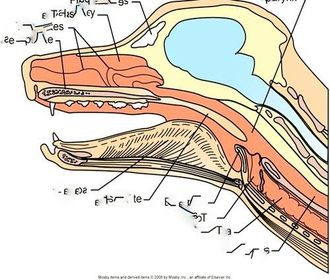
| Epiglottis | Covers larynx during swallowing, also covers trachea, preventing food from entering the trachea and lungs |
| Locations for blood collection | Jugular - neck Cephalic - cranial aspect of front limb Saphenous vein (dog) - lateral aspect of pelvic limbs Femoral Vein (cat) - Medial aspect of pelvic limbs Coccygeal (cow) - base of tail |
| Separates the right and left pumping ventricles from each other | Septum |
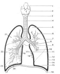
| 1) Epiglottis 2) Larynx 4) Trachea Main braches of trachea: bronchus/bronchi Branches off of bronchi: bronchioles Sacs at end of bronchioles: Alveoli | |
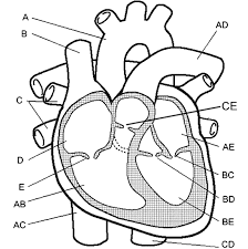
| Right side (A-AC) | A) Aorta B) Cranial/superior Vena Cava C) Pulmonary Veins D) Right Atrium E) Tricuspid Valve AB) Right Ventricle AC) Caudal/Inferior Vena Cava |
| Left side (AD-CD) | AD) Pulmonary Atery CE) Pulmonary Valve AE) Left Atrium BC) Mitral/bicuspid Valve BD) Aortic Valve BE) Left Ventricle CD) Descending Aorta |
| Start at bottom right, move clockwise: Esophagus Trachea Larynx Epiglottis Soft Palate Tongue Hard Palate Nasal Turbinates Nasal Cavity Frontal Sinus Pharynx | |
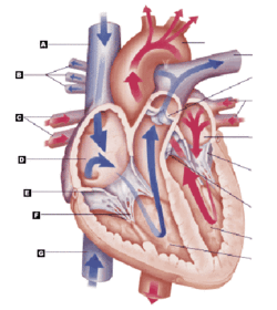
| Identify the structure at letter 'F' | Chordae Tendineae |
| These two blood vessels do not contain valves to control blood flow when entering the heart | Inferior (caudal) and Superior (cranial) Vena Cava |
| This slender gap between the parietal and visceral surfaces is filled with fluid to reduce friction between layers as the heart pumps | Pericardial Cavity |
| Where is the majority of the myocardium located? | The lower two chambers of the heart (ventricles) |
| What are the long fibres of connective tissue that attach the tricuspid valve to the papillary muscles of the heart? | Chordae Tendineae |
| Explain the process that leads to the closing of the tricuspid valve | When the heart begins to contract, ventricular pressure increases until it is greater than the pressure in the atrium |
| How many leaflets does the tricuspid valve have? | 3 |
| How many leaflets does the mitral valve have? | 2 |
| Where is the coronary sinus located? | This is a hole in the wall of the aorta that leads into the coronary artery |
| How many chambers does a mammalian heart have? | 4 chambers |
¿Quieres crear tus propias Fichas gratiscon GoConqr? Más información.