
Mapa mental
Heart
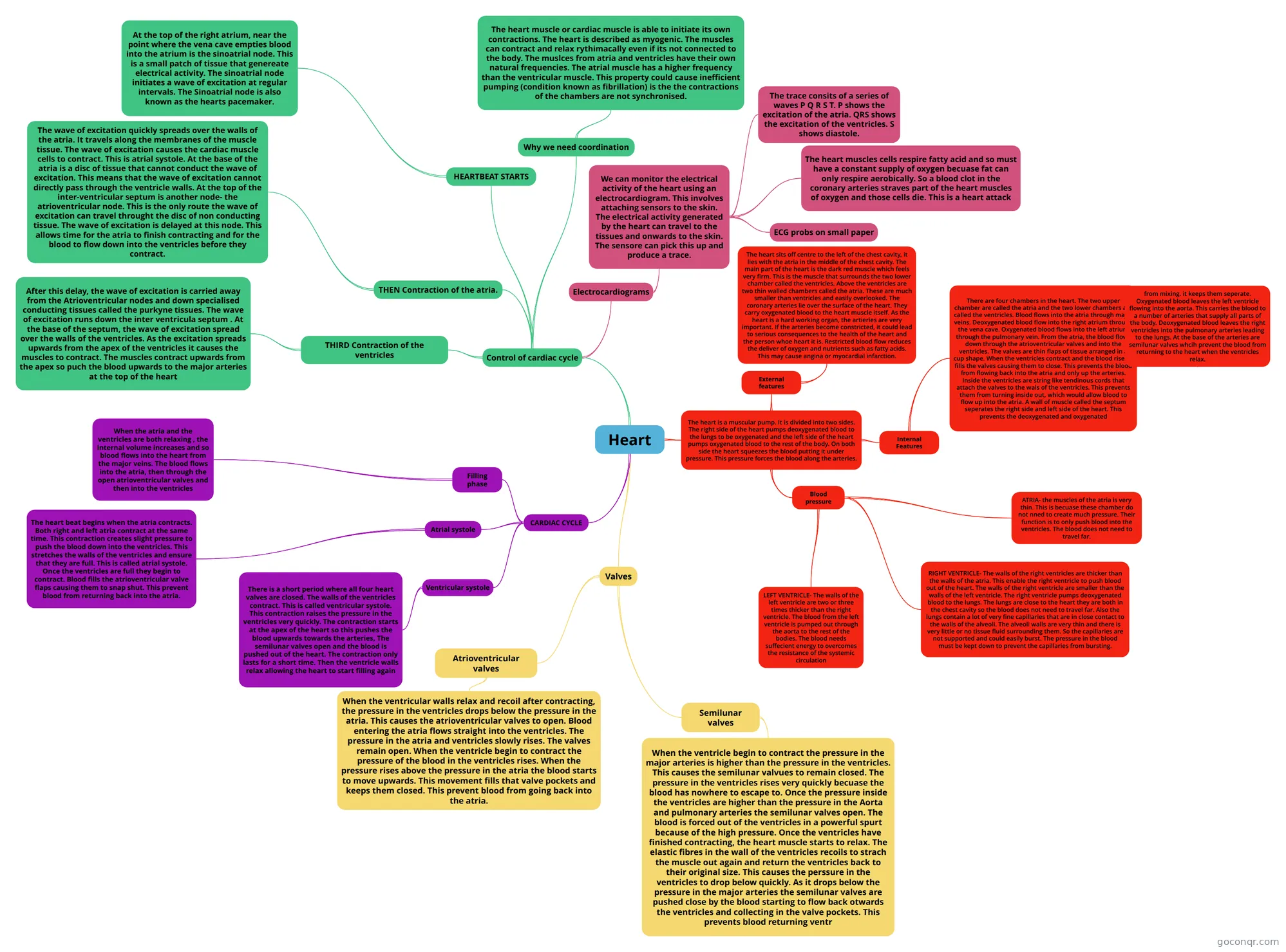
Mind map: the heart and cardiac cycle
Contenido del mapa
Heart
The heart is a muscular pump. It is divided into two sides. The right side of the heart pumps deoxygenated blood to the lungs to be oxygenated and the left side of the heart pumps oxygenated blood to the rest of the body. On both side the heart squeezes the blood putting it under pressure. This pressure forces the blood along the arteries.
- External features
- The heart sits off centre to the left of the chest cavity, it lies with the atria in the middle of the chest cavity. The main part of the heart is the dark red muscle which feels very firm. This is the muscle that surrounds the two lower chamber called the ventricles. Above the ventricles are two thin walled chambers called the atria. These are much smaller than ventricles and easily overlooked. The coronary arteries lie over the surface of the heart. They carry oxygenated blood to the heart muscle itself. As the heart is a hard working organ, the artieries are very important. If the arteries become constricted, it could lead to serious consequences to the health of the heart and the person whoe heart it is. Restricted blood flow reduces the deliver of oxygen and nutrients such as fatty acids. This may cause angina or myocardial infarction.
- Internal Features
- There are four chambers in the heart. The two upper chamber are called the atria and the two lower chambers are called the ventricles. Blood flows into the atria through major veins. Deoxygenated blood flow into the right atrium through the vena cave. Oxygenated blood flows into the left atrium through the pulmonary vein. From the atria, the blood flows down through the atrioventricular valves and into the ventricles. The valves are thin flaps of tissue arranged in a cup shape. When the ventricles contract and the blood rises it fills the valves causing them to close. This prevents the blood from flowing back into the atria and only up the arteries. Inside the ventricles are string like tendinous cords that attach the valves to the wals of the ventricles. This prevents them from turning inside out, which would allow blood to flow up into the atria. A wall of muscle called the septum seperates the right side and left side of the heart. This prevents the deoxygenated and oxygenated
- from mixing. it keeps them seperate. Oxygenated blood leaves the left ventricle flowing into the aorta. This carries the blood to a number of arteries that supply all parts of the body. Deoxygenated blood leaves the right ventricles into the pulmonary arteries leading to the lungs. At the base of the arteries are semilunar valves whcih prevent the blood from returning to the heart when the ventricles relax.
- There are four chambers in the heart. The two upper chamber are called the atria and the two lower chambers are called the ventricles. Blood flows into the atria through major veins. Deoxygenated blood flow into the right atrium through the vena cave. Oxygenated blood flows into the left atrium through the pulmonary vein. From the atria, the blood flows down through the atrioventricular valves and into the ventricles. The valves are thin flaps of tissue arranged in a cup shape. When the ventricles contract and the blood rises it fills the valves causing them to close. This prevents the blood from flowing back into the atria and only up the arteries. Inside the ventricles are string like tendinous cords that attach the valves to the wals of the ventricles. This prevents them from turning inside out, which would allow blood to flow up into the atria. A wall of muscle called the septum seperates the right side and left side of the heart. This prevents the deoxygenated and oxygenated
- Blood pressure
- ATRIA- the muscles of the atria is very thin. This is becuase these chamber do not nned to create much pressure. Their function is to only push blood into the ventricles. The blood does not need to travel far.
- RIGHT VENTRICLE- The walls of the right ventricles are thicker than the walls of the atria. This enable the right ventricle to push blood out of the heart. The walls of the right ventricle are smaller than the walls of the left ventricle. The right ventricle pumps deoxygenated blood to the lungs. The lungs are close to the heart they are both in the chest cavity so the blood does not need to travel far. Also the lungs contain a lot of very fine capillaries that are in close contact to the walls of the alveoli. The alveoli walls are very thin and there is very little or no tissue fluid surrounding them. So the capillaries are not supported and could easily burst. The pressure in the blood must be kept down to prevent the capillaries from bursting.
- LEFT VENTRICLE- The walls of the left ventricle are two or three times thicker than the right ventricle. The blood from the left ventricle is pumped out through the aorta to the rest of the bodies. The blood needs suffecient energy to overcomes the resistance of the systemic circulation
- External features
CARDIAC CYCLE
- Filling phase
- When the atria and the ventricles are both relaxing , the internal volume increases and so blood flows into the heart from the major veins. The blood flows into the atria, then through the open atrioventricular valves and then into the ventricles
- Atrial systole
- The heart beat begins when the atria contracts. Both right and left atria contract at the same time. This contraction creates slight pressure to push the blood down into the ventricles. This stretches the walls of the ventricles and ensure that they are full. This is called atrial systole. Once the ventricles are full they begin to contract. Blood fills the atrioventricular valve flaps causing them to snap shut. This prevent blood from returning back into the atria.
- Ventricular systole
- There is a short period where all four heart valves are closed. The walls of the ventricles contract. This is called ventricular systole. This contraction raises the pressure in the ventricles very quickly. The contraction starts at the apex of the heart so this pushes the blood upwards towards the arteries, The semilunar valves open and the blood is pushed out of the heart. The contraction only lasts for a short time. Then the ventricle walls relax allowing the heart to start filling again
- Filling phase
Valves
- Atrioventricular valves
- When the ventricular walls relax and recoil after contracting, the pressure in the ventricles drops below the pressure in the atria. This causes the atrioventricular valves to open. Blood entering the atria flows straight into the ventricles. The pressure in the atria and ventricles slowly rises. The valves remain open. When the ventricle begin to contract the pressure of the blood in the ventricles rises. When the pressure rises above the pressure in the atria the blood starts to move upwards. This movement fills that valve pockets and keeps them closed. This prevent blood from going back into the atria.
- Semilunar valves
- When the ventricle begin to contract the pressure in the major arteries is higher than the pressure in the ventricles. This causes the semilunar valvues to remain closed. The pressure in the ventricles rises very quickly becuase the blood has nowhere to escape to. Once the pressure inside the ventricles are higher than the pressure in the Aorta and pulmonary arteries the semilunar valves open. The blood is forced out of the ventricles in a powerful spurt because of the high pressure. Once the ventricles have finished contracting, the heart muscle starts to relax. The elastic fibres in the wall of the ventricles recoils to strach the muscle out again and return the ventricles back to their original size. This causes the perssure in the ventricles to drop below quickly. As it drops below the pressure in the major arteries the semilunar valves are pushed close by the blood starting to flow back otwards the ventricles and collecting in the valve pockets. This prevents blood returning ventr
- Atrioventricular valves
Control of cardiac cycle
- Why we need coordination
- The heart muscle or cardiac muscle is able to initiate its own contractions. The heart is described as myogenic. The muscles can contract and relax rythimacally even if its not connected to the body. The muslces from atria and ventricles have their own natural frequencies. The atrial muscle has a higher frequency than the ventricular muscle. This property could cause inefficient pumping (condition known as fibrillation) is the the contractions of the chambers are not synchronised.
- HEARTBEAT STARTS
- At the top of the right atrium, near the point where the vena cave empties blood into the atrium is the sinoatrial node. This is a small patch of tissue that genereate electrical activity. The sinoatrial node initiates a wave of excitation at regular intervals. The Sinoatrial node is also known as the hearts pacemaker.
- THEN Contraction of the atria.
- The wave of excitation quickly spreads over the walls of the atria. It travels along the membranes of the muscle tissue. The wave of excitation causes the cardiac muscle cells to contract. This is atrial systole. At the base of the atria is a disc of tissue that cannot conduct the wave of excitation. This means that the wave of excitation cannot directly pass through the ventricle walls. At the top of the inter-ventricular septum is another node- the atrioventricular node. This is the only route the wave of excitation can travel throught the disc of non conducting tissue. The wave of excitation is delayed at this node. This allows time for the atria to finish contracting and for the blood to flow down into the ventricles before they contract.
- THIRD Contraction of the ventricles
- After this delay, the wave of excitation is carried away from the Atrioventricular nodes and down specialised conducting tissues called the purkyne tissues. The wave of excitation runs down the inter ventricula septum . At the base of the septum, the wave of excitation spread over the walls of the ventricles. As the excitation spreads upwards from the apex of the ventricles it causes the muscles to contract. The muscles contract upwards from the apex so puch the blood upwards to the major arteries at the top of the heart
- Electrocardiograms
- We can monitor the electrical activity of the heart using an electrocardiogram. This involves attaching sensors to the skin. The electrical activity generated by the heart can travel to the tissues and onwards to the skin. The sensore can pick this up and produce a trace.
- The trace consits of a series of waves P Q R S T. P shows the excitation of the atria. QRS shows the excitation of the ventricles. S shows diastole.
- The heart muscles cells respire fatty acid and so must have a constant supply of oxygen becuase fat can only respire aerobically. So a blood clot in the coronary arteries straves part of the heart muscles of oxygen and those cells die. This is a heart attack
- ECG probs on small paper
- We can monitor the electrical activity of the heart using an electrocardiogram. This involves attaching sensors to the skin. The electrical activity generated by the heart can travel to the tissues and onwards to the skin. The sensore can pick this up and produce a trace.
- Why we need coordination
Información
- Descripción
- Mapa Mental sobre Heart, creado por sramzan123 el 20/05/2015.
- Creado por
- sramzan123
- Visitas
- 1
- Valoración
Más sobre Heart Anatomy and Function