
Mapa mental
Musculoskeletal system
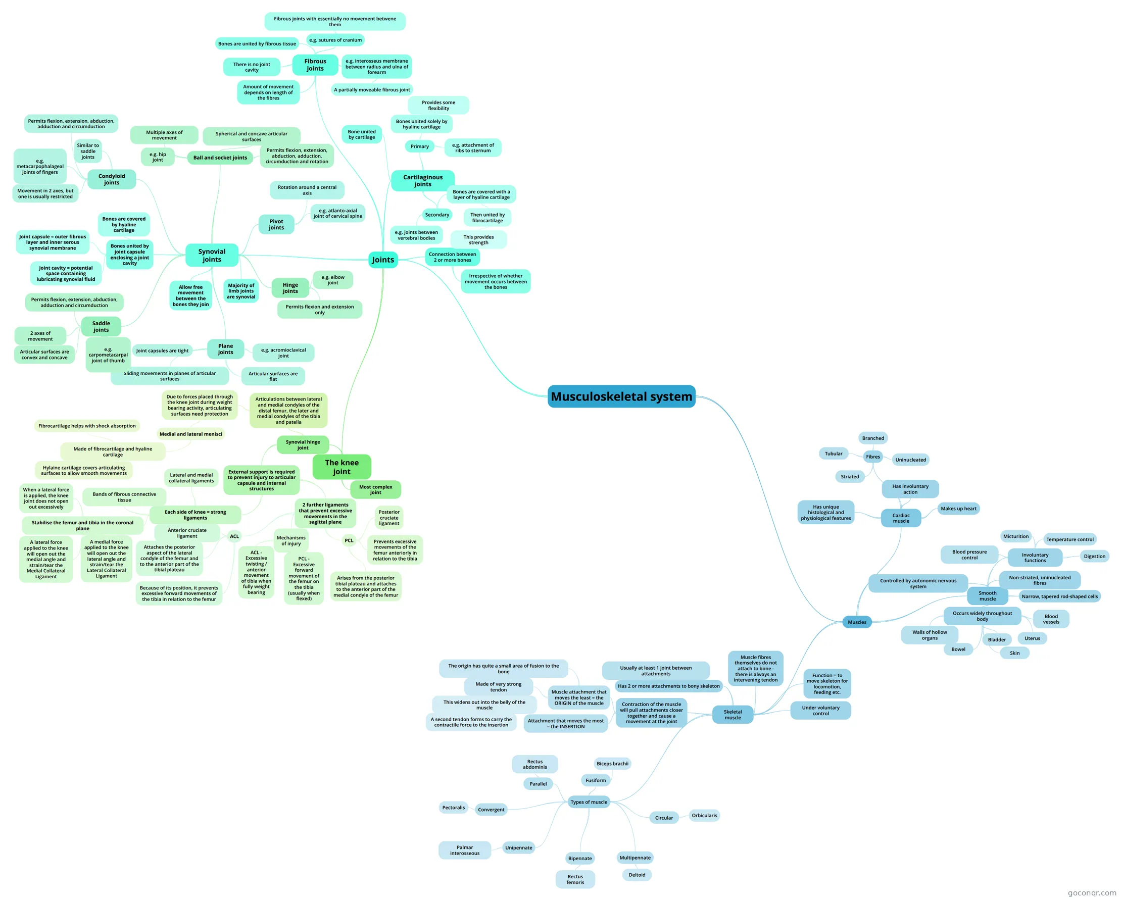
Mind map: Musculoskeletal system
Contenido del mapa
Musculoskeletal system
Joints
- Connection between 2 or more bones
- Irrespective of whether movement occurs between the bones
- Synovial joints
- Plane joints
- e.g. acromioclavical joint
- Articular surfaces are flat
- Joint capsules are tight
- Sliding movements in planes of articular surfaces
- Hinge joints
- e.g. elbow joint
- Permits flexion and extension only
- Saddle joints
- e.g. carpometacarpal joint of thumb
- Articular surfaces are convex and concave
- 2 axes of movement
- Permits flexion, extension, abduction, adduction and circumduction
- Condyloid joints
- Similar to saddle joints
- Movement in 2 axes, but one is usually restricted
- Permits flexion, extension, abduction, adduction and circumduction
- e.g. metacarpophalageal joints of fingers
- Ball and socket joints
- e.g. hip joint
- Spherical and concave articular surfaces
- Multiple axes of movement
- Permits flexion, extension, abduction, adduction, circumduction and rotation
- Pivot joints
- e.g. atlanto-axial joint of cervical spine
- Rotation around a central axis
- Bones united by joint capsule enclosing a joint cavity
- Joint capsule = outer fibrous layer and inner serous synovial membrane
- Joint cavity = potential space containing lubricating synovial fluid
- Bones are covered by hyaline cartilage
- Allow free movement between the bones they join
- Majority of limb joints are synovial
- Plane joints
- Fibrous joints
- Bones are united by fibrous tissue
- There is no joint cavity
- Amount of movement depends on length of the fibres
- e.g. sutures of cranium
- Fibrous joints with essentially no movement betwene them
- e.g. interosseus membrane between radius and ulna of forearm
- A partially moveable fibrous joint
- Cartilaginous joints
- Bone united by cartilage
- Primary
- Bones united solely by hyaline cartilage
- Provides some flexibility
- e.g. attachment of ribs to sternum
- Bones united solely by hyaline cartilage
- Secondary
- Bones are covered with a layer of hyaline cartilage
- Then united by fibrocartilage
- This provides strength
- Then united by fibrocartilage
- e.g. joints between vertebral bodies
- Bones are covered with a layer of hyaline cartilage
- The knee joint
- Most complex joint
- Synovial hinge joint
- Articulations between lateral and medial condyles of the distal femur, the later and medial condyles of the tibia and patella
- Due to forces placed through the knee joint during weight bearing activity, articulating surfaces need protection
- Medial and lateral menisci
- Made of fibrocartilage and hyaline cartilage
- Fibrocartilage helps with shock absorption
- Hylaine cartilage covers articulating surfaces to allow smooth movements
- Made of fibrocartilage and hyaline cartilage
- Medial and lateral menisci
- Due to forces placed through the knee joint during weight bearing activity, articulating surfaces need protection
- External support is required to prevent injury to articular capsule and internal structures
- Each side of knee = strong ligaments
- Lateral and medial collateral ligaments
- Bands of fibrous connective tissue
- Stabilise the femur and tibia in the coronal plane
- When a lateral force is applied, the knee joint does not open out excessively
- A lateral force applied to the knee will open out the medial angle and strain/tear the Medial Collateral Ligament
- A medial force applied to the knee will open out the lateral angle and strain/tear the Lateral Collateral Ligament
- 2 further ligaments that prevent excessive movements in the sagittal plane
- ACL
- Anterior cruciate ligament
- Attaches the posterior aspect of the lateral condyle of the femur and to the anterior part of the tibial plateau
- Because of its position, it prevents excessive forward movements of the tibia in relation to the femur
- PCL
- Posterior cruciate ligament
- Arises from the posterior tibial plateau and attaches to the anterior part of the medial condyle of the femur
- Prevents excessive movements of the femur anteriorly in relation to the tibia
- Mechanisms of injury
- ACL - Excessive twisting / anterior movement of tibia when fully weight bearing
- PCL - Excessive forward movement of the femur on the tibia (usually when flexed)
- ACL
- Each side of knee = strong ligaments
- Articulations between lateral and medial condyles of the distal femur, the later and medial condyles of the tibia and patella
- Connection between 2 or more bones
Muscles
- Cardiac muscle
- Makes up heart
- Has unique histological and physiological features
- Fibres
- Striated
- Tubular
- Branched
- Uninucleated
- Has involuntary action
- Smooth muscle
- Involuntary functions
- Digestion
- Blood pressure control
- Micturition
- Temperature control
- Occurs widely throughout body
- Walls of hollow organs
- Bowel
- Bladder
- Uterus
- Blood vessels
- Skin
- Controlled by autonomic nervous system
- Narrow, tapered rod-shaped cells
- Non-striated, uninucleated fibres
- Involuntary functions
- Skeletal muscle
- Under voluntary control
- Function = to move skeleton for locomotion, feeding etc.
- Has 2 or more attachments to bony skeleton
- Usually at least 1 joint between attachments
- Contraction of the muscle will pull attachments closer together and cause a movement at the joint
- Muscle attachment that moves the least = the ORIGIN of the muscle
- The origin has quite a small area of fusion to the bone
- Made of very strong tendon
- This widens out into the belly of the muscle
- A second tendon forms to carry the contractile force to the insertion
- This widens out into the belly of the muscle
- Attachment that moves the most = the INSERTION
- Muscle attachment that moves the least = the ORIGIN of the muscle
- Muscle fibres themselves do not attach to bone - there is always an intervening tendon
- Types of muscle
- Fusiform
- Biceps brachii
- Parallel
- Rectus abdominis
- Convergent
- Pectoralis
- Unipennate
- Palmar interosseous
- Bipennate
- Rectus femoris
- Multipennate
- Deltoid
- Circular
- Orbicularis
- Fusiform
- Cardiac muscle
Información
- Descripción
- Mapa Mental sobre Musculoskeletal system, creado por Alice Patterson el 11/10/2015.
- Creado por
- Alice Patterson
- Visitas
- 1
- Valoración