
Mapa mental
DNA profiling
Contenido del mapa
DNA profiling
Tandem repeats are repetitive segments of DNA that do not code for proteins. The number of tandem repeats shows a family resemblance.
- In DNA profiling methods short tandem repeats are compared. They are separated by electrophoresis.
- STR are polymorphic - ask adams about this
- In DNA profiling methods short tandem repeats are compared. They are separated by electrophoresis.
Procedure
- DNA obtained from individual e. mouth swab
- DNA digested with restriction enzymes which cut the DNA at specific recognition sites into fragments of varying size
- Fragments are separated by electrophoresis and stained. A banding pattern can be seen.
- The DNA to which the individual is being compared to also undergoes the same process
- Banding patterns can then be compared
- The DNA to which the individual is being compared to also undergoes the same process
- Fragments are separated by electrophoresis and stained. A banding pattern can be seen.
- DNA digested with restriction enzymes which cut the DNA at specific recognition sites into fragments of varying size
- DNA obtained from individual e. mouth swab
Uses
- Forensic science
- Maternity and paternity disputes
- Analysis of disease
PCR
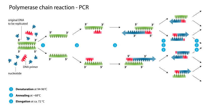
- Used to amplify DNA
- It differs from DNA replication because it can only amplify short sequences, it requires a PRIMER, and a cycle of heating and cooling is needed
- A cyclic reaction
- The DNA polymerase is used from Thermophilus Aquaticus (it is called taq polymerase and can withstand high temperatures)
- Process
- 0. Sample is mixed with DNA nucleotides, primers, Mn2+, and tan DNA polymerase
- 1. Heat to 94-96 to break H bonds (DNA is denatured into 2ssDNA)
- 2. Cool to 68, primers can then anneal. small section of double strand is then on the ssDNA. This is so the taq polymerase can bind to it.
- 3. Temperature raised to 72 (optimum for taq!), DNA remain as single strands, taq can then catalyse the addition of DNA nucleotides to the single strands.
- It starts at the primer and moves in the 5' to 3' direction
- The whole process repeats and is repeated for many cycles. The amount of DNA increases exponentially: 1 2 4 8 16 etc
- 3. Temperature raised to 72 (optimum for taq!), DNA remain as single strands, taq can then catalyse the addition of DNA nucleotides to the single strands.
- 2. Cool to 68, primers can then anneal. small section of double strand is then on the ssDNA. This is so the taq polymerase can bind to it.
- 1. Heat to 94-96 to break H bonds (DNA is denatured into 2ssDNA)
- 0. Sample is mixed with DNA nucleotides, primers, Mn2+, and tan DNA polymerase
- Applications
- Tissue typing prior to transplantation
- detection of oncogenes
- Detecting mutations (parents, fetal cells etc.)
- Identifying viral infections in host cells genome (HIV)
- Monitoring the spread of infectious disease
- Research (extinct animals)
- Used to amplify DNA
Electrophoresis
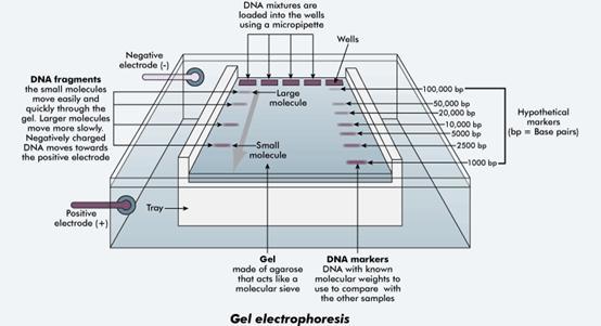
- Process used to separate different sized fragments of DNA
- Uses an agarose gel plate covered by a buffer solution
- Process
- 1. DNA samples cute at recognition sites by restriction enzymes. This is carried out at 35-40 and may take 1 hour
- 2. Electrophoresis tank: agarose gel, combs at one end, buffer solution added. Removing the comb leaves wells.
- 3. Loading dye is added to the digested DNA, this is then added to wells in the electrophoresis gel (pipette). Loading dye helps carry the DNA down into the well.
- 4. Electrodes then put into place, connected to a 18v battery, this is left to run (higher voltage and high current can risk electric shock)
- At the end, buffer solution is removed and a dye is added which adheres to the DNA and stains the fragments
- 4. Electrodes then put into place, connected to a 18v battery, this is left to run (higher voltage and high current can risk electric shock)
- 3. Loading dye is added to the digested DNA, this is then added to wells in the electrophoresis gel (pipette). Loading dye helps carry the DNA down into the well.
- 2. Electrophoresis tank: agarose gel, combs at one end, buffer solution added. Removing the comb leaves wells.
- 1. DNA samples cute at recognition sites by restriction enzymes. This is carried out at 35-40 and may take 1 hour
- Separating proteins
- carried outings the presence of sodium dodecyl sulphate (SDS)
- SDS is charged, it equalises the surface charge on the molecule and allows proteins to separate
- This technique can be used to analyse haemoglobin proteins to diagnose conditions like sickle cell anemia, aplastic anaemia, thalassaemia, leukaemia
- DNA PROBES
- A DNA probe is a short single stranded length of DNA, complementary to a section of DNA being investigated
- They can be labelled
- radioactive marker (32 P) revealed by exposure to photographic film
- fluorescent marker, emits colour on exposure to UV light
- They usually anneal by complimentary base pairing
- Uses
- Locate a gene for use in genetic engineering
- Genome comparison studies
- to identify the presence/absence of an allele for a genetic disease
- Micro arrays
- Number of probes a fixed surface
- Sample DNA anneals to complimentary fixed probes, this can reveal the presence of mutated alleles
- Steps
- 1. Sample DNA broken into fragments
- 2. amplified using PCR
- 3. Reference and test DNA samples are labelled with fluorescent markers
- ADD
- 3. Reference and test DNA samples are labelled with fluorescent markers
- 2. amplified using PCR
- 1. Sample DNA broken into fragments
- Process used to separate different sized fragments of DNA
Imágenes y archivos del mapa
Información
- Descripción
- GOD Biology Mapa Mental sobre DNA profiling, creado por Sophie Cooper el 21/03/2017.
- Creado por
- Sophie Cooper
- Visitas
- 1
- Valoración