
Mapa mental
Circulatory System
Contenido del mapa
Circulatory System
Vessels
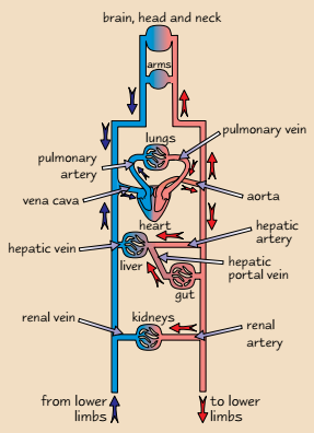
- Arteries
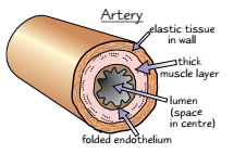
- Carry blood AWAY from the heart
- Thick and muscular walls-resists damage due to high pressure
- Elastic tissue to stretch and recoil-maintains high pressure
- Folded endothelium layer-allows stretching to maintain high pressure
- Also smooth to reduce friction
- Arterioles
- Contract to restrict flow and relax to aloow flow to organs
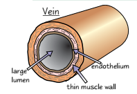
- Veins
- Take blood INTO heart
- Low pressure
- Wide lumen
- Little elastic and muscle tissue
- Valves stop back flow of blood
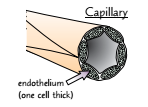
- Capillaries
- One cell thick-increases rate of diffusion
- Large number to increase surface area
- Numerous and Branched- increases surface area
- Narrow diameter- ensures RBC in contact with wall
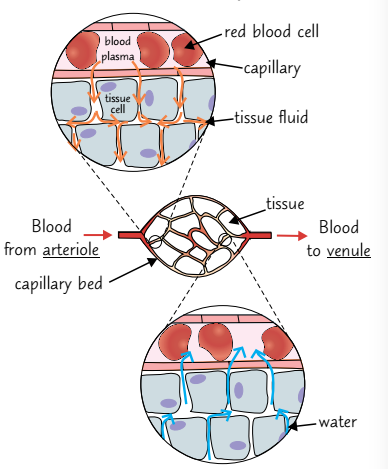
- Wall spaces- gaps between cells allow rapid formation of tissue fluid
- Arteries
Tissue Fluid
- Surrounds cells in tissues
- Made from small molecules (oxygen, water and nutrients)
- No big proteins- too large to push through capillary walls
- Cells take in O2 and nutrients from T.F. and release metabolic waste into it
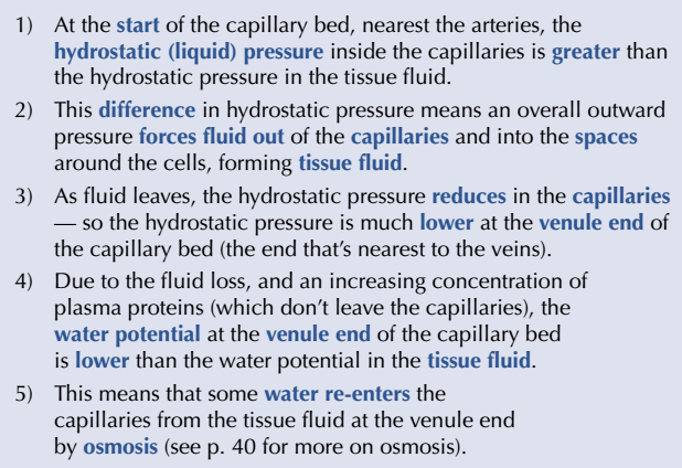
- Fluid leaves capillaries at arteriole end but large proteins remain-due to larger force pushing out than forces pulling in
- Lower water potential in venule end than in tissue fluid
- Force pulling in is stronger than force pushing out
- Water moves into capillary via osmosis
- Excess T.F. is drained into LYMPHATIC SYSTEM
- Water moves into capillary via osmosis
- Force pulling in is stronger than force pushing out
- Lower water potential in venule end than in tissue fluid
- High blood pressure = High hydrostatic pressure
Cardiac Cycle
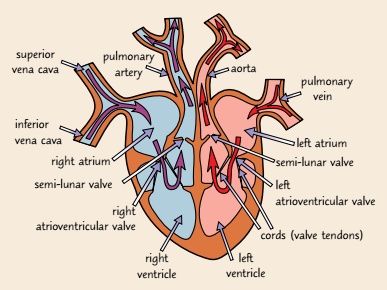
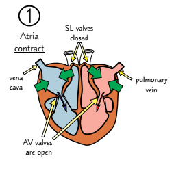
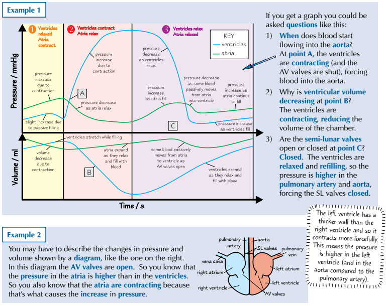
- 1. When ventricle relaxes, the atria contracts-decreasing volume and increasing pressure. This pushes blood into the ventricles. AV valve opens and SL valves close.
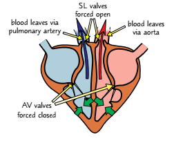
- 2. Atria now relax. Ventricles contract (decreasing volume and increasing pressure). Pressure is higher in ventricles than atria so AV valves shut (stops back flow). Pressure in ventricles higher than Aorta so SL valves open
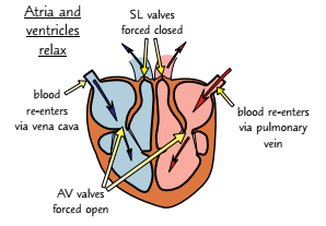
- 3. Both ventricles and atria relax. Higher pressure in Aorta (than ventricle) close SL valves (stops back flow). Atria fill again due to high pressure in Vena Cava. Increases pressure in atria. Ventricles relax so pressure falls below that in atria so AV valves open.
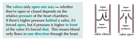
- AV vales OPEN when pressure in Atria HIGHER than Ventricle
- AV valves CLOSE when pressure in Atria LOWER than Ventricle
- SL valves OPEN when pressure in Ventricle HIGHER than Aorta
- SL valves CLOSE when pressure in Ventricle LOWER than Aorta
- 2. Atria now relax. Ventricles contract (decreasing volume and increasing pressure). Pressure is higher in ventricles than atria so AV valves shut (stops back flow). Pressure in ventricles higher than Aorta so SL valves open
- 1. When ventricle relaxes, the atria contracts-decreasing volume and increasing pressure. This pushes blood into the ventricles. AV valve opens and SL valves close.
Imágenes y archivos del mapa
Información
- Descripción
- AS level (Gas Exchange) Biology Mapa Mental sobre Circulatory System, creado por Molly Barton el 29/03/2017.
- Creado por
- Molly Barton
- Visitas
- 12
- Valoración