
Mapa mental
Vision
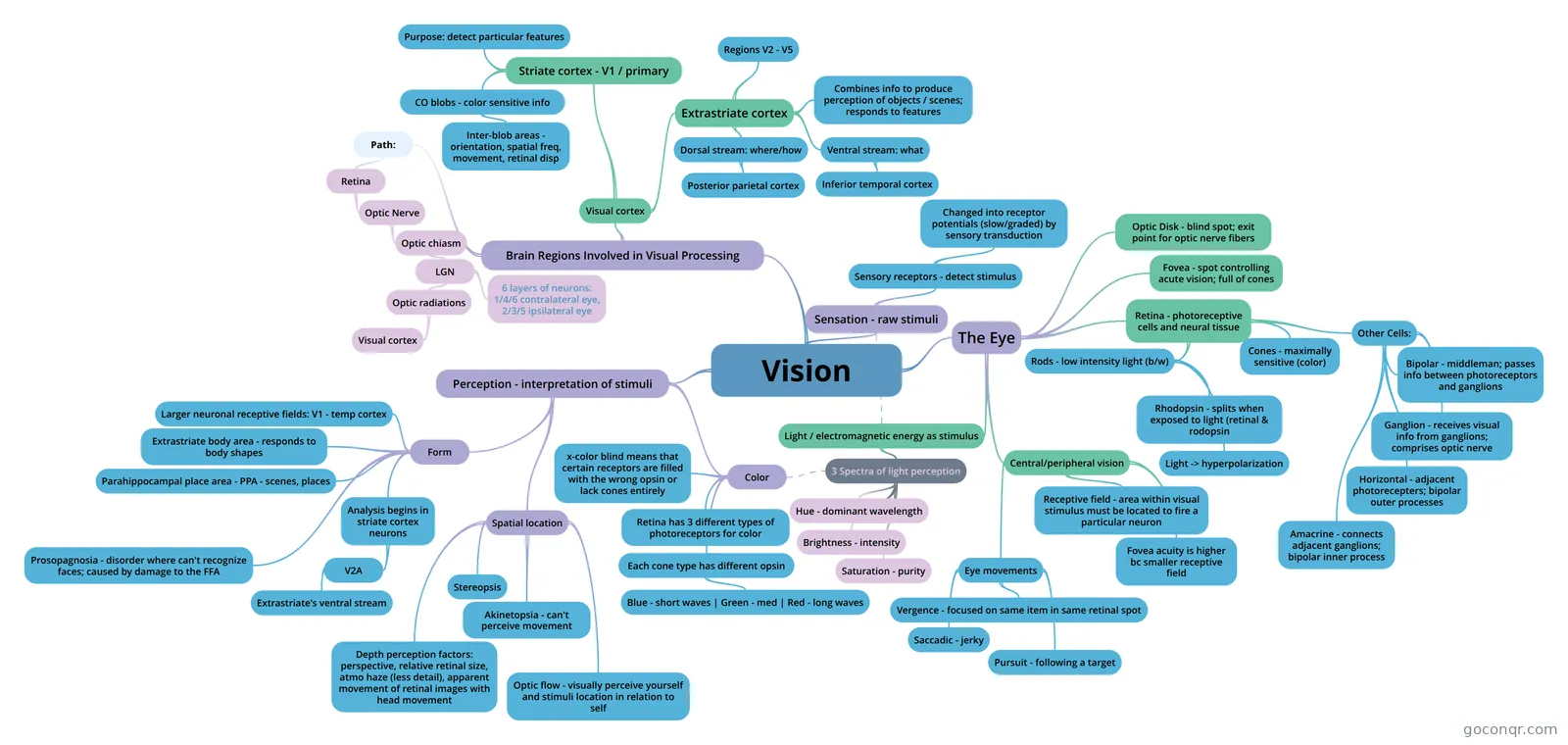
Mind map: Vision
Contenido del mapa
Vision
The Eye
- Light / electromagnetic energy as stimulus
- 3 Spectra of light perception
- Brightness - intensity
- Saturation - purity
- Hue - dominant wavelength
- 3 Spectra of light perception
- Retina - photoreceptive cells and neural tissue
- Rods - low intensity light (b/w)
- Rhodopsin - splits when exposed to light (retinal & rodopsin
- Light -> hyperpolarization
- Cones - maximally sensitive (color)
- Other Cells:
- Bipolar - middleman; passes info between photoreceptors and ganglions
- Ganglion - receives visual info from ganglions; comprises optic nerve
- Horizontal - adjacent photorecepters; bipolar outer processes
- Amacrine - connects adjacent ganglions; bipolar inner process
- Rods - low intensity light (b/w)
- Fovea - spot controlling acute vision; full of cones
- Optic Disk - blind spot; exit point for optic nerve fibers
- Central/peripheral vision
- Receptive field - area within visual stimulus must be located to fire a particular neuron
- Fovea acuity is higher bc smaller receptive field
- Eye movements
- Vergence - focused on same item in same retinal spot
- Saccadic - jerky
- Pursuit - following a target
- Light / electromagnetic energy as stimulus
Brain Regions Involved in Visual Processing
- Path:
- Retina
- Optic Nerve
- Optic chiasm
- LGN
- Optic radiations
- Visual cortex
- 6 layers of neurons: 1/4/6 contralateral eye, 2/3/5 ipsilateral eye
- Optic radiations
- LGN
- Optic chiasm
- Optic Nerve
- Retina
- Visual cortex
- Striate cortex - V1 / primary
- CO blobs - color sensitive info
- Inter-blob areas - orientation, spatial freq, movement, retinal disp
- Purpose: detect particular features
- CO blobs - color sensitive info
- Extrastriate cortex
- Combines info to produce perception of objects / scenes; responds to features
- Regions V2 - V5
- Dorsal stream: where/how
- Posterior parietal cortex
- Ventral stream: what
- Inferior temporal cortex
- Striate cortex - V1 / primary
- Path:
Perception - interpretation of stimuli
- Color
- Retina has 3 different types of photoreceptors for color
- Each cone type has different opsin
- Blue - short waves | Green - med | Red - long waves
- x-color blind means that certain receptors are filled with the wrong opsin or lack cones entirely
- Form
- Analysis begins in striate cortex neurons
- V2A
- Extrastriate's ventral stream
- V2A
- Larger neuronal receptive fields: V1 - temp cortex
- Prosopagnosia - disorder where can't recognize faces; caused by damage to the FFA
- Extrastriate body area - responds to body shapes
- Parahippocampal place area - PPA - scenes, places
- Analysis begins in striate cortex neurons
- Spatial location
- Stereopsis
- Depth perception factors: perspective, relative retinal size, atmo haze (less detail), apparent movement of retinal images with head movement
- Optic flow - visually perceive yourself and stimuli location in relation to self
- Akinetopsia - can't perceive movement
- Color
Sensation - raw stimuli
- Sensory receptors - detect stimulus
- Changed into receptor potentials (slow/graded) by sensory transduction
- Sensory receptors - detect stimulus
Información
- Descripción
- Mapa Mental sobre Vision, creado por Emily McNeil el 27/07/2017.
- Creado por
- Emily McNeil
- Visitas
- 0
- Valoración