
Fichas
Biology F212 - Biological molecules 1
Fichas del conjunto (30)
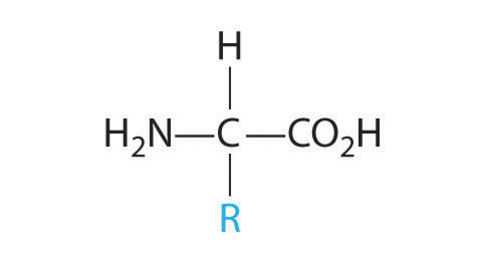
- 1Structure of an amino acid
- 2Primary StructureThe sequence of amino acids in a protein molecule
- 3Secondary structureCoiling or folding of the chain due to formation of
hydrogen bonds.
Alpha-helix and Beta-pleated sheet - 4Tertiary StructureOverall 3D structure of the molecule. Formation of hydrogen bonds, disulphide bridges and hydrophobic & hydrophillic interactions
- 5Quarternary structureProtein structure where it consists of more than 1 polypeptide chain.
E.g Haemoglobin has 4 polypeptide chains - 6Structure of a collagen molecule- 3 polypeptide chains ( 3 syllables=3 chains)
- 1000 amino acids long
- Hydrogen and covalent bonds form
- Cross links are staggered for strength - 7Structure of haemoglobin- Globular protein
-Soluble in water
-Wide range of amino acids
-Contains 4 haem groups with Fe+ ions
-Alpha helix - 8Structural difference between alpha and beta glucoseIn alpha glucose the OH group on carbon 1 is above the plane of the ring.
In beta glucose the OH group on carbon 1 is below the plane of the ring. - 9Structure of starch (amylase)-Made of alpha glucose
- Straight chain
- tends to coil up - 10Structure of cellulose-Made of beta glucose, in a chain alternate glucose subunits are inverted
-Forms straight chains
-Forms plant cell walls
- The beta-glycosidic bonds can only be broken by a cellulose enzyme (humans don't have this) - 11Structure of glycogen- Mostly like amylase
- many 1-4 glycosidic bonds
- 9% 1-6 branches - 12Structure of a triglyceride- Glycerol and 3 fatty acids
-Insoluble in water does not affect cell water potential
-Compact energy store - 13Structure of a phospholipid-Glycerol plus 2 fatty acids and a phosphate group
-Part hydrophobic, part hydrophillic (ideal for cell surface membranes) - 14Cholesterol-Small, thin molecule
-Fit into the lipid bilayer giving strength and stability - 15Chemical test for protein - Biuret testIf a protein is present the solution changes colour from pale blue to lilac
- 16Chemical test for reducing sugars -Benedict's testAdd benedict's solution, heat to 80 degrees, if a reducing sugar is present the mixture will change colour from blue to orange/red
- 17Chemical test for non-reducing sugars -benedict's testIf the reducing test is negative, boil with hydrochloric acid, cool and neutralise with sodium hydrogencarbonate and repeat benedict's test
- 18Chemical test for starch -Iodine solutionColour changes from yellow to blue/black if starch is present
- 19Chemical tests for lipids -emulsion testMix with ethanol, pour into water, if an emulsion forms then a lipid is present
- 20How the concentration of glucose can be determined by colorimetry-Benedict's test reveals reducing sugars (if there is an orange/brown precipitate)
-The more reducing sugar present the more precipitate will form and the more benedicts solution used up.
-Once precipitate has been filtered out, the concentration of remaining solution can be measured
-This tells you how much benedicts was used up and can be used to estimate the the concentration of reducing sugar in original sample - 21Preparing a calibration curve- Zero the device using a blank (water)
- Take a range of known concentrations of reducing sugars
-carry out benedicts test on each and filter out precipitate
-Use a colorimeter to get readings of the amount light passing through the solutions
-Plot the readings on a graph to show %transmission against concentration
-Measure the %transmission of the unknown in the colorimeter
-Use this to read the equivalent reducing sugar concentration from %transmission - 22Deoxyribonucleic acid (DNA)-Is a polynucleotide
-Usually double stranded
-Made up of nucleotides containing the bases Adenine (A), Thymine (T), Guanine (G), Cytosine (C) - 23Ribonucleic acid (RNA)-Is a polynucleotide
-Usually single stranded
- Made up of nucelotides containing the bases Adenine (A), Uracil (U), Cytosine (C), Guanine (G) - 24DNA replication-Double helix is untwisted
- DNA 'unzipped' and hydrogen bonds between bases are broken
-Free DNA nucleotides are H-bonded onto their exposed complementary bases
-DNA polymerase catalyses formation of covalent bonds between phosphates and sugars
-This continues till there are 2 identical strands
-These are then 'proof read' by DNA polymerase to prevent mistakes - 25GeneA gene is a sequence of DNA nucelotides that code for a polypeptide
- 26Protein Synthesis-Required gene exposed by splitting H-bonds holding the helix together in the region
- RNA nucleotides form a complementary strand (mRNA) which is a copy of the coding strand
-mRNA peels off DNA and leaves via nuclear pores
-mRNA attaches to a ribosome
-tRNA molecules bring amino acids to the ribosome in the correct order according to base sequence on the mRNA
-Amino acids are joined together by peptide bonds to form a protein with a specific tertiary structure - 27Enzymes-Globular Proteins
-Specific tertiary structure
-Catalyse metabolic reactions in living organisms
-Can be intracellular or extracellular - 28Enzyme specificityThe active site is a specific shape depending on the reaction it catalyses, meaning that other molecules wont fit the active site.
- 29Lock and Key hypothesisThe theory of enzyme action in which the enzyme active site is complementary to the substrate molecule
- 30Induced fit hypothesisThe theory of enzyme action in which the enzyme active site changes shape to fit the substrate molecule more closely as it binds.
Información
- Descripción
- as level Chemistry Fichas sobre Biology F212 - Biological molecules 1, creado por scarlettcain97 el 30/05/2014.
- Creado por
- scarlettcain97 · may 2014
- Actualizado
- 17 ene 2017
- Visitas
- 636
- Etiquetas
- Valoración