3894499
| Question | Answer |
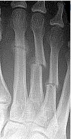
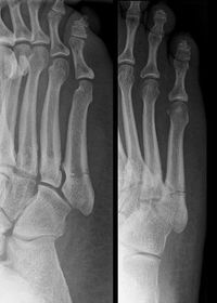
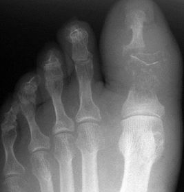
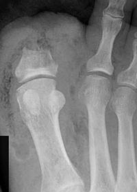
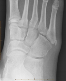
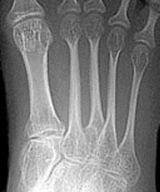
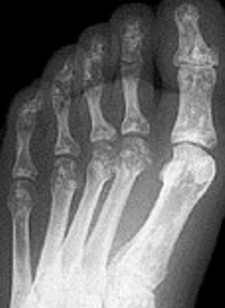
| Describe the alignment of metatarsal 2 | There is no angulation nor displacement of the distal segment relative to the proximal segment. |
| Describe the alignment of metatarsal 3 | The distal segment is displaced laterally (50% apposition) and angulated medially. |
| Describe the alignment of metatarsal 4 | The distal segment is displaced laterally (75% apposition) but there is no apparent angulation. |
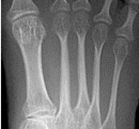
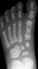
| Describe the tubulation of the metatarsals | Metatarsals are overtubulated - decreased girth. |
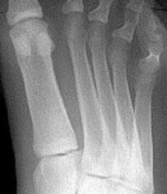
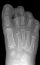
| Describe the tubulation of the metatarsals | Metatarsals are undertubulated - increased girth. |
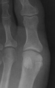
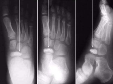
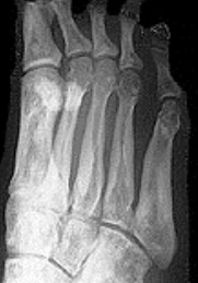
| Describe the normal variation of the sesamoid bones | Tibial sesamoid is bipartite |
| What is this? | Bipartite medial cuneiform |
| What is this? | Bipartite navicular |
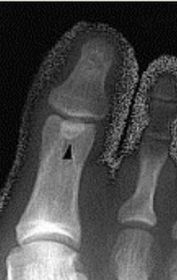
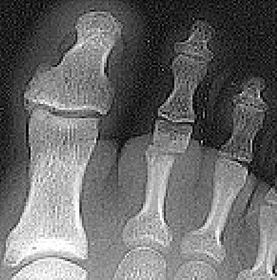
| What is this? Where is it typically found? | Os interphalangeus. Typically found on the inferior aspect of the hallux IPJ. It is rare to see this ossicle at the IPJs of the lesser toes. |
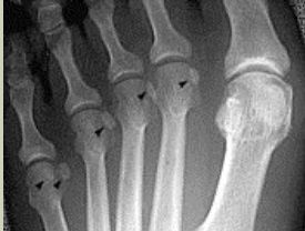
| What is this? | Accessory sesamoids. |
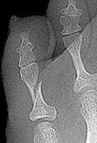
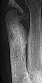
| What is this? | Os Intermetatarseum |
| What is this? | Tibialis Anterior Tendon Sesamoid |
| What is this? | Os Vesalianium |
| What is this? What tendon is it found in? | Os Peroneum. Sesamoid bone found in peroneus longus tendon. |
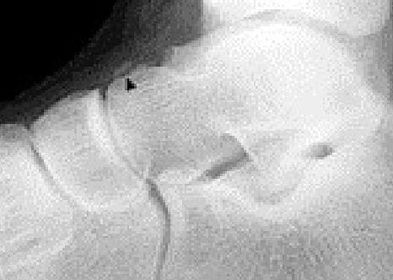
| What is this? | Os Infranaviculare |
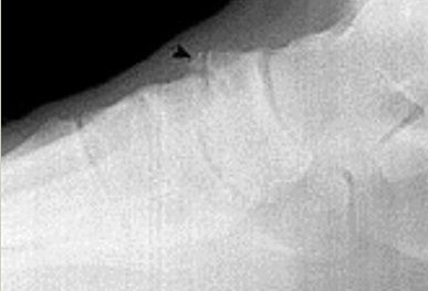
| What is this? | Os Supranaviculare |
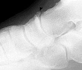
| What is this? | Os Supratalare |
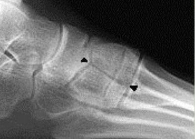
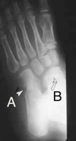
| What is this? What type is it? | Accessory navicular (Os tibiale externum) Type 1: Sesamoid in the tendon |
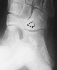
| What is this? What type is it? | Accessory navicular (Os tibiale externum) Type 2: Articulating accessory ossification centre |
| What is this? What type is it? | Accessory navicular (Os tibiale externum) Type 3: Fused accessory ossification centre |
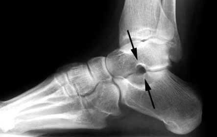
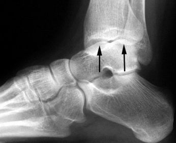
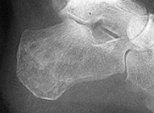
| What is this? What is it called when fused to the talus? | Os trigonum Called trigonal (Steida's) process when fused to the talus |
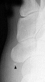
| What is this? | Os calcaneus secundarius |
| What is this? | Os supracalcaneum |
| What is this? | Os subtibiale |
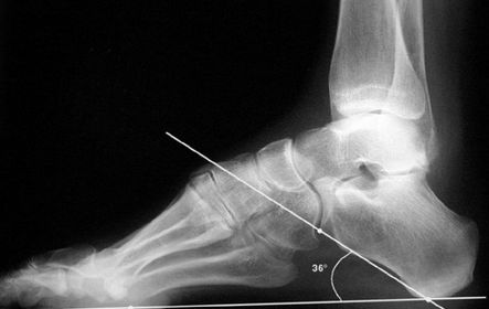
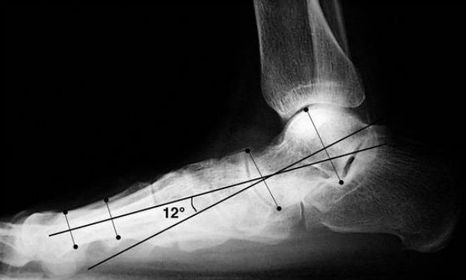
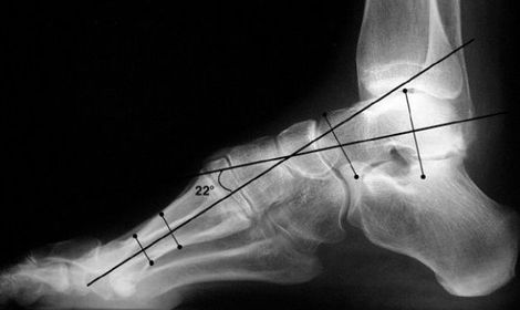
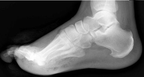
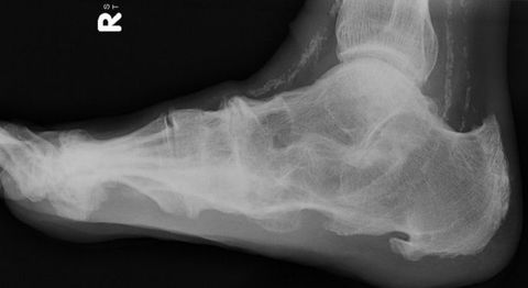
| What foot type does this calcaneal inclination angle indicate? | Pes cavo-varus |
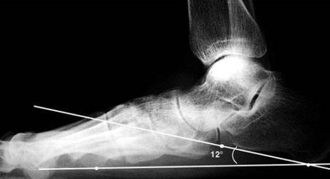
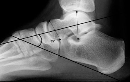
| What foot type does this calcaneal inclination angle indicate? | Pes plano-valgus |
| What is this angle called? What foot type does it indicate? | Talar - 1st MT or Meary's Angle Pes plano-valgus |
| What is this angle called? What foot type does it indicate | Talar - 1st MT or Meary's Angle Pes cavo-varus |
| What is this angle called? What is normal? What does an increased angle indicate? | Talo-calcaneal angle. 25-45 degrees. Increased angle = hindfoot valgus |
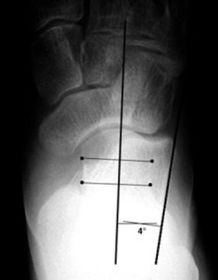
| What is this angle called? Is this normal or abnormal? | AP - Talo-calcaneal angle This is normal |
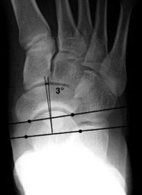
| What is this angle called? Is this normal or abnormal? | AP-Talo-calcaneal angle This is abnormal. The angle is reduced indicating pes cavo-varus |
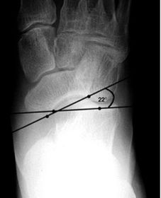
| What is this angle called? Is this normal or abnormal? | Talo-navicular coverage. This is normal. |
| What is this angle called? Is this normal or abnormal? | Talo-navicular coverage. This is abnormal as the talus is laterally deviated with medial overhang of navicular indicating pes cavo-varus |
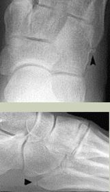
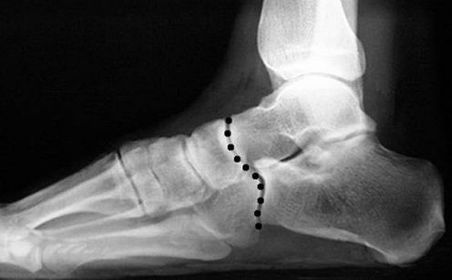
| What is this called? What does an anterior break indicate? What does a posterior break indicate? | Cyma Line/Midtarsal joint. Anterior break = pronated foot type Posterior break = supinated foot type |
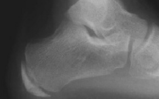
| What is this? What foot type is this visible in? | Sinus tarsi/bullet hole sign. Pes cavo-varus |
| What is this? What foot type is this visible in? | Double talar dome sign. Pes cavo-varus |
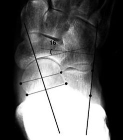
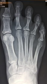
| What angle is this? Is this normal or abnormal? | 1st intermetatarsal angle Abnormal. Greater than 9 degrees is abnormal. Usually due to metatarsus primus varus |
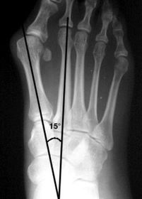
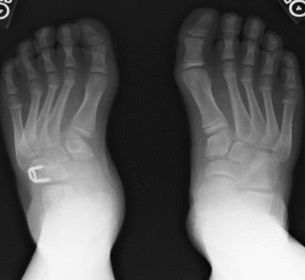
| What deformity is this? | Metatarsus Adductus |
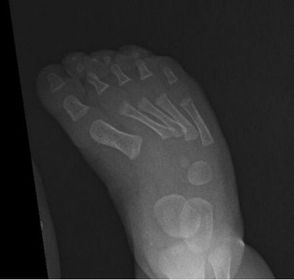
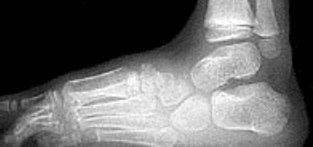
| What deformity is this? | Metatarsus Adductovarus (ladder like arrangement of the metatarsals in lateral view) |
| What deformity is this? | Skew foot |
| What disease is this? | Osteoarthritis |
| What disease is this? | Rheumatoid arthritis |
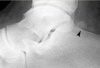
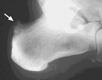
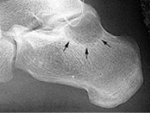
| What disease is this? What does the arrow indicate? | Rheumatoid arthritis. Arrow = Bone erosion secondary to inflammation of retrocalcaneal bursa. |
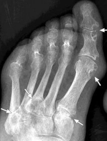
| What disease is this? | Gout |
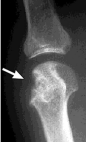
| What disease is this? What do the arrows indicate? | Gout Arrows = 'punched out' erosions |
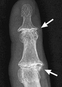
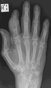
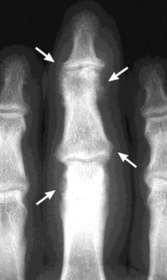
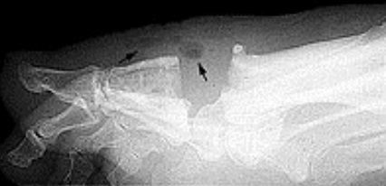
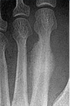
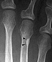
| What disease is this? What indicates this? | Tophaceous gout. Soft tissue swelling surrounding the index finger PIPJ, with associated erosion and bone resorption |
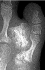
| What disease is this? | Tophaceous gout. |
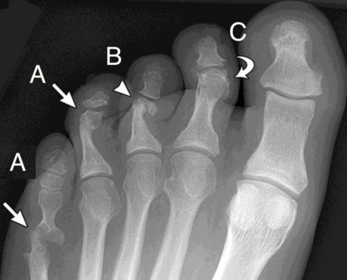
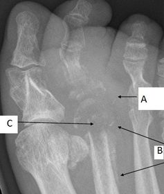
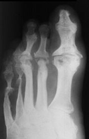
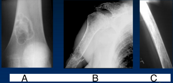
| What disease is this? What is shown by A, B and C? | Psoriatic arthritis. A = destructive changes B = Pencil in cup deformity C = IPJ fusion |
| What disease is this? | Psoriatic arthritis |
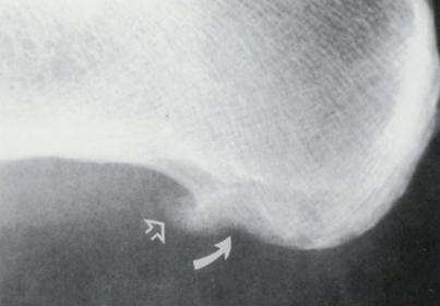
| What disease is this? What does the arrow show? | Non-articular psoriatic arthritis Arrow = 'fluffy spur' |
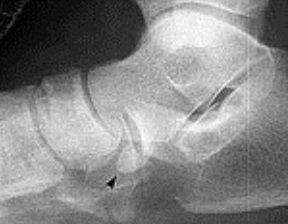
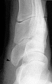
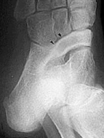
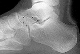
| What is this? | Calcaneonavicular coalition (AKA calcaneonavicular bar or anteater's nose) |
| What is this? | Calcaneonavicular coalition (AKA calcaneonavicular bar or anteater's nose) |
| What is this? | Calcaneonavicular coalition (AKA calcaneonavicular bar or anteater's nose) |
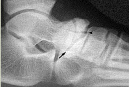
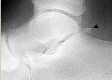
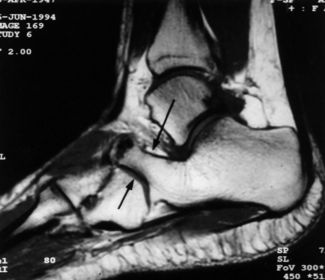
| What is this? What is shown by A and B? | STJ coalition A = talar neck spurring B = Halo or 'C' sign |
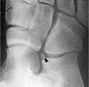
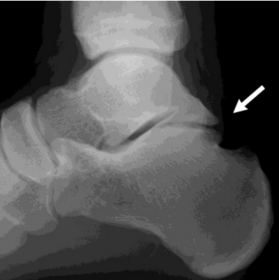
| What is this? | Posterior talocalcaneal coalition |
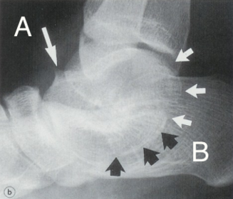
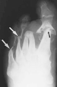
| What is shown by A and B? | A = Talonavicular coalition B = Calcaneocuboid coalition |
| What is this? | Cuboid-navicular coalition |
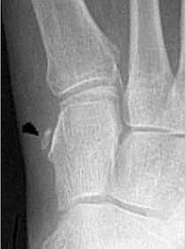
| What type of fracture is shown at the 2nd metatarsal? | Transverse fracture |
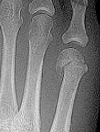
| What type of fracture is this? | Oblique fracture |
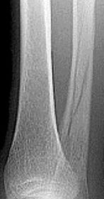
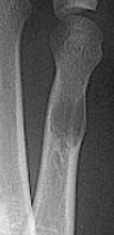
| What type of fracture is this? | Spiral fracture |
| What type of fracture is this? | Comminuted fracture |
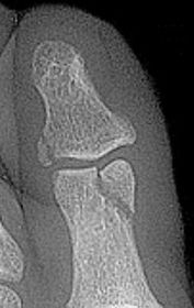
| What type of fracture is this? | Osteochondral fracture |
| What type of fracture is this? | Impaction fraction |
| What type of fracture is this? | Intra-articular fracture |
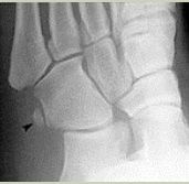
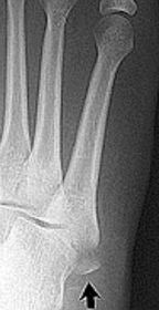
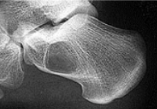
| What type of fracture is this? | Avulsion fracture |
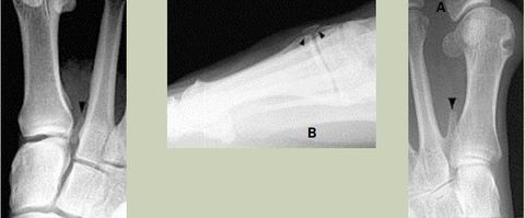
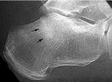
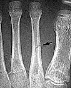
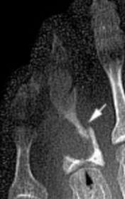
| What do the arrows show? | Stress fracture |
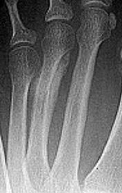
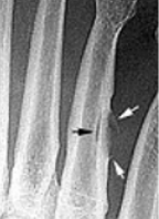
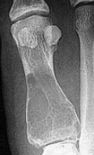
| Describe what has happened at the shaft of the 3rd met | Exuberant periosteal new bone production. May have resulted if a stress fracture was not treated and the patient continued weight bearing activities. |
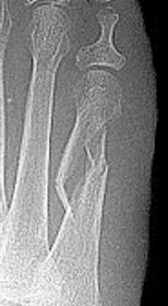
| What type of fracture is this? | Greenstick fracture (common in paediatric patients) |
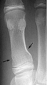
| Describe what the arrows are showing | Torus fracture of the 1st met. "Buckling" of the proximal one third diaphysis is seen both medially and laterally. Common in paediatric patients |
| What is the Salter Harris classification used for? What is the mnemonic for the 5 types? | Used to classify fractures involving the growth plate. S - slipped = type I A - above = type II L - lower = type III T - through = type IV R - ruined/rammed = type V |
| What type of fracture is this? | Salter-Harris Type II |
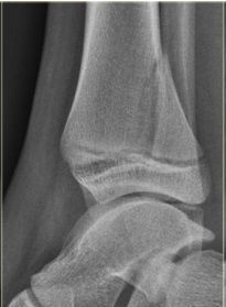
| What type of fracture is this? | Salter-Harris Type III |
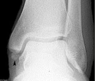
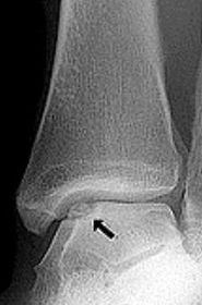
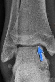
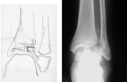
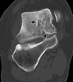
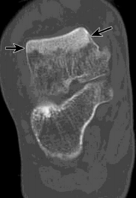
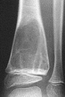
| What type of fracture is this? How does it occur? | Tillaux fracture - a fracture of the anterolateral tibial epiphysis that is commonly seen in adolescents. The fragment is avulsed due to the strong ATF ligament in an external rotation injury of the foot. |
| What type of fracture is this? | Jones fracture - fracture of the diaphysis of the 5th metatarsal. More distal than an avulsion fracture. |
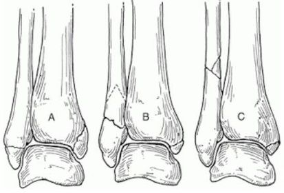
| What classification system is this? Describe the 3 classifications | Weber Ankle Fracture Classification A = Fracture below syndesmosis B = Fracture begins at joint level & extends proximally in an oblique fashion C = Fracture above the joint line |
| What do the arrows indicate? | Infectious periostitis |
| What disease is this? What signs indicate this? | Osteomyelitis. Osteolysis (fragmentation), sequestra (dead/necrotic bone fragments floating in pus), rarefacation (loss of bone density). |
| What does the arrow show? | Soft tissue emphysema |
| What disease is this? What do A, B & C show? | Osteomyelitis. A = Soft tissue swelling B = Periosteal reaction C = Bony destruction |
| What disease is this? | Gas gangrene |
| What disease is this? | Gas gangrene |
| What disease is this? What causes it? | Brodie's abscess - chronic bone abscess caused by organisms of low virulence. Patient most likely did not go through an acute phase (just chronic osteo) |
| What disease is this? | Monckeberg’s arteriosclerosis |
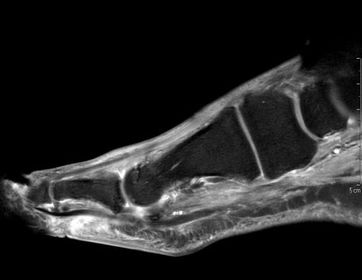
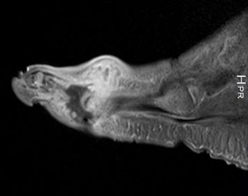
| What does this indicate? | Increased signal and enhancement of the soft tissue plantar to the proximal phalanx suggestive of subcutaneous infection without abscess |
| What does this indicate? | Increased signal and soft tissue distension dorsally representing subcutaneous abscess communicating with associated with plantar ulcerative defect. |
| What disease is this? | Atrophic neuroarthropathy |
| What disease is this? | Atrophic forefoot neuropathy (diabetic osteolysis) |
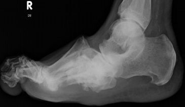
| What disease is this? | Charcot Neuroarthropathy |
| This patient has taken a short course of prednisone. What does the image show? | Osteonecosis of the talus |
| What does this image show? | Avascular necrosis of the talus, secondary to a talar neck fracture |
| What do these images show? What is this disease called? How does this differ from Meuller Wiess Syndrome? | Osteochondrosis of the navicular. Kohler's disease. Kohler's disease only appears in children, Meuller Wiess Syndrome only appears in adults and is a spontaneus osteonecrosis of the navicular, not a true osteochondrosis. |
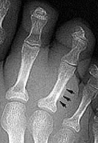
| Describe what is occurring at the head of the 3rd met | Freiberg’s infarction (avascular necrosis of the metatarsal head) |
| What disease is this? | Sever's disease |
| What is this? What else could it be? | Iselin’s disease Traction apophysis, Os peronei, avulsion fraction, lis francs dislocation |
| Describe the patterns of destruction in A, B, C. Which is more malignant? | A = geographic destruction B = moth eaten destruction C = permeative destruction Malignancy = C>B>A |
| What type of lesion is this? | Geographic - slow growing lesion |
| What type of lesion is this? | Moth eaten and permeative (mixed increased and decreased density throughout the entire calcaneus) |
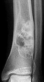
| Describe this lesion | A lytic lesion of the proximal phalanx that appears quite aggressive due to bone loss; however, the well-defined margins of the bone suggests a soft tissue tumor causing pressure atrophy against adjacent bone |
| What type of lesion is this? What type of periosteal reaction is this? | Geographic. Continuous shell |
| What type of periosteal reaction is this? | Interrupted |
| What type of periosteal reaction is this? | Complex |
| What type of lesion is this? | Unicameral bone cyst |
| What type of lesion is this? | Giant cell tumour |
| What type of lesion is this? | Aneurysmal bone cyst |
| What type of lesion is this? | Chondromyxoid fibroma |
| What type of lesion is this? | Osteogenic |
| What type of lesion is this? | Chondrogenic |
| What disease is this? What are the signs of this? | Osteogenesis imperfecta (“Brittle bone disease”) Diffuse cancellous osteopenia (coarse primary trabeculae), diminished tubular bone girth |
| What disease is this? What are signs of this? | Osteopoikilosis Defined homogeneous circular foci of increased density (bone islands), cortical-like densities in cancellous bone |
| What disease is this? What is it caused by? What indicates this? | Osteopetrosis ("marble bone disease") Diffuse bone sclerosis due to osteoclasts dysfunction. "Bone within bone" appearance |
| What disease is this? | Osteochondromatosis (multiple cartilaginous exostoses) |
| What disease is this? | Melorheostosis |
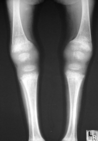
| What disease is this? | Rickets |
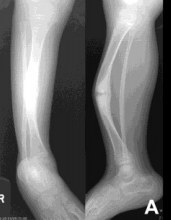
| What disease is this? | Osteogenesis Imperfecta |
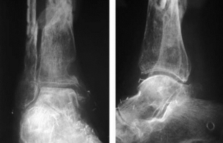
| What does this image show? What causes it? | Regional bone loss Disuse, immobilisation, paralysis |
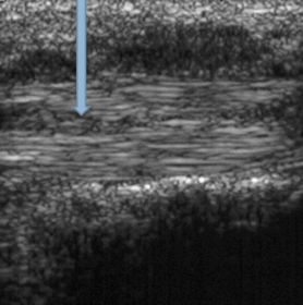
| In an ultrasound, what colour is bone, tendon and water (oedema)? | Bone = white Tendon = grey Water = black |
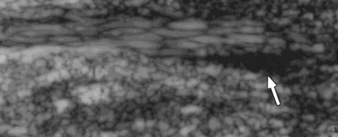
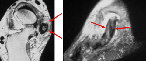
| What does the arrow indicate? | A cleft (tear) in the tendon |
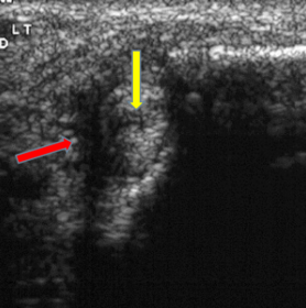
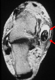
| What does the red arrow show? What does the yellow arrow show? | Red arrow = fluid Yellow arrow = tendon tear |
| What does the arrow indicate? | Fluid |
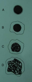
| Describe the tendon pathology as seen in an MRI of A-D | A = normal tendon B = Tenosynovitis C = Tendonitis D = Tendon Dysfunction (Functional Rupture) |
| What do the arrows indicate? | Tenosynovitis |
| What do the arrows indicate? | Tendonitis |
| What do the arrows indicate? | Tendon Dysfunction – Functional Rupture |
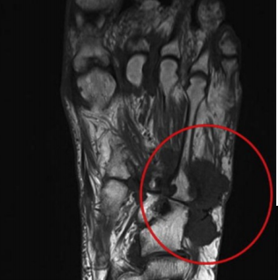
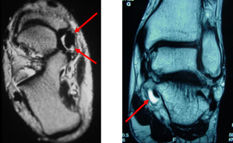
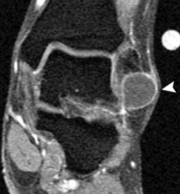
| What does this show? | Ganglion in lateral ankle soft tissue |
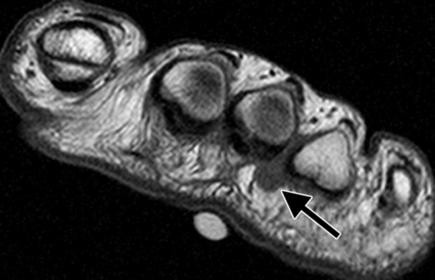
| What does the arrow show? What could this be? | Teardrop-shaped hypointense lesion in third interspace. Could be morton's neuroma |
Want to create your own Flashcards for free with GoConqr? Learn more.