10385606
| Question | Answer |
| 3 meninges layers: | Dura, arachnoid, pia |
| T/F: all three meninge layers have their own innervation. | F. Only dura. |
| Which meninge layer has its own blood supply? | Dura. |
| _____ folds on itself to separate the L/R hemispheres. This structure is called the ______. | Dura. Falx cerebri or cerebral falx |
| The horizontal fold of dura is called _______ | Tentorium cerebelli or cerebellar tentorium |
| What are the two layers of the dura? | 1. periosteal layer 2. meningeal layer |
| T/F: Both layers of dura folds to form the falx cerebri. | F. only meningeal layer. |
| The space between the invaginated meningeal layer and periosteal layer is the _________. | Superior sagittal sinus. |
| Which structures can be found in the sinus that acts as a pathway for CSF to meet the venous blood? | Arachnoid granulation |
| At the bottom of cerebral falx where the meningeal layers separate slightly is the ____ sinus. | Inferior sagittal sinus |
| T/F: the sinus contains both venous blood and CSF. | T. |
| Another space that contains CSF and blood vessels at the same time is the ______ . | Subarachnoid space |
| Parenchyma means _______. | Nerve tissue |
| The Pia mater invaginates together with cerebral arteries to form the _______. | Perivascular space |
| Epidural hematoma occurs between _______ while subdural hematoma occurs between________. | Epidural: between skull and dura Subdural: between dura and arachnoid mater |
| Which is more likely to cause hernia of cingulate gyrus? Epidural or subdural hematoma? | Subdural. |
| On the spinal nerve level, the dura is referred to as _______, arachnoid becomes _______ while the pia becomes _______. | Dura: epineurium Arachnoid: perineurium (lining of each fascicle) Pia: endoneurium (between fascicles) |
| The anterior circulatory pathway supplies __% of blood cerebral supply while the posterior circulatory provides blood supply to ______________. | Anterior: 80%. Posterior: 20% of cerebrum, brainstem, and cerebellum. |
| The vertebral artery enters the skull via: | Foramen magnum |
| The (internal/external) carotid artery enters the skull via __________. | Internal carotid. Enters via the carotid canal. |
| Where does the anterior, middle, and posterior cerebral artery come off of? | Anterior and middle come off of internal carotid artery. Posterior cerebral artery comes off of vertebral artery. |
| What are the 5 branches of internal carotid, from anterior to posterior? | 1. opthalamic artery (optic nerve, retina) 2. anterior cerebral artery 3. middle cerebral artery 4. posterior communicating artery 5. anterior choroidal artery (blood supply to internal capsule) |
| Infarction of internal capsule is very dangerous because: | It is the pathway of many nerve fibers going to other areas. |
| Name the arteries from most inferior/posterior to the most anterior/superior, starting from the vertebral arteries. | 1. vertebral artery 2. posterior spinal artery 3. posterior inferior cerebellar artery 4. anterior spinal artery 5. anterior inferior cerebellar artery 6. basillar artery 7. superior cerebellar artery 8. posterior cerebral artery 9. posterior communicating artery |
| What are the branches coming off of the basilar artery? | circumferential pontine arteries |
| Which structure is between the optic chiasm and the mammary glands? | infundibulum |
| The anterior cerebral artery supplies the _____ lobe and the ____ lobe on the ___ surface. | Frontal lobe, parietal lobe on the medial surface. |
| The posterior cerebral artery supplies the _____ lobe including areas _____ and ______. | Occipital lobe. cuneas and lingual. |
| The ACA wraps around the superior portion of the cerebrum, sharing the perfusion with __________ . | Middle cerebral artery. |
| What functions are affected if the MCA is blocked? | 1. UE sensory and motor, 2. language 3 .orientation in space |
| Based on sensory homunculus, the ACA supplies sensory of _________ (body regions) while the MCA supplies __________. | ACA: genitalis, LE and trunk. MCA: UE and face, teeth, tongue, pharynx, intra-abdominal. |
| The MCA passes the _____ sulcus and gives off very small branches called _______ which supplies the ________. | The MCA passes the [lateral] sulcus and gives off very small branches called [lenticulostriate arteries] which supplies the [basal ganglia]. |
| The lenticulostriate arteries are very prone to blood clot because they are ______. | Very narrow. |
| Major and supporting blood supply to the rostral midbrain: | Major: Posterior cerebral artery Supporting: Post. communicating, Superior cerebellar artery. |
| Major and supporting blood supply to the caudal midbrain: | Major: Superior cerebellar artery Supporting artery: PCA, basillar |
| Most important blood supply for the pons comes from _________. A rostral supporting artery is _______ and a caudal supporting artery is _________. | Most important: basilar. Rostral supporter: superior cerebellar artery Caudal supporter: Anterior inferior cerebellar artery |
| The main blood supply for medulla comes from the _______, while the rostral medullar also has support from _______ and _______, the caudal medullar has support from ______ and _________. | The main blood supply for medulla comes from the [vertebral artery], while the rostral medullar also has support from [PICA] and [anterior spinal artery], the caudal medullar has support from [anterior spinal artery] and [posterior spinal artery]. |
| Three key structures for blood supply to the cerebellum. | 1. superior cerebellar artery 2. anterior cerebellar artery 3. posterior cerebellar artery |
| The posterior spinal artery supplies the _________ part of the spine. | Dorsal columns, dorsal roots, and dorsal horns. |
| The anterior spinal artery supplies _____________ of the spine. | 1. lateral columns, 2. ventral horns 3. intermediate zone 4. ventral column |
| Sensory defects are usually associated with blood supply shortage of the _______ artery while motor defects are usually associated with _________. | Sensory: posterior spinal artery and maybe anterior also. Motor: anterior spinal artery |
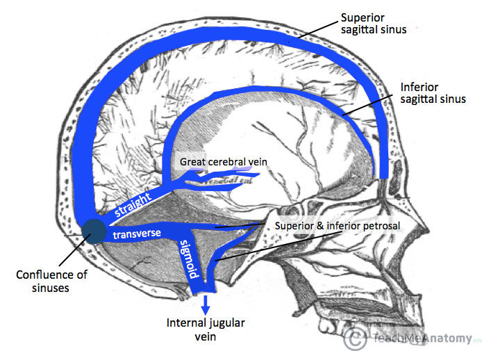
| Lable the sinuses of the brain. | See picture |
| The sinus intersects at _________. | the confluence of sinuses. |
| The superior sagittal sinus and inferior sagittal sinus drains to the _________, where they separates to go through L and R _______ sinuses, turning into the ________ and finally enters the _______. | Confluence of sinuses. L and R transverse sinuses Turning into sigmoid sinuses Drains into internal jugular vein |
| There are _____ around the blood capillaries to prevent the diffusion of larger substances, called the ____. However some chemicals are able to get through, such as ____, _____ and _____. | Tight junctions forming Blood-brain-barriers. Alcohol, nicotine, and caffeine can get pass. |
| Which organs do NOT have a BBB? | 1. choroid plexus 2. vascular organ of the lamina terminalis 3 neurohypophysis 4. pineal gland 5. subcommisural organ 6. Area postrema |
| What are the 3 layers of the choroid plexus? | 1. capillary endothelial wall 2. fragmented pia and collagen 3. ependymal choroid epithelium |
| Which cells are the filter for CSF? | Ependymal cells |
| What are the 3 key components of CSF production? | 1. vascular fenestration 2. tight junction formed by ependymal cells 3. choroid fonds and villi |
| How much CSF is produced per day? | about 500ml |
| How much space is available for CSF? | 150 ml. (25 ml in ventricular system, 125 in subarachnoid space) therefore drainage is important! |
| Outline the pathway of CSF from arterioles to veins. | arterioles -- choroid plexus -- lateral ventricles -- interventricular foramen -- 3rd ventricle -- cerebral aqueduct -- 4th ventricle -- Luschka foramina and Megendie foramen -- subarachnoid space (with cisterns) -- arachnoid granulation -- superior sagittal sinus -- confluence of sinuses -- transverse sineses -- sigmoid sinuses -- internal jugular vein |
| Lable the major cisterns | 1. Interpeduncular cistern 2. pontine cistern 3. quadrageminal cistern 4. Cisterna magna 5. Lumbar cistern |
| Virchow-Robin space is also called the ________. They expand when we are sleep, allowing CSF to wash our brain. | Perivascular space |
Want to create your own Flashcards for free with GoConqr? Learn more.