
Flashcards
5. The Respiratory System
Cards in this deck (45)
- 13 components of the Upper Respiratory Tract1. Nasal + Oral Cavity
2. Pharynx
3. Larynx - 22. PharynxCommonly known as the throat.
Used for both the digestive + respiratory system.
Divided into 3 sections:
1. Nasopharynx
2. Oropharynx
3. Laryngopharynx - 33. LarynxAlso referred to as the voice box as it contains the vocal cords.
Protects the trachea -> selects what can pass through - stops bigger items (i.e. food) from entering lungs.
Directs air and food to their appropriate tubes.
Made up of cartilage, muscle + other tissues. - 45 Components of the Lower Respiratory System1. Trachea
2. Bronchi
3. Lungs
4. Bronchioles
5. Alveoli - 51. TracheaCommonly known as the windpipe.
Connects the larynx to the bronchi.
Composed of C-shaped rings of cartilage.
Filters the air before it enters the lungs.
Flexible, unlike other structures in the neck.
Lined with cilia + mucus to move particles up to the throat to be swallowed. - 62. BronchiThe Trachea branches into 2 primary bronchi - left & right.
Right bronchus = more wide, short + vertical than the left - because the left must accommodate the heart.
Primary bronchi then divide into secondary bronchi -> which divide into tertiary bronchi.
Approximately 23 successive branches lead to the bronchioles. - 73. BronchiolesContinue tree-like branching pattern - split into even smaller parts - terminal bronchioles -> lead to respiratory bronchioles.
- 84. Lungs2 of the largest organs in the body.
Made up of airway tubes and alveoli.
Contain elastic connective tissues which allow them to expand and recoil.
Due to a large amount of surface area, can efficiently support the exchange between oxygen and carbon dioxide. - 95. AlveoliThe lungs contain millions of small alveolar sacs called Alveoli - found at the end of terminal bronchioles.
Allow oxygen and carbon dioxide to move between the lungs and the bloodstream.
Have thin walls allowing gases to pass through + surrounded by capillaries which blood passes through. - 10Path of air/oxygen into the body & blood1. Air enters the body through the Nasal + Oral cavity.
2. Air passes through the pharynx-> larynx -> trachea.
3. The air travels down 1 of the 2 bronchi as the windpipe splits in 2.
4. Air then enters the lungs by travelling through the bronchioles. - 11Gases involved in gaseous exchangeOxygen + Carbon Dioxide
- 12The process of gaseous exchangeTakes place in the alveoli (tiny air sacs in the lungs).
Takes place through process of diffusion - where gases move from area of high concentration to low concentration.
-> Oxygen moves out of the alveoli (high conc.) into the blood (low conc.) down a concentration gradient.
-> Carbon Dioxide moves out of the blood (high conc.) into the alveoli (low conc.) in order to be exhaled. - 133 efficiency features of the gaseous exchange1. Millions of alveoli in lungs -> creates large surface area - allows quicker diffusion/exchange.
2. Short distance/diffusion pathway (from alveoli to blood) - the alveolar membrane is only 1 epithelial cell thick + the capillary membrane is only 1 squamous cell thick -> therefore gases only have to travel the distance of 2 cells.
3. Maintenance of concentration gradients = the concentration of oxygen remains high in the alveoli & the concentration of Co2 remains high in the blood -> allows constant exchange of gases. - 14Structures that protect the lungs1. Nasal Cavity
2. Mucous
3. Cilia
4. Length of tract
5. Larynx - 15How the Nasal Cavity protects the lungs- Opening of nose (nares) are lined with hairs to aid filtration of air - traps pathogens.
- Covered by mucus membrane
- Nasal cavity splits into right & left by the nasal septum - 3 pairs of C-shaped structures (Conchae) extend from this.
- Conchae increase surface area of nasal cavity.
- Mucus glands & blood vessels aid in humidifying and warming the air.
- Within the bones surrounding the nasal cavity are paranasal sinuses (hollow areas) which make the skull lighter and moisten and warm the air. - 16How the Larynx protects the lungsDirects food + air to the appropriate tubes - stops food from entering the lungs.
- 17How Mucus protects the lungsLines the walls of the respiratory system to trap bacteria, solids & harmful material.
Mucus is secreted by goblet cells. - 18How Cilia protects the lungsCilia = hair-like structures.
Waft trapped particles in the mucus back up to the mouth to be swallowed.
Combination of mucus + cilia motion = Mucociliary Escalator.
-These particles then go to the stomach and are destroyed by high acidity - don't reach the lungs. - 19How the length of the respiratory tract protects the lungs.Warms + humidifies the air before it reaches the lungs -> this prevents cold air from damaging the fragile alveoli or inhibiting enzyme functions (which could lead to decreased gas diffusion).
- 20Structure of the lungsOccupy a considerable portion of the thoracic cavity.
Each lung is enveloped in a double sac of serous membrane (pleura).
Parietal pleura = attached to the chest wall.
Visceral pleura = reflected onto the surface of the lung.
Pleural cavity = the space in between the parietal and visceral pleura.
- Right lung divided into 3 lobes (superior, middle & inferior)
- Left lung divided into 2 lobes (upper and lower). - 21Surrounding entities of the lungs1. Rib cage
2. Diaphragm
3. Visceral/Pulmonary & Parietal Pleurae
4. Pleural Cavity
5. Pleural Fluid - 221. Rib CageAn arrangement of bones in the thorax -> also called the thoracic cage.
Formed by the vertebral column, ribs and sternum.
Encloses the heart and lungs. - 232. DiaphragmA sheet of internal skeletal muscle - separates the thoracic cavity from the abdominal cavity.
Extends across the bottom of the thoracic cavity (under the lungs).
Important function in respiration -> as the diaphragm contracts, the volume of the thoracic cavity increase & air is drawn into the lungs. - 243. Visceral, Pulmonary & Parietal Pleurae- Pleurae = A serous membrane which folds back on itself to form a 2-layered membranous pleural sac.
- Pulmonary Pleurae = the 2 pleurae surrounding each lung.
- Visceral Pleurae = the delicate serous membrane that covers the surface of each lung.
Parietal Pleurae = outer membrane which is attached to the inner surface of the thoracic cavity. - 254. Pleural CavityThe thin fluid-filled anatomic space between the 2 pulmonary pleurae.
Contains Pleural fluid. - 265. Pleural FluidA serous fluid produced by serous membranes.
Allows the pleurae to slide effortlessly against each other during ventilation. - 27How the surrounding entities cause the lungs to expand and contract during breathing.During the process of inspiration (breathing in):
1. The diaphragm + intercostal muscles contract -> this causes the diaphragm to flatten and drop and the rib cage to move upwards and outwards.
2. This increases the volume of the thorax and the lungs are pulled outward due to the surface tension of the pleural cavity.
3. This leads to the interpleural pressure decreasing below the atmospheric pressure and the lungs expanding as air is pulled into them.
During Expiration (breathing out):
1. Diaphragm and intercostal muscles relax and move back to their original position -> causes the diaphragm to become curved and the rib cage to move downwards and inwards, compressing the chest cavity.
2. This decreases the volume of the thorax + increases the intrepleural pressure -> leading to the lungs contracting and the air being forced out of them. - 28Inspiration= Breathing In.
Crucial for survival - the way we draw the oxygen our body needs in from the atmosphere to the respiring cells.
Lung volume must increase -> decreases the internal air pressure, so air from the outside rushes in to equalize the pressure. - 29Muscles of Inspiration (4)1. Sternocleidomastoid
2. Scalenes
3. External Intercostals
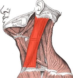
4. Diaphragm - 301. Sternocleidomastoid MucleAccessory Muscle.
Originates in the Sternum + Clavicle and inserts to the mastoid process of the temporal bone of the skull.
Contracts pulling the sternum and clavicle up towards the skull. - 312. Scalene MusclesAccessory Muscle.
3 muscles in the lateral neck - anterior, posterior + medial.
Originates from the transverse processes of C2-C7 and insert onto the first and second ribs.
Contracts pulling the ribs upwards towards the neck. - 32Accessory Muscle of RespirationMuscles that assist, but do not play a primary role in breathing.
- Sternocleidomastoid muscles
- Scalene muscles - 333. External Intercostal MusclesPrimary Muscles of Respiration.
Origin = lower border of ribs -> insert onto the upper border of the rib below.
Contracts - causes rib cage to move up and outwards - increasing the size of the thoracic cavity.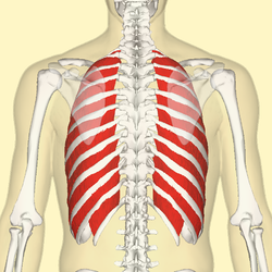
- 344. DiaphragmPrimary Muscle of Respiration.
Under the lungs - base of the thoracic cavity.
Contracts downwards - flattens & pulls the lungs with it -> increases the size of the thoracic cavity. - 35Primary muscles of RespirationMuscles that contribute to inhalation and exhalation by aiding in the expansion and contraction of the thoracic cavity.
- Diaphragm
- Intercostal Muscles - 36Expiration= Breathing Out.
Needed to get rid of any waste products that can harm the body e.g. carbon dioxide.
Need to decrease the volume within the lungs -> increases internal air pressure. - 37Muscles of Expiration (5)1. Internal Intercostals
2. External Oblique
3. Internal Oblique
4. Rectus Abdominis
5. Transversus Abdominis - 381. Internal Intercostal MusclesAccessory Muscle.
Skeletal muscles located between the ribs -> lie deep to the external intercostal muscles.
Contracts - pulls the rib cage down and inwards -> decreasing the size of the thoracic cavity.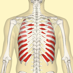
- 392. External Oblique MusclesAbdominal muscle.
Largest and most superficial (outermost) of the 3 flat muscles of the lateral anterior abdomen.
Origin = lower 8 ribs. Inserts various places in the hip.
Pulls rib cage downwards - compresses the abdominal cavity -> pushes the diaphragm up. - 403. Internal Oblique MusclesMuscle in the abdominal wall - lies below the external oblique and just above the tranversus abdominis.
Accessory Muscle of Respiration - 414. Rectus AbdominisAlso known as the "Abdominals" or "Abs".
Origin = pelvis.
Inserts - on the sternum + costal cartilages.
Accessory Muscle.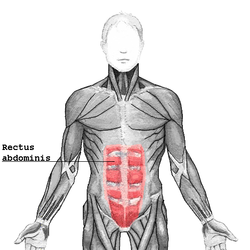
- 425. Transversus AbdominisMuscle layer on the front + side of the abdominal wall.
Layered below the internal oblique muscle. - 43Resting BreathingUnconscious passive process.
Only uses the primary muscles of respiration -> diaphragm & external intercostal muscles.
Elastic recoil of the lungs. - 44Active BreathingConscious process.
Uses the primary + accessory muscles.
Further increases the volume in the thoracic cavity -> when our body requires higher level of oxygen. - 45Clavicular BreathingType of active breathing.
Body requires more oxygen - when exercising or under repiratory dysfunction (asthma attack) -> uses the scalene + sternocleidomastoid muscles.
Information
- Description
- Masters Medicine (Intro to Medicine) Flashcards on 5. The Respiratory System, created by t.whittingham on 06/01/2016.
- Created by
- t.whittingham
- Views
- 10
- Rating