5735720
Description
Flashcards by Amelia Claire, updated more than 1 year ago
|
|
Created by Amelia Claire
almost 8 years ago
|
|
| Question | Answer |
| THE CARDIOVASCULAR SYSTEM: functions to transport materials to and from cells. connects every cell in the body ¯\_(ツ)_/¯ | PUMP= heart MEDIUM = blood CONDUCTING SYSTEM = vessels |
| Pulmonary Circuit (LOW Pressure) *Carries blood to and from the lungs for gas exchange.* | R Ventricle -> Pulmonary Artery -> Lungs -> Pulmonary Vein -> L Atrium -->> SYSTEMIC CIRCUIT |
| Systemic Circuit (HIGH pressure) *carries oxygenated blood from heart to the rest of the body* | L Ventricle -> Aorta -> body -> superior / inferior vena cava // coronary sinus -> R Atrium ---> PULMONARY CIRCUIT |
| Heart <3 | Heart rests within the thoracic cavity, on diaphragm in the MEDIASTINUM. Heart is a socialist, sits LEFT. |
| Pericardial Sac/Pericardium | Double membrane fibrous sac. Filled with serous fluid to reduce friction. Anchors and cushions heart, keeps it in place. OUTER LAYER = parietal pericardium INNER LAYER = visceral layer / epicardium. This layer is attached to the outer layer of heart. |
| The outer layer of the Pericardium | Parietal Pericardium |
| The inner layer of the pericardium | visceral / epicardium attached to OUTER layer of the heart |
| Heart Wall (3 Layers) | epicardium (= visceral layer of serous pericardium) myocardium (=cardiac muscle tissue - pump action) endocardium (= endothelial lining, continuous with great vessels) |
| myocardium | cardiomyocytes / cardiac muscle single central nuclear connected via gap junctions & desmosomes. high aerobic capacity, intercalated discs maintain structure, enhance connections, conduct action potentials. |
| heart anatomy | Great veins and arteries at top of heart (the flat "base") Bottom of heart is pointy apex. |
| Coronary Sulcus | Divides Atria fromVentricles |
| Interventricular Sulcus | Separates the ventricles |
| Atria | Receives blood into the heart (top chamber) |
| Ventricles | Pumps blood from heart (bottom chamber) L Ventricle => blood to body R Ventricle => blood to lungs |
| Interatrial Septum | Separates atria |
| Connective Tissue Septum Separates muscles of atria and ventricles - why? | To isolate the electrical impulses and to ensure the impulse runs on the right track |
| Fossa Ovalis | a shallow depression where the foramen oval was located |
| Auricle | Flap of expandable atrium visible when not filled |
| Blood flow through heart: BLOOD ENTERS via Right Atrium from: Superior/Inferior Vena Cava + Coronary Sinus. | R Atrium -> AV Valve-> R Ventricle -> Pulmonary Trunk -> Pulmonary Arteries -> LUNGS -> pulmonary veins -> L Atrium -> AV Valve -> L Ventricle -> BODY - > S&I Vena Cava/Coronary Sinus-> R Atrium |
| heart Valves prevent back flow of blood. (AV ventricle to aorta, SL artery o heart) (NO VALVES BETWEEN VEINS AND ATRIA) | Atrioventricular Valves (AV) sit between atria and ventricle Right = Tricuspid Left = Bicuspid Semilunar Valves sit at base of aorta and pulmonary artery between ventricles and vessels. |
| How is the opening and closing of the valves controlled? | Valve opens when pressure in Atria is greater than pressure in Ventricle. Valve close when Pressure in Ventricles is greater than pressure in Atria. (pA>pV=OPEN => pV>pA=CLOSE) |
| CORONARY CIRCULATION - how do we get oxygen and nutrients to the heart itself? coronary arteries spread over the heart the Coronary Sinus brings deoxygenated blood back to the atria ... | Coronary arteries branch of ascending aorta. they FILL on diastole (ventricles relaxed) Carry oxygenated blood to myocardium. Pulsatile blood flow, little flow in systole. Arteries recoil when ventricles relax. Mediates pressure, blood keeps moving. |
| Autorhythmic Fibres (found in SA node, AV node, AV Bundle, Bundle Branches and Purkinje Fibres) | Autorhythmic Fibres spontaneously generate action potentials, conduct action potentials along system pass on to contractile fibres. |
| Contractile Fibres Atria contract and relax together Ventricles contract and relax together | 99% of all muscle fibres do the mechanical work. Heart tissue is excitable -> the spread of electrical activity through intercalated discs-desmosomes and gap junctions enables contraction in unison (Syncytium) |
| Conducting System | Sinoatrial node (SA Node) Atrioventricular Node (AV Node) AV Bundle R&L Bundle Branches Purkinje Fibres |
| Sinoatrial Node | SA Node - Pace Maker Spontaneously depolarises sets the basic rhythm (80-100 bp/m) can be modified by neurotransmitters |
| Atrioventricular Node | AV Node - slight delay spontaneously depolarises (40-60 bp/m) the delay enables the atria to fully contract in order to fill ventricle before electrical activity arrives at ventricle and causes ventricular contraction. |
| Autorhythmic Cells | Autorhythmic cells have special ion channels for the action potentials. There is a spontaneous depolarisation and the membrane is constantly drifting towards threshold. There is a long refractory period (plateau) and then depolarisation. prevents tetany |
| Sequence of Events | 1. SA node mass of cells (pacemaker) generates AP spontaneously 2. Stimulus spreads across atria and reaches AV node 3. AV Nodal delay = atria contracts before ventricle 4. AP spreads along AV bundle fibres and Purkinje Fibres 5. AP relayed across ventricles, Ventricles contract |
| Criteria for Efficient Pumping organised by intertribal pathway, internal pathway and AV nodal pathway | Atria Must Contract before Ventricles Coordinate excitation so that each heart chamber contracts as a syncytium Atria contract together, Ventricles contract together |
| Cardiac Cycle | Atrial contraction (atrial systole) -> Atrial relaxation (Atrial diastole) -> Ventricular Contraction (ventricular systole) -> Ventricular Relaxation *Ventricular diastole) Atrial diastole is quite lengthly to allow atria to fill as no valves from vein-> atria Ventricular systole shorter than diastole. |
| Electrocardiogram - summation of APs occurring in heart | P Wave = Atrial depolarisation PR= AV nodal delay, atria contracting QRS = Ventricular Depolarisation ST= Ventricles contract and empty T Wave = Ventricular depolarisation TP= heart at rest, atria filling |
| end diastolic volume (EDV) | volume of blood in each ventricle at the end of ventricular diastole. affected by filling time, duration of ventricular diastole, venous return (blood flow during ventricular diastole). |
| end systolic volume (ESV) | Volume of blood remaining in each ventricle at the end of ventricular systole (~40% of EDV) Preload -> ↑ preload = ↓ ESV contractility -> force during contraction at given preload (influenced by hormones, PSNS, SNS) |
| Stroke Volume (SV) Most important factor in the Cardiac Cycle | Volume of blood pumped out of each ventricle during a beat. SV = EDV - ESV |
| Ejection Fraction | % of EDV represented by SV |
| Cardiac Output (CO) | volume pumped by left ventricle in one minute (CO = HR x SV) Dynamic - can be affected by ANS and hormones |
| heart rate = autonomic innervation of the heart | both SNS and PNS Medulla oblongata Cardioaccelatory Centre ↑HR SNS Cardioinhibitory Centre ↓HR PNS Reflex pathways, baroreceptors, chemoreceptors (CNS) brain stem has both ↑↓ centres |
| Autonomic Tone | Resting heart rate is slightly slower than rate set by node (pacemaker) because PNS slows it slightly - changes the membrane potential of the autorhythmic cell. |
| membrane potential | acetolcholyne means membrane repolarises for longer => takes longer to get to next threshold. membrane potential may be brought closer to threshold ↑ rate of depolarisation |
| HORMONES | adrenaline, noradrenaline and thyroid hormones ↑ HR (SA node)// contractile cells |
| Atrial Reflex + Venous Return | adjustment to heart rate, depending on venous return. Stretch on right atrium walls triggers reflect to ↑ sympathetic activity => ↑ HR Venous return affects nodal cells to ↑ HR |
| Preload and Afterload | Preload =degree of ventricular stretching. ↑ EDV = ↑ preload. After load = tension ventricle produces to open semilunar valve to eject blood. Restricted by high BP, stiff arteries. ↑ force required = ↑ ESV |
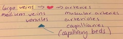
| Blood Vessels | Arteries Arterioles Capillaries Venues Veins |
| which is what? veins? | |
| Arterial walls have three tunics: Tunica Interna - Inner Tunic | Tunica Interna - inner layer Endothelium Basement Membrane Internal Elastic Lamina |
| Arterial Walls have three tunics: Tunica Media - middle tunic | Tunica Media - middle layer - thickest layer Elastic Fibres Smooth Muscle External Elastic Lamina (muscular arteries) |
| Arterial Walls have three tunics: Tunica Externa - outer tunic | Tunica Externa Elastic and Collagen Fibres |
| About Arteries | Arteries are elastic - they stretch and recoil Largest diameter Carry blood away from the heart Tunica Media contains a large number of elastic fibre and store elastic energy, move blood during ventricular diastole |
| Muscular Arteries | Medium sized tunica media has a lot of smooth muscle - constrict and dilate. Superficial Muscular arteries form pressure point to control bleeding or measure pulse. |
| Arterioles | -small arteries deliver blood to capillaries -active in vasoconstriction and vasodilation (SNS/Smooth Muscle) regulators of systemic vascular resistance |
| Metarterioles | supply capillary beds, no smooth muscle at distal end "thoroughfare" can bypass capillary sphincter |
| Capillaries | endothelial cells & basement membrane Enormous surface area and thin barrier for effective diffusion. Exchange nutrients and waste via interstitial fluid |
| True Capillary | Emerge from arterioles or metarterioles, flow regulated by a sphincter |
| Continuous Capillary | uninterrupted lining |
| Fenestrated Capillary | Have pores fond in kidneys (kidney filters, found in choroid plexus (CSF) of brain (blood brain barrier). |
| Sinusoidal Capillary | Large fenestrations, specialised capillaries Found in liver, spleen, bone marrow. |
| Venules | Small veins formed from merging capillaries. They go on to merge with other venues, and form veins. |
| Veins have 3 tunics. | Tunica Interna - valves to prevent backflow Tunica Media Tunica Externa (thickest layer - collagen and elastic fibres) Veins larger in diameter and have a thinner wall than arteries. Do not have the same recoil, "capacitance vessels" |
| blood flow | a volume of blood that flows through a tissue p/unit time. it is dynamically regulated and and determined by blood pressure and resistance. |
| Flow is proportional to change in pressure over resistance | Flow is inversely proportional to resistance |
| Arterial Pressure | 120mmHg at Aorta 35mmHg at Capillary (arteriole end) 35-18mmHg at Capillary (venous end) ΔP=~100mmHg |
| Pressure V Resistance | For flow to occur, circulatory pressure must be > than total peripheral resistance TPR is determined by vascular resistance, viscosity, turbulence. |
| vascular resistance | opposition to blood flow due to friction between blood and vessel wall. =>↑ length ↑ resistance ↑ diameter of vessel ↓ resistance inversely proportional to the 4th power of the radius of the lumen. |
| peripheral resistance | highest at arterioles, as they have an actively controlled radius due to vasoconstriction and vasodilation |
| Turbulence | sudden change in diameter of vessel (plaque deposits, clot) slows blood flow |
| Viscosity | ratio of RBCs (haematocrit/ packed elements?) to plasma |
| Velocity of Blood Flow is Inversely Related to Cross Sectional Area | Velocity decreases as blood flows from aorta to capillaries and increases as it flows from capillaries to veins up to the heart. It is slowest in the capillary for effective nutrient delivery. |
| blood pressure measures pressures during ventricular contraction and relaxation | ~120 Systolic Pressure / 80 Diastolic Pressure |
| Diastole | Passive process; the heart spends more time in diastole than systole |
| systole | ventricular contraction - active process |
| Venous Pressure & Venous Return | Venous Pressure is low Resistance is low (veins have large diameter) and blood flow velocity is increasing as blood is returning to the heart. |
| Venous Return | Volume of blood returning to the heart from systemic veins is maintained by Pressure Gradient established y the Heart the Skeletal Muscle Pump Respiratory Pump Valves |
| Capillary Pressure and Exchange Methods | Diffusion Transcytocis (vesicular transport) Bulk Flow (filtration and absorption) |
| Bulk Flow Capillary Exchange: | Important for regulation of volumes of blood and interstitial fluid osmotic and hydrostatic pressure balance determines how much fluid moves in or out of capillary |
| Hydrostatic Pressure | Pressure blood exerts on the walls of the vessels (pushes fluid OUT) |
| Osmotic Pressure | Opposes hydrostatic pressure. Proteins in plasma not present in interstitial fluid draw water into plasma/capillary from the interstitial fluid (water moves from low c solute to high). |
| What does pressure do at the arteriole end of the capillary ? | Pushes fluid out |
| What does pressure do at the venue end of the capillary? | Pushes fluid IN |
| Oedema | Fluid accumulating in interstitial fluid due to an imbalance; related to R heart failure and valves not working. Venous Pressure increases, fluid not returning to capillaries. |
| Homeostatic mechanisms maintain adequate blood flow to all tissues (PERFUSION) | Primary homeostatic variable is MEAN ARTERIAL PRESSURE MAP = COxTPR => ↑CO ↑TPR ↑MAP |
| Cardiovascular Regulation is regulated by Heart Rate and Stroke Volume | 3 Mechanisms; Autoregulation Neutral Mechanisms Hormonal Mechanisms |
| Autoregulation: | Local factors alter blood flow through capillaries via local dilators/constrictors act on pre capillary sprinters to control blood flow through capillary bed exercise muscle: ↑CO2. Local vasodilation in muscle to ↑ blood flow to get rid of CO2 and access O2. |
| Vasodilators | Lactic acid histamine nitric oxide |
| vasoconstrictors | prostaglandins from platelets |
| Autoregulation Homeostasis Example | Stress, trauma, chemical change ↑ tissue activity. Inadequate local pressure and blood flow Local ↓ in resistance and ↑in blood flow. Homeostasis! |
| Neural Mechanisms | Control via cardiac centres and vasomotor centres in medulla oblongata; vasomotor centres are areas that change blood vessel diameter, changing resistance. |
| Cardiovascular Centre | input from all over the body, higher brain regions can change cardiovascular output. |
| Proprioceptors | detect movement and pressure in joints - linked to afferent system |
| Baroreceptors | detect blood pressure - located in aorta and carotid sinus |
| Chemoreceptors | detect changes in CO2 and O2 |
| Sympathetic Nervous System | Cardioacellerator Nerves ↑ Heart rate Increases contractility Vasomotor nerves => can cause vasoconstriction or vasodilation |
| Parasympathetic Nervus System | Vagus Nerve ↓ heart rate |
| Baroreceptor Reflex | Works second by second to change blood pressure depending on what we are doing --> send impulses to CV centre when BP falls; BP receptors stretched less. CV Centre will ↑ CO, thus, BP will ↑ |
| Sympathetic System (can constrict or dilate vessels) For vasodilation... | homeostasis disturbed baroreceptors stimulated cardioinhibitory centre stimulated // cardioaccelertory system inhibited Vasodilation occurs ↓ Cardiac output HOMEOSTASIS |
| Sympathetic System (can constrict or dilate vessels) For vasoconstriction | Homeostasis disturbed baroreceptors inhibited vasomotor centre stimulated // cardioaccelatory system stimulated vasoconstriction ↑ Cardiac output HOMEOSTASIS |
| Hormones & Cardiovascular Regulation | Endocrine system provides long and short term regulation of Cardiovascular system. adrenaline & noradrenaline from adrenal medulla increase cardiac output and peripheral vasoconstriction |
| homeostatic mechanisms regulate cardiovascular activity to ensure adequate perfusion to tissues: | Determined by CO, TPR and MAP. Regulated by local factors, neural and hormonal mechanisms. |
| Over time, CV system changes with long term training | heart size ↑ Heart rate more efficient SV and EDV ↑ CO ↑ Blood flow more effective Blood Volume ↑ |
| During Exercise | Cardiac output ↑ and blood flow %% changes to various organs and tissues. While the overall % to brain reduces, it still gets the same AMOUNT of blood. |
Want to create your own Flashcards for free with GoConqr? Learn more.