6301299

| Question | Answer |
| 3.6 Organisms respond to changes in their internal and external environments | N/A |
| 3.6.1 Stimuli, both internal and external, are detected and lead to a response | N/A |
| 3.6.1.1 Survival and response | N/A |
| How do organisms benefit from reacting to their environment? | Increased chances of survival if they respond to changes |
| How is growth regulated in plants? | Movement of specific growth factors from growing regions to other tissues, causing growth responses to directional stimuli |
| What is a tropism? | Growth movement of part of plant in response to directional stimulus - can be positive (towards stimulus) or negative (against it) |
| What are some examples of tropism? | - Phototropism - growth response to light e.g. shoot shows positive phototropism growing towards light - Hydrotropism - growth response to water e.g. root shows positive hydrotropism growing towards moisture - Geotropism - growth response to force of gravity e.g. shoot shows negative geotropism growing upwards against gravity |
| Explain the advantages of these tropisms | - Phototropism - allows shoots to grow towards light sources and increase rate of photosynthesis - Hydrotropism - allows roots to grow towards water in the soil to acquire water and minerals for use in photosynthesis and other processes - Geotropism - allows shoots to grow away from soil into the open air and reach sunlight for photosynthesis |
| What is a plant growth factor? | Hormone-like coordinated plant response |
| What is IAA? | Specific growth factor involved in tropism - Indoleacetic acid, belongs to a group of substances called auxins |
| Explain the process of phototropism | Positive phototropism - cells in shoot tip produce IAA, which is then transported down the shoot, initially evenly distributed - light causes movement of IAA from the light side to the shaded side of the shoot - Greater concentration of IAA builds up on the shaded side of the shoot than on the light side - IAA causes elongation of shoot cells beneath the tip and cells on the shaded side elongate more due to greater concentration of IAA - Greater elongation on the shaded side causes the shoot tip to bend towards the light |
| Explain the process of geotropism | Positive geotropism - cells in root tip produce IAA, which is then transported along the root, initially evenly distributed - gravity influences the movement of IAA from the upper side to the lower side of the root - Greater concentration of IAA builds up on the lower side of the root than the upper side - IAA inhibits the elongation of root cells and as there is a greater concentration of IAA on the lower side, the cells on this side elongate less - Greater elongation of cells on the upper side compared to the lower side causes the root to bend downwards in the direction of gravity |
| What is a taxis? | Movement of a cell, organism in response to stimulus |
| What are some examples of taxes | - algae moving towards light (positive phototaxis) - earthworms moving away from light (negative phototaxis) - sperm cells of moss plant attracted towards chemical produced by female reproductive organ (positive chemotaxis) |
| What is kinesis? | Random movement of a cell, organism to search for favourable conditions |
| What are some examples of kinesis? | - shrimp moving randomly to find shade |
| What is a reflex reaction? | Unconcious reaction to environmental stimulus, causing automatic response, bypassing the brain |
| What are the stages of a reflex arc? | - Stimulus detected by receptor e.g. temperature receptors in skin - Impulse carried along sensory neurone to spinal cord - Impulse taken up by intermediate neurone and passed on to motor neurone - Impulse carried by motor neurone to effector e.g. muscle - Effector carries out response |
| What are the advantages of a reflex arc? | - Quick response - More efficient in situations to which the response is always the same - Frees processing power of the brain for other activity |
| What are some examples of reflex responses? | - Earthworm digging upward in response to soil surface vibration - Infant rooting response (baby turning head towards direction of contact) - complex - Human hand withdrawing from hot object |
| 3.6.1.2 Receptors | N/A |
| What is true of receptors? | - They are specific (detect only one form of stimulus) - They are transducers (energy input of stimulus converted into nervous impulse known as GENERATOR POTENTIAL) |
| What is a Pacinian Corpuscle? | Receptor specific to mechanical pressure - found beneath the surface of the skin |
| Diagram of a Pacinian corpuscle | |
| How does pressure lead to a generator potential in a Pacinian corpuscle? | Stretch-mediated Sodium ion channels are deformed by pressure, causing the establishment of a generator potential |
| Human eye diagram | |
| What are the receptors in the retina of the human eye? | Detect light - Rod cells - Cone cells |
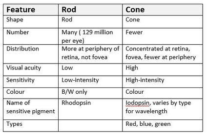
| Features of rod & cone cells | |
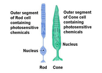
| Structure of rod & cone cells | |
| How does light stimulate rod & cone cells? | Breaks down photosensitive pigment, causes ions to enter |
| What effect do the structural differences of rod & cone cells have on function? | - Rod cells are attached in threes to each bipolar neurone, giving them a sensitivity to lower light as generator potentials are summative, thus are more likely to exceed the threshold to cause an impulse. This gives lower visual acuity however, as only one impulse is transmitted to the brain per three, rather than individually. - Three different variants of pigment in cone cells gives sensitivity to three different ranges of visible light wavelength, allowing colour vision |
| 3.6.1.3 Control of heart rate | N/A |
| What does it mean when the heart is described as myogenic? | Self-stimulating, signal initiated in muscle tissue rather than by nerve impulses from outside (neurogenic) |
| What are the stages of myogenic stimulation? | - wave of electrical exciting spreads out from the sinoatrial node (SAN) across both atrial muscles, which contract - layer of non-conductive tissue (atrioventricular septum) prevents the wave entering the ventricles, the atrioventricular node (AVN) is stimulated by the wave of excitation - after a short delay, the AVN conveys a wave of excitation along Purknye tissue which make up the Bundle of His - the bundle of His conducts the wave through the atrioventricular septum into ventricular muscles, branching into smaller Purknye tissue - wave of excitation released from Purknye tissue, causing the ventricles to contract from the bottom upwards |
| What is the autonomic nervous system? | Involuntary nervous system - sympathetic nervous system stimulates effectors to speed up responses e.g. increasing heart rate - parasympathetic nervous system inhibits effectors to slow down responses e.g. slowing heart rate to conserve energy |
| What is the role of chemoreceptors in regulating heart rate? | Found in the walls of carotid arteries (supply the brain with blood), sensitive to changes in PH - increased carbon dioxide concentration of the blood reduces the PH, caused by increased muscular/metabolic activity - reduced carbon dioxide concentration of the blood increases the PH, caused by reduced muscular/metabolic activity |
| How is heart rate increased/decreased? | When the PH of the blood is too high/low, this is detected by chemoreceptors in the wall of the carotid arteries - chemoreceptors increase the frequency of nervous impulses to the centre in the medulla oblongata that increases/decreases heart rate - this centre increases the frequency of nerve impulses via the sympathetic/parasympathetic nervous system to the SAN - SAN increases/reduces frequency of waves of electrical excitation, thus heart rate increases Faster/slower heart rate causes more/less carbon dioxide to be removed by the lungs, thus blood PH returns to normal in this negative feedback loop (chemoreceptors send fewer impulses) |
| What is the role of pressure receptors in regulating heart rate? | Found in the walls of carotid arteries (supply the brain with blood) and the aorta, sensitive to changes in pressure - when blood pressure is higher than normal, pressure receptors transmit more nerve impulses to the centre in the medulla oblongata that decreases heart rate. This centre sends impulses via the parasympathetic nervous to the SAN, leading to reduced heart rate - when blood pressure is lower than normal, pressure receptors transmit more nerve impulses to the centre in the medulla oblongata that increases heart rate. This centre sends impulses via the sympathetic nervous to the SAN, leading to increased heart rate |
| How is cardiac output generated? | Cardiac output (CO) = Heart rate (R) x Volume (V) |
| 3.6.2 Nervous coordination | N/A |
| 3.6.2.1 Nerve impulses | N/A |
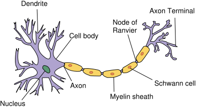
| What is the structure of a myelinated motor neurone like? | - cell body - associated with the production of proteins and neuron transmitters - dendrons - extensions of cell body which subdivide into fibres called dendrites that carry nerve impulses towards the cell body - axon - single long fibre that carries nerve impulses away from the cell body - Schwann cells - surround the axon, protecting it & providing electrical insulation. Carry out phagocytosis and are involved in nerve regeneration. Schwann cells wrap themselves around the axon - myelin sheath - covering to axon made up of membranes of Schwann cells, rich in lipid myelin - nodes of Ranvier - constrictions between schwann cells |
| Diagram of a myelinated motor neurone | |
| What is a resting potential? | Axon membrane contains many channel proteins: - some ion channels have gates to allow or prevent sodium and potassium ions from moving through them by facilitated diffusion - sodium & potassium ions can only move through their own gated channels, however ions can travel though any ungated channels freely The inside of the axon membrane is negative relative to its outside - said to be POLARISED 65mv |
| How is a resting potential established? | Active transport of ions - Sodium ions actively transported out of the axon via the sodium potassium pump - Potassium ions actively transported into the axon via the sodium potassium pump Sodium: potassium 2:1 ratio - although both ions are positive, the outward movement of Na+ is greater than the inward movement of K+ ions. Thus there are more Na+ ions in the tissue fluid surrounding the axon than in the cytoplasm, the opposite is true for K+. This creates an electrochemical gradient, thus the ions begin to diffuse naturally down the concentration gradient (but most of the Na+ gates are closed and the K+ gates open) |
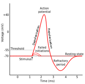
| What is an action potential? | When a stimulus of sufficient size is detected by a receptor in the nervous system, its energy causes a temporary reversal of the charges either side of this point at the axon membrane - the stiumulus inverts the charge across the axon membrane, it is said to be DEPOLARISED +40mv |
| How is an action potential generated? | At resting potential some potassium voltage-gated channels are open (permanently open ones) but sodium gates are closed - energy of the stimulus causes some sodium voltage-gated channels to open, therefore Na+ ions diffuse into the axon along the electrochemical gradient. Being positive they trigger a reversal in the potential difference across the membrane - more Na+ gated channels open during the influx, causing a greater influx - action potential of around +40mv is established, Na+ gates close and K+ voltage gates open - electrochemical gradient is reversed, more K+ channels open and K+ ions flow out of the axon by faciliated diffusion - electrochemical gradient is overshot as the axon is HYPERPOLARISED (inside is more negative relative to outside). Closeable K+ gates close and sodium potassium pump begins REPOLARISATION |
| Graph of an action potential | |
| How does an action potential travel along an unmyelinated axon? | Localised electrical current established by influx of sodium ions causes sodium voltage-gated channels further along the axon to open - nerve impulse propagated along the axon |
| How does an action potential travel along a myelinated axon? | Myelin sheath acts as an electrical insulator preventing action potentials from forming. Each schwan cell has a localised current - action potentials can only occur at nodes of Ranvier, thus action potentials travel by jumping between adjacent nodes in a process called SALTATORY CONDUCTION |
| What is a nerve impulse? | Transmission of an action potential along the axon of a neurone is a nerve impulse - action potential is the same size at the end as when it started |
| What is the all-or-nothing principle? | A stimulus must exceed the threshold value in order to trigger an action potential - any stimulus below this value will not trigger an action potential - all action potentials triggered are roughly the same size The size of a stimulus can be perceived by: - the frequency of impulses generated in a given time - by having different neurones with different threshold values |
| What is the refractory period? | Once an action potential has been created in any region of an axon, there is a period afterwards when sodium ions can not flow in because the sodium voltage gates are closed. - no action potentials can be generated in this time |
| What is the importance of the refractory period? | - Ensures that action potentials are propagated in one direction only - as depolarisation cannot occur backwards as the region is in refractory - Produces discrete impulses - due to the refractory period action potentials are separated, as they cannot occur straight after each other - Limits the number of action potentials - limits the number of action potentials in a given time and thus the strength of stimulus detected, as there is a time in which there can be no impulses |
| How does myelination affect the speed of conductance? | Saltatory conduction increases the speed of conductance, as the nerve impulse jumps along the axon - increase from 30ms^-1 to 90ms^-1 |
| How does axon diameter affect the speed of conductance? | Greater diameter of an axon increases the speed of conductance, as this reduces leakage of ions which make membrane potentials more difficult to maintain |
| How does temperature affect the speed of conductance? | Higher temperature increases the speed of conductance, as this increases the rate of diffusion of ions (due to them having more kinetic energy) - repolarisation is also faster at higher temperatures, as the rate of respiration is increased due to faster enzyme action, thus active transport can occur faster - risk of denaturation of enzymes if temperatures are too high, which would cause the tertiary structures of transport proteins and enzymes to deform |
| 3.6.2.2 Synaptic transmission | N/A |
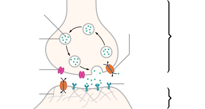
| What is the structure of a synapse? | - presynaptic neurone ends with a synaptic bulb - synaptic bulb contains mitochondria, RER and vesicles containing the neurotransmitter acetylcholine - membrane of presynaptic neurone contains many calcium ion channels - synaptic cleft is the space between the presynaptic neurone and the postsynaptic neurone - membrane of postsynaptic neurone contains many sodium ion channels with receptors for acetylcholine |
| Diagram of a synapse | |
| What are the stages in transmission across a cholinergic synapse? | Excitory Synapse: - action potential reaches synaptic bulb - action potential causes calcium ion channels to open and Ca2+ ions to flood into the synaptic bulb by FACILITATED DIFFUSION - this causes vesicles containing acetylcholine to bind to the presynaptic membrane, where they release acetylcholine into the synaptic cleft - acetylcholine moves via DIFFUSION & binds to receptors on sodium ion channels in the membrane of the postsynaptic neurone, causing the channels to open - sodium ions flow in, causing depolarising and generation of an action potential - acetylcholinesterase hydrolyses acetylchloline into choline and ethanoic acid (causing sodium channels to close), which diffuse back into the presynaptic neurone - ATP is used to recombine choline and ethanoic acid into acetylcholine |
| How does the structure of the synapse ensure unidirectional nerve impulse transmission? | - neurotransmitter is produced only in the presynaptic neurone - receptor proteins are found only on the postsynaptic membrane - acetylcholine is hydrolysed and recylced once and action potential is generated |
| What is spatial summation? | Multiple presynaptic neurones collectively release enough neurotransmitter to exceed the threshold value of the postsynaptic neurone - necessary in instances of low-frequency action potentials |
| What is temporal summation? | A single presynaptic neurone releases enough neurotransmitter in many bursts over a short period of time to exceed the threshold value of the postsynaptic neurone |
| How do inhibitory synapses inhibit nerve impulse transmission? | Inhibitory Synapse: - presynaptic neurone releases a type of neurotransmitter that binds to chloride ion protein channels on the postsynaptic neurone - neurotransmitter causes chloride ion channels to open - Chloride ions (Cl-) move via facilitated diffusion into postsynaptic neurone - binding of the neurotransmitter causes potassium (K+) protein channels nearby to open - potassium ions move via facilitated diffusion out of the postsynaptic neurone into the synaptic cleft - combined effect of Cl- ions moving in & K+ ions moving out causes the inside of the postsynaptic neurone to be very negative relative to the outside - this is called hyperpolarisation and makies it much less likely that an action potential will be generated as many more sodium ions would be needed to cause depolarisation |
| What are the differences between transmission a cholinergic synapse and a neuromuscular junction? | - neuromuscular junction ends an action potential whereas a cholingergic synapse does not - neuromuscular junctions only involve motor neurones and muscle tissue, whereas cholinergic synapses can involve motor, sensory and intermediate neurones - neuromuscular junctions end with muscle tissue whereas cholinergic synapses end with a postsynaptic neurone |
| How can drugs affect transmission across a synapse? | - stimulate the nervous system by causing more action potentials in postsynaptic neurones - a drug may have a shape complementary to neurotransmitters and thus bind to sodium ion channels on the postsynaptic membrane, or may inhibit the enzyme that breaks down neurotransmitter - inhibit the nervous system by creating fewer action potentials in postsynaptic neurones - a drug may inhibit the release of neurotransmitter or block receptors on sodium/potassium channels on the postsynaptic neurone |
| 3.6.3 Skeletal muscles are stimulated to contract by nerves and act as effectors | N/A |
| What are antagonistic muscles? | Muscles that work in antagonistic partnership to cause movement - one contracts while the other relaxes, the muscles switch role to reverse movement - skeleton is incompressible so this is the only way to move |
| What is a skeletal muscle? | Muscle used for movement - attached to the skeleton |
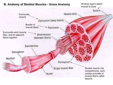
| What are the subdivisions of a skeletal muscle? | - Whole muscle - Bundle of muscle fibres - Muscle fibre - Microfibrils |
| Diagram of subdivisions of skeletal muscle | |
| What is the structure of a microfibril? | Bundle of protein filaments in parallel to each other, appear striated because of banding - made up of two types of protein filament: - thinner actin filaments - thicker myosin filaments |
| What is the banding structure of a microfibril like? | Z line separating each sarcomere - I band consisting of actin filaments - A band consisting of myosin filaments and overlap with actin filaments - H zone consisting of A band not including the overlap |
| Diagram of the microfibril band structure | |
| What are the stages of muscle contraction? | - Action potential reaches the neuromuscular junction, depolarises the muscle fibre membrane and travels into the muscle through t-tubules - sarcoplasmic reticulum opens calcium ion channels, releasing Ca+ ions into the muscle cytoplasm, where they bind to tropomyosin on actin filaments - Tropomyosin moves & uncovers myosin head binding sites, which myosin heads bind to, forming a cross-bridge - Myosin head changes its angle, moving the actin filament along. Myosin head loses ADP molecule, which is replaced by ATP, causing the head to detach from the binding site - ATP is hydrolysed into ADP & Pi, releasing energy for the head to return to its normal position. Myosin head can bind to the next binding sit and continue contraction. - When muscle relaxes, Ca+ ions actively transported back into sarcoplasmic reticulum, returning tropomyosin to its usual place |
| What is the role of phosphocreatine in muscle contraction? | Phosphocreatine donates inorganic phosphate (Pi) to ADP molecules to form ATP, for use in breaking the myosin-actin cross-bridge, forming ATP & creatine |
| What are the types of muscle fibre? | - Slow twitch - Fast twitch |
| What are the characteristics of slow twitch muscle fibres? | - Contract slowly - High proportion in muscles used for posture e.g. back - Good for endurance activities e.g. posture - Can work for a long time without tiring - Energy released slowly through aerobic respiration. Lots of mitochondria and blood vessels to supply oxygen - Reddish because rich in myoglobin (protein storing oxygen) |
| What are the characteristics of fast twitch muscle fibres? | - Contract quickly - High proportion in muscles used for fast movement e.g. eyes, legs - Good for short bursts of speed e.g. sprinting - Tire very quickly - Energy released slowly through anaerobic respiration using glycogen. Few mitochondria and blood vessels - Whitish due to lack of myoglobin |
| 3.6.4 Homeostasis is the maintenance of a stable internal environment | N/A |
| 3.6.4.1 Principles of homeostasis and negative feedback | N/A |
| What is homoeostasis in mammals? | Processes involving physiological control systems that maintain the internal environment within restricted limits |
| What is the importance of maintaining a stable core temperature? | Ensures optimum temperature for enzyme action - too hot and enzymes will denature - too cold and enzymes catalysed reactions will be slower |
| What is the importance of maintaining a stable blood PH? | Ensures optimum PH for enzyme action (varies by enzyme) - extreme PH difference will cause enzymes to denature |
| What is the importance of maintaining a stable blood glucose concentration? | Ensures blood glucose concentration is at normal levels - too low glucose concentration and there will be insufficient respiratory substrate for cells - too high glucose concentration and the water potential of blood will increase, causing water to leave cells |
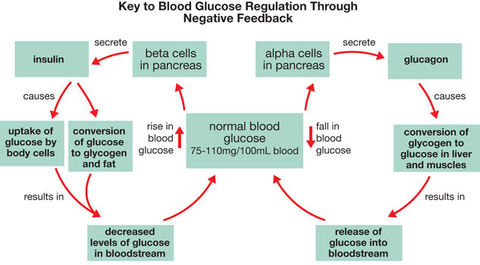
| What is negative feedback? | A change is detected, which causes a response to act against the change - restores system to normality e.g. blood glucose concentration |
| What is positive feedback? | When stimulus is detected, response is continued e.g. oxytocin causes contractions during labour, contractions continue as long as oxyctocin is present - allows response to continue |
| What is the importance of separate feedback mechanisms? | Allows response to stimulus in different directions from original state - greater degree of control |
| 3.6.4.2 Control of blood glucose concentration | N/A |
| What are some factors that influence blood glucose concentration? | - Diet - glucose from the hydrolysis of carbohydrates such as starch, maltose, lactose, sucrose - hydrolysis of glycogen in the small intestine (glycogenolysis) - gluconeogenesis - production of glucose from other substances e.g. fats and protein |
| Why must the concentration of blood glucose be regulated? | - If is too high, the water potential of the blood will fall, cause movement of water out of cells into the blood, dehydrating them - If it is too low, cells will have insufficient substrate for respiration and die due to being unable to carry out vital processes |
| What is glycogenesis? | Formation of glycogen from glucose - takes place in cells e.g. liver and muscle cells when glucose is abundant in the blood and must be stored |
| What is glycogenolysis? | Breakdown of glycogen to form glucose - takes place in cells e.g. liver and muscle cells when glucose is needed for respiration |
| What is gluconeogenesis? | Production of glucose from the hydrolysis of non-carbohydrate substances e.g. fats & proteins - takes place in muscle cells when glucose is not available for respiration and it cannot be absorbed from the blood in sufficient quantities - causes weight loss |
| What is the role of the liver in glycogenesis, glycogenolysis, gluconeogenesis? | These processes all occur in the liver |
| What is the role of insulin in regulation of blood glucose? | When blood glucose concentration is too high, this is detected by receptors on β cells on islets of Langerhans in the pancreas - β cells secrete Insulin into the blood - Insulin binds to glycoprotein receptors on the cell surface membranes of liver and muscle cells (and others) - This causes a change to the tertiary structure of glucose transport carrier proteins, changing their shape and opening them, thus more glucose enters cells by facilitated diffusion - Vesicles containing carrier proteins fuse with the cell surface membrane, increasing the number and thus more glucose enters cells - Insulin also stimulates the enzyme that carriers out glycogenesis - Increases the respiratory rate of cells - Blood glucose concentration is reduced - Insulin is no longer secreted |
| What is the role of glucagon in regulation of blood glucose? | When blood glucose concentration is too low, this is detected by receptors on α cells on islets of Langerhans in the pancreas - α cells secrete glucagon into the blood - Glucagon binds to protein receptors on the cell surface membrane of liver cells - this stimulates the activation of enzymes that carry out glycogenolysis in liver cells - this also stimulates the enzymes that carry out gluconeogenesis in liver & muscle cells - Blood glucose concentration is increased - Glucagon is no longer secreted |
| What is the role of adrenaline in regulation of blood glucose? | In times of stress or excitement, Adrenaline is released by the Adrenal glands (above kidneys) - attach to protein receptors on cell surface membrane of target cells - activate enzymes that cause glycogenolysis in the liver |
| What is the second messenger model of adrenaline and glucagon action? | Hormone attaches to transmembrane protein (embedded in plasma membrane) - receptor shape changes, activating an enzyme called adenylate cyclase inside the membrane - activated adenylate cyclase converts ATP to cyclic AMP, which acts as the SECOND MESSENGER - cyclic AMP activates the protein kinase enzyme - protein kinase enzyme catalyses glycogenolysis/gluconeogenesis |
| Diagram of control of blood glucose negative feedback loops | |
| What is Type I Diabetes? | Caused by genetic factors - pancreas no longer produces insulin, may be the result of an autoimmune response targeting β cells - Symptoms include weight loss, tiredness, glucose in urine, blurred vision - Managed by injections of insulin that match glucose intake |
| What is Type II Diabetes? | Caused by lifestyle factors such as obesity - Glycoprotein receptors on the liver and muscle cells are lost or lose responsiveness to Insulin/inadequate insulin supply - Symptoms include weight loss, tiredness, glucose in urine, blurred vision - Management of carbohydrate in diet and matching this to the amount of exercise taken, may be supplemented by insulin injections or drugs that stimulate insulin production |
| 3.6.4.3 Control of blood water potential | N/a |
| What is osmoregulation? | Control of the water potential of the blood |
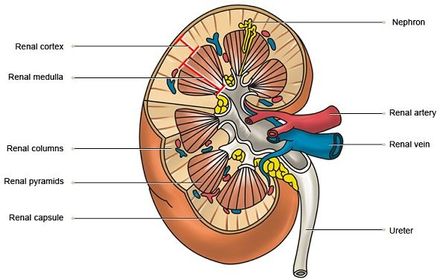
| What the structure of the kidney like? | - Fibrous capsule - membrane surrounding the kidney - Cortex - lighter region made up of Bowman's capsules, convoluted tubules and blood vessels - Medulla - darker region made up of loops of Henle, collecting ducts and blood vessels - Renal pelvis - funnel shaped cavity that collects urine into the ureter - Ureter - tube that carries urine to the bladder - Renal artery - supplies the kidney with blood - Renal vein - returns blood to the heart |
| Diagram of the kidney structure | |
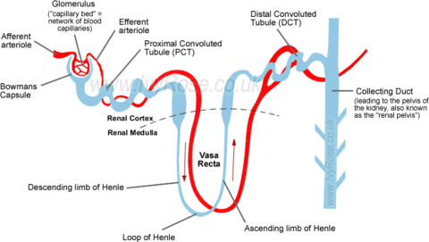
| What is a nephron? | Functional unit of the kidney that filters the blood and produces urine. Made up of: - Bowman's capsule - cup-shaped, surrounds mass of capillaries called glomerulus, inner layer made up of podocytes - Proximal convoluted tubule - loops surrounded by blood vessels, epithelial cells have microvilli - Loop of Henle - loop surrounded by blood vessels - Distal convoluted tubule - loops surrounded by fewer blood vessels than PCT, epithelial cells have microvilli - Collecting duct - tube into which multiple DCT emptu, lined with epithelial cells, becomes wider into pelvis |
| What are the blood vessels relevant to the Nephron? | - Afferent arteriole - tiny vessel that supplies the nephron with blood. Enters the Bowman's capsule - Glomerulus - knot of capillaries in the Bowman's capsule from which fluid is filtered - Efferent arteriole - tiny vessel leaving the Bowman's capsule. smaller diameter than the afferent arteriole, which causes high hydrostatic pressure in the glomerulus - Blood capillaries - network surrounding the PCT, loop of Henle and DCT, where they reabsorb mineral salts, glucose and water, merge to form renal vein |
| Diagram of the nephron structure | |
| What is the role of the nephron in osmoregulation? | - formation of glomerular filtrage by the Bowman's capsule - reabsorption of water and glucose by the proximal convoluted tubule - maintenance of a gradient of sodium ions in the medulla by the loop of Henle - reabsorption of water by the distal convoluted tubule & collecting duct |
| What is the role of the Bowman's capsule? | Formation of glomerular filtrate - blood enters the kidney through the renal artery, into the afferent arteriole, and into the glomerulus - efferent arteriole carries blood away from the glomerulus, and as its diameter is less than the afferent arteriole, this creates hydrostatic pressure in the glomerulus - water, glucose and mineral ions are forced out of the capillary through the gaps between specialised porous cells lining the inside of the Bowman's capsule called podocytes. Protein & blood cells are too large to be filtered - the blood flows out of the glomerulus Movement of substances into the Bowman's capsule must overcome the resistance of: - low water potential of the glomerulus - hydrostatic pressure of fluid in Bowman's capsule - epithelial cells Gaps between podocytes allow movement against this resistance in between rather than through cells |
| What is the role of the proximal convoluted tubule? | Reabsorption of glucose and water, nearly 85% of the filtrate is reabsorbed - sodium ions are actively transported out of epithelial cells lining the PCT into blood capillaries which carry them away. This lowers the Na+ concentration - Sodium ions diffuse down a concentration gradient from the lumen of the PCT into the epithelial cells via co-transport through carrier proteins - glucose, amino acids, chloride ions, water are co-transported into epithelial cells along with Na+ ions - these substances move via facilitated diffusion back into the blood Epithelial cells lining the PCT are adapted for absorption with - microvilli to provide a large surface area - infoldings at bases give a large surface area - high density of mitochondria provide energy for active transport |
| What is the role of the loop of Henle? | Maintaining a gradient of sodium ions in the medulla so that water potential is lower than in the collecting duct, reabsorbing some water - sodium ions are actively transported out of the Ascending limb (impermeable to water) using ATP produced by the many mitochondria, lowering the water potential of the interstitial region - water moves via osmosis from the Descending limb (permeable to water) into the interstitial region, into the blood and is reabsorbed - filtrate water potential decreases down the Descending limb as more water is lost, reaching its lowest at the bend of the loop of Henle - at the bend of the loop, sodium ions move via facilitated diffusion out of the filtrate into the interstitial region, and ions are pumped out in the Ascending limb, so filtrate water potential increases going up - this creates a gradient of water potential, with water potential decreasing from the cortex down into the medulla, as ions are more concentrated further down. Water potential is furthered lowered in the interstitial region by the absorption of water by capillaries. |
| What is the role of the distal convoluted tubule? | Reabsorption of water, make final adjustments to water and salts that are absorbed, control the PH of the blood by selecting which ions to absorb - permeability of walls altered by hormones |
| What is the role of the collecting ducts? | Reabsorption of water - counter-current multiplier created by the loop of Henle ensures that although the water potential of the filtrate going further down the collecting duct decreases, the water potential of the interstitial region is always lower further into the medulla. This ensures constant, steady absorption of water. |
| What is the role of the hyperthalmus in osmoregulation? | Detects change in blood water potential, produces ADH, stimulates pituitary gland to secrete ADH - osmoreceptor cells in the hypothalamus of the brain detect a fall in water potential. These cells shrink due to water loss when the water potential of the blood supplying them is lower than normal. - Shrinking of osmoreceptors causes hypothalamus to produce a hormone called Anti-Diuretic hormone (ADH), which passes to the posterior pituitary gland - when water potential of the blood increases, the hypothalamus sends fewer impulses to the pituitary gland thus less ADH is secreted |
| What is the role of the posterior pituitary in osmoregulation? | Secretes ADH into the blood - posterior pituitary gland secretes ADH into the capillaries - stimulated by impulses from the hypothalamus |
| What is the role of antidiureutic hormone (ADH) in osmoregulation? | Increases reabsorption of water in the kidney - ADH travels in the blood to the kidney, where it binds to protein receptors on the cell surface membranes of epithelial cells of the DCT and collecting duct - binding of ADH activates phosphorylase enzyme in the cells, which causes vesicles containing sections of plasma membrane to bind to the cell surface membrane - these sections of plasma membrane are studded with water channel proteins called aquaporins, thus when the vesicles bind the permeability of epithelial cells to water is greatly increased by increasing the number of aquaporins. - increased permeability of membranes to water causes water to move via osmosis into the interstitial space at a greater rate - ADH also increases the permeability of cells to urea, so if water potential of the blood is extremely low, urea can be reabsorbed to lower the water potential of the interstitial space and cause absorption of more water - when water potential of the blood increases, less ADH binds thus the membrane becomes less permeable |
| How does ADH affect the concentration of urine? | - increased ADH makes urine more concentrated by causing more water to be absorbed |
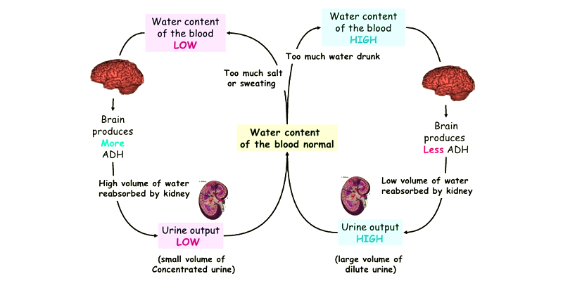
| Diagram of osmoregulation negative feedback loop |
Image:
Osmoregulation (image/png)
|
Want to create your own Flashcards for free with GoConqr? Learn more.