6503193
Description
Flashcards by Ashutosh Kumar, updated more than 1 year ago
|
|
Created by Ashutosh Kumar
over 7 years ago
|
|
| Question | Answer |
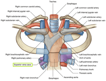
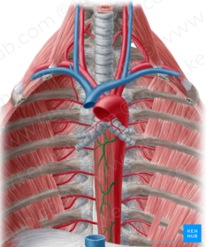
| Name the structures in relation to the SVC and right brachiocephalic vein that are Anterior: Posterior: Right: Left: | Anterior: Sternum Posterior: Azygos vein, vagus nerve and trachea Right: Right lung, Right phrenic nerve and internal thoracic artery Left: Ascending arota, brachiocephalic trunk and left brachiocephalic vein |
| The brachiocephalic veins unite to form the SVC behind the right first costal cartilage but what is the surface marking for their origin? | The brachiocephalic veins are formed by the union of the internal jugular and subclavian veins posterior to the sternoclavicular joint. |
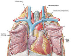
| Describe the relation of the phrenic and vagus nerve at the thoracic inlet: | The phrenic nerves are lateral to the vagus nerves at the thoracic inlet and pass anteriorly in the superior mediastinum. |
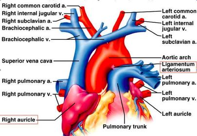
| What is the ligamentum arteriosum? | The ligamentum arteriosum is the remnant of the ductus arteriosus, a vessel that shunted blood from the pulmonary trunk to the aorta in the fetus in order to bypass the nonfunctioning lungs. The ligamentum arteriosum passes from the bifurcation of the pulmonary trunk to the inferior surface of the arch of the aorta. |
| What is the surface marking for the arch of the aorta? | The arch of the aorta begins and ends (i.e. joins with the ascending and descending aorta, respectively) just below the level of the sternal angle (T4/5 vertebra). |
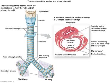
| Describe the relations of the trachea Anterior: Posterior: Left: Right: | Anterior: Arch of the aorta, brachiocephalic trunk and left brachiocephalic vein Posterior: Esophagus and recurrent laryngeal nerves Left: Arch of the aorta, descending aorta and left vagus nerve Right: Azygos vein, right brachiocephalic and SVC |
| What is the surface marking of the tracheal bifurcation? | Anatomy texts state that this is at the level of the sternal angle (T4/5 vertebral level) but recent research from our department indicates that is below this level. |
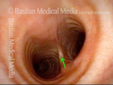
| Name: Demarcates: | Carina; an anteroposterior ridge which demarcates the internal division of the left and right main bronchus easily seen during bronchoscopy |
| Why might widening of the carina seen at bronchoscopy be a potentially sinister sign in the evaluation of a patient with lung cancer? | Widening of the carina at bronchoscopy can indicate enlarged (sub-carinal) lymph nodes at the tracheal bifurcation from metastatic spread. |
| Describe the constituents of the trachea: | |
| What clinical relevance does the diameter and orientation of the right main bronchus have to accidental inhalation of a foreign body? | The right bronchus is usually wider and more vertical than the left. Also, the carina tends to lie slightly to the left of the midline of the trachea. This means that an inhaled foreign body more often enters the right main bronchus than the left. |
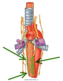
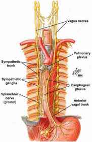
| Name: Constituents: | The left vagus nerve descends down onto the esophagus to form the esophageal plexus (formed by branches of the left vagus nerve and visceral branches of the sympathetic trunk). |
| Name: Source: | Esophageal plexus of arteries Arise as visceral branches of the thoracic aorta |
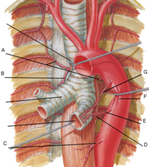
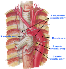
| Name G, E and B: | Besides the esophageal arteries, the bronchial arteries are the other visceral branches of the thoracic aorta G: Left superior bronchial artery E: Left inferior bronchial artery B: Right bronchial artery |
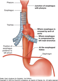
| If you were examining the oesophagus with an endoscope, why might it appear indented at three main sites? | The oesophagus may appear indented: 1. At the junction with the pharynx (the cricopharyngeus muscle) 2. Where it is crossed by the left main bronchus 3. Where it passes through the oesophageal hiatus in the diaphragm. A swallowed foreign body such as an accidentally ingested coin can be temporarily held up at these sites |
| Name: Course: | The sympathetic trunks lie anterior to the neck of the ribs in the upper thorax and on the lateral aspect of the vertebral bodies lower down. |
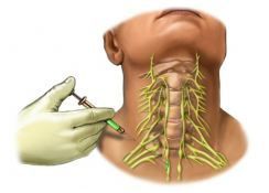
| Name the structure being anesthetized: Location: | Inferior (stellate) cervical ganglion Location: Anterior to the transverse process of C7 and neck of the first rib. |
| Drugs were given to try and encourage the ductus to close but this was unsuccessful and so the baby had the Patent ductus arteriosus tied off surgically via a thoracotomy through the left side of the chest. The baby’s heart failure improved but he remained breathless and a chest X-ray showed a raised left hemidiaphragm due to paralysis of the left side of the diaphragm. What nerve supplies the diaphragm and why might it have been injured during this operation? | The diaphragm receives its motor innervation from the phrenic nerve (formed by the C3,4, and 5 nerve roots) (“C345 keeps the diaphragm alive”). When the surgeons closed the patent ductus arteriosus they accidentally bruised the left phrenic nerve, which runs anteriorly under the mediastinal pleura. This caused temporary paralysis of the left hemidiaphragm. Paralysis of the diaphragm prevents it from contracting (flattening) in inspiration and so it is pulled upwards passively by negative intrathoracic pressure. |
| Describe the effect of a patent ductus arteriosus (PDA): | A patent ductus arteriosus (PDA) allows a proportion of oxygenated blood ejected from the left ventricle into the aorta to flow back to the lungs via the pulmonary arteries (because of the pressure gradient from the aorta to the pulmonary arteries). If this shunt is substantial, the infant becomes short of breath because there is extra blood going to the lungs through the PDA, causing pulmonary congestion and increasing the infant’s work of breathing. |
Want to create your own Flashcards for free with GoConqr? Learn more.