6629338
Description
Flashcards by Ashutosh Kumar, updated more than 1 year ago
|
|
Created by Ashutosh Kumar
over 7 years ago
|
|
| Question | Answer |
| List the 3 distinguishing features of the large intestine: | 1. Tenia coli (3 distinct bands of smooth muscle fibres running longitudinally): Omental tenia Mesocolic tenia Free tenia 2. Haustra (Lengthwise contraction, sacculations between the tenia coli, internally seen as semilunar folds). 3. Epiploic appendages (omental appendices) (small fatty projections of the omentum). |
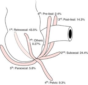
| Describe the appendix: List the positions of the appendix in order of prevalence: | The appendix is a 6-10 cm blind diverticulum containing lymphoid tissue. The ostium of the appendix lies on the posteromedial cecum. The Appendix can lie in a number of different positions: 1. Retrocecal 2. Subcecal 3. Postileal 4. Pelvic (can be considered the same as subcecal) 5. Preileal. |
| Describe how the vagal trunks arise: | On the anterior aspect of the esophagus the esophageal plexus can be found. This is contributed to by the PNS via right and left vagal nerves, and the SNS via the greater splanchnic nerve (T5-9). From the esophageal plexus, the left vagus nerve will form the anterior vagal trunk whereas the right vagus nerve will form the posterior vagal trunk. |
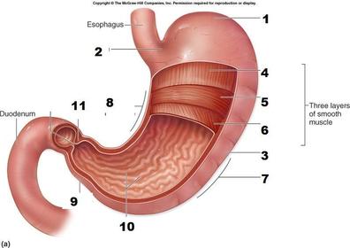
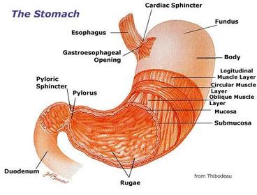
| 1.Fundus, where air collects 2.Cardia (T11) 3.Body, where food accumulates 4. Outer longitudinal layer 5. Middle circular layer 6. Inner oblique layer 7. Greater curvature 8. Lesser curvature (gastric canal; a furrow, forms during swallowing) 9. Pylorus 10. Gastric rugae; longitudinal contractions of the gastric mucosa, temporary or permanent. 11. Pyloric sphincter; circular smooth muscle band around pyloric orifice; controls delivery of chyme to duodenum. | |
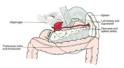
| List the structures which contribute to the stomach bed: | 1. Upper pole of the left kidney. 2. Left adrenal gland. 3. Body and tail of pancreas. 4. Diaphragm. 5. Transverse mesocolon. |
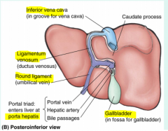
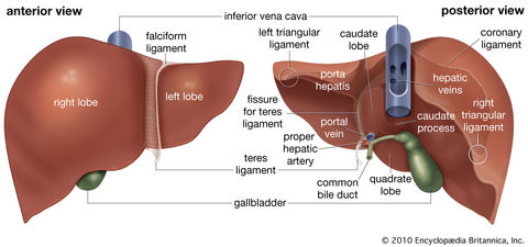
| Describe the ligaments of the liver: | Falciform ligament- a peritoneal ligament which runs in the sagittal plane from the anterior surface of the liver to the anterior abdominal wall as far inferior as the umbilicus. In the free inferior margin of the falciform ligament is the ligamentum teres/round ligament. This is the obliterated umbilical vein. The falciform ligament will then extend up the anterior surface of the liver to the superior surface, dividing to form the coronary ligaments, which at the margins of the liver are called the right and left triangular ligaments. The coronary ligaments join superiorly at the diaphragm, resulting in the liver moving with respiration. In addition, on the liver, where the coronary ligaments are yet to join with each before they ascend, there is a ‘bare area’ where the liver is not covered by peritoneum. On the posterior aspect the ligamentum venosum can be found. This is a remnant of the obliterated ductus venosus, which joined the left portal vein with the IVC, thereby bypassing the liver and increasing the oxygenated blood supply to the brain. |
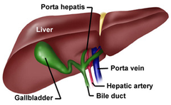
| Describe the porta hepatis: | Structures enter and leave the liver via the porta hepatis (the point of entry to the liver): The right and left hepatic arteries. Portal vein. The right and left hepatic ducts. |
| Describe the two surfaces of the liver: | 1. Diaphragmatic surface; right and left triangular ligaments, coronary ligaments and 'bare area'. 2. Visceral surface; right, left, quadrate and caudate lobes. |
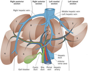
| Describe the divisions of the liver: | The liver consists of 4 anatomical lobes; Right, left, caudate and quadrate lobes. The liver can also be divided into 8 functional segments; each served by subdivisions of the portal vein, hepatic artery and bile duct. |
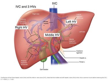
| Describe the venous drainage of the liver: | The liver is drained by left, right and middle hepatic veins. |
Want to create your own Flashcards for free with GoConqr? Learn more.