6635975
| Question | Answer |
| CHAPTER 3: MOVEMENT OF SUBSTANCES | ... |
| 1. Explain using the terms ‘concentration gradient’ and stating the two regions that it is between in appropriate situations. - defn of conc gradient - defn of water potential -defn of permeable membrane -defn of partially permeable membrane | Concentration gradient ➔ difference in concentration of substances between two regions. Water potential ➔ the tendency of water to move from 1 place to another Permeable membrane ➔ allows both solvent & solutes to pass through it Partially permeable membrane ➔ allows some substances, but not others, to pass through it e.g. the cell surface membrane |
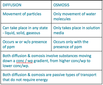
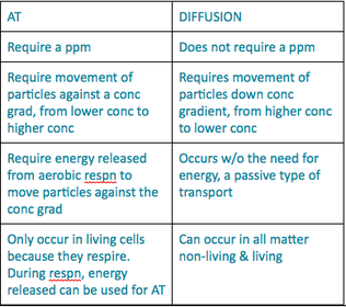
| 2. Define diffusion, osmosis and active transport. | Diffusion ➔ the net movement of particles from higher conc to lower conc, down a conc gradient, without the need for energy. Osmosis ➔ the net movement of water molecules from higher wp to lower wp, down a wp grad, across a ppm, a passive type of transport w/o the need for energy Active transport ➔ movement of particles against a conc grad, from a lower conc to higher conc, across a ppm, with the help of energy released from respn |
| 3. Describe the importance of diffusion in nutrient uptake in humans and gaseous exchange in plants and humans. (a) Nutrient uptake in humans | In the small intestine, when the conc of glucose & a.a in the lumen of the small intestine is higher than in the blood capillaries of the villi, glucose & a.a are absorbed by diffusion into the blood capillaries of the villi. Glycerol & fatty acids are absorbed by diffusion into the epithelium & then combine to form minute fat globules which then enter the lacteals. |
| 3. (b) Gaseous exchange in humans | - Gaseous exchange in alveoli of lungs occur by diffusion. Blood entering the lungs has a lower conc of O2 & higher conc of CO2 than atmospheric air entering the alveoli in lungs - Conc gradient for O2 & CO2 is set up btwn the blood in the blood capillaries & the atmospheric air in the alveoli - Oxygen dissolves in the moisture lining the alveolar walls & diffuses into the blood capillaries. - CO2 diffuses out of RBCs, out of the blood capillaries & into the alveoli, where it is expelled out when exhaling |
| 3. (c) Gaseous exchange in plants | - In daylight when photosynthesis occurs, the CO2 in leaf is rapidly used up. The CO2 conc in the leaf becomes lower than that in the atmospheric air, so a diffusion gradient exists. Therefore, CO2 diffuses from the surrounding air through the stomata into the air spaces in the leaf - The surfaces of the mesophyll cells are always covered by a thin film of water so that CO2 can dissolve in it - The dissolved CO2 then diffuses into cells During the night, O2 diffuse in the same way as how CO2 diffuse in during daytime. |
| 4. Describe the effects of osmosis on plant and animal tissues. | Refer to 10: Effect of osmosis on plant & animal tissues *Slightly different: - When plant tissues are placed in soln of higher wp, the tissues become TURGID (same as cells) - When plant tissue is placed in soln of lower wp, tissues become flaccid (DIFF from cells - plasmolysed) |
| 5. Describe the importance of active transport as in ion uptake by root hairs and uptake of glucose by cells in the villi. | ION UPTAKE BY ROOT HAIRS: When the conc of ions in the soil soln is lower than that in the root hair cell sap, the root hairs have to absorb the mineral ions by active transport, against a conc gradient. The energy for this process comes from the cellular respn in the root hair cells. UPTAKE OF GLUCOSE BY CELLS IN THE VILLI: When the conc of glucose in the lumen of the small intestine is lower than that in the blood capillaries in the villi of the small intestine, glucose is absorbed by active transport into the blood capillaries in the villi of the small intestine. Energy for this process is released by the aerobic respn by the cells in the villi. |
| 6. State the similarities and differences between diffusion and osmosis. | |
| 7. State the differences between active transport and diffusion. | |
| 8. State and explain the factors (e.g. surface area to volume ratio, temperature, concentration gradient and particle size) that affect the rate of diffusion, osmosis and active transport. | (1) Temperature ➔ the higher the temp, the higher the KE of the particles, the faster they move & thus faster rate of diffusion (2) Particle size ➔ the smaller the particle size, the greater the SA:V of the particles the faster the rate of diffusion (3) SA:V ➔ the higher the SA:V, the faster the rate of diffusion (4) Conc grad ➔ the steeper the conc grad, the faster the rate of diffusion & osmosis, BUT for AT, the steeper the conc gradient, the slower the rate of AT (5) Thickness of barrier ➔ the thicker the barrier, the greater the diffusion distance & the slower the rate of diffusion |
| 9. Use the terms hypotonic, hypertonic and isotonic solutions in appropriate situations. | Hypotonic solution: It is when the water potential outside the cell is higher than the water potential inside the cell. (in relation to animal cell only) Hypertonic solution: It is when the water potential inside the cell is higher than the water potential outside the cell. (in relation to animal cell only) Isotonic solution: It is when the water potential inside and outside the cell is the same. Water then moves in and out of the cell at equal rates, therefore there is no net movement of water molecules. |
| 10. Describe fully, what happens, when... (a) A plant cell is placed in a soln of higher wp | When a plant cell is placed in a soln of higher wp, the wp of the soln is higher than than the cell sap of the plant cell. By osmosis, the water molecules move from the solution into the cell, through the pp cell surface membrane, down a wp gradient. As water molecules enter the cell, the vacuole increases in size & pushes the cytoplasm against the cell wall. The cell does not burst due to the inelastic cell wall. The turgidity of the cell with water is called turgor. The pressure exerted by water in the vacuole on the cell wall is the turgor pressure. |
| (b) a plant cell is placed in a solution of lower water potential? | When a plant cell is placed in a soln of lower wp, the wp of the cell sap is higher than the wp of the soln. By osmosis, water molecules move out of the cell, through the pp cell surface membrane, down a wp gradient. As water molecules leave the plant cell, the vacuole decreases in size & causes the cytoplasm & the cell membrane to shrink away from the cell wall. Shrinkage of cytoplasm & cell membrane away from cell wayy is called plasmolysis. The cell is said to be plasmolysed (& the tissue is flaccid) |
| (c) an animal cell is placed in a hypotonic solution? | When an animal cell is placed in a hypotonic solution, the wp of the soln is higher than the wp of the cytoplasm inside the cell. By osmosis, water molecules move into the cell, through the pp cell surface membrane, down a wp gradient. As more water enter the cell, the animal cell swells in size & may burst because it does not have an inelastic cell wall to protect it. |
| (d) an animal cell is placed in a hypertonic solution? | When an animal cell is placed in a hypertonic soln, the qp of the soln is lower than the cytoplasm inside the cell. By osmosis, water molecules move out of the cell into the soln, through the pp cell surface membrane down a wp gradient. As the cell loses water, the cell shrinks & little spikes appear on the cell surface membrane of the cell & this process is called crenation. Animal cell becomes crenated. |
| 10. (e) What is turgor pressure & what are the advantages & disadvantages of it? | * Turgor pressure ➔ the pressure exerted by the water in the vacuole on the cell wall Advantages: - Plays an impt role in maintaining the shape of soft tissues in plants. Most leaves & young stems, esp those of herbaceuos & non-woody plants, are able to remain firm & erect because of turgor pressure within cells. Turgor pressure allows young plants to stay upright & absorb light for photosynthesis. - The turgor pressure in the leaf mesophyll cells helps to support the leaf & keep it firm & spread out widely to absorb sunlight for photosynthesis. |
| CHAPTER 9: TRANSPORT IN PLANTS | ... |
| 1. State the structure and functions of xylem and phloem. (ai) Functions of xylem | (1) Conducting water & dissolved mineral salts from the roots to the stems & leaves (2) Providing mechanical support for the plant |
| 1. (aii) Structure of xylem tissue | Xylem tissue consists mainly of: Xylem vessels ➞ a long hollow tube extending from roots to leaf The xylem vessel is a structure made up of many dead cells. The inner walls are strengthened by deposits of lignin, in the form of rings,spirals or the whole vessel is lignified except in pits. |
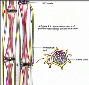
| 1. (bi) Function of phloem | Conducts manufactured food (sucrose & a.a) from the green parts of the plant, esp the leaves, to other parts of the plant. |
| 1. (bii) Structure of phloem tissue | Phloem tissue consists of sieve tubes + companion cells - A sieve tube ➞ consists of columns of elongated, thin-walled living cells called sieve tube cells or sieve tube elements. - The ‘end-walls’ separating the cells have a lot of minute pores. The cross walls are called sieve plates - A mature sieve tube cell has only a thin layer of cytoplasm inside the cell. This cytoplasm is connected to cells above & below through sieve plates. - Each sieve tube cell has lost its large central vacuole, nucleus & most organelles. - Each sieve tube also has a companion cell beside it, which carries out the metabolic processes needed to keep the sieve tube cell alive. Each companion cell is a narrow, thin-walled cell with many mitochondria, cytoplasm and a nucleus. Companion cells provide nutrients & help sieve tube cells to transport manufactured food. |
| 2. Describe how the xylem and phloem are adapted to its functions. (a) Xylem | (1) The xylem vessel has an empty lumen w/o protoplasm or ‘end-walls’ ➞ will not obstruct the flow of water & mineral salts/ reduce resistance to water & mineral salts flowing through xylem & can move easier (2) Walls of the xylem vessel are thickened with lignin ➞ Lignin is a hard & rigid substance, this prevents the collapse of the vessel as all the xylem vessels together provide mechanical support for the plant |
| 2. (b) | (1) Companion cells have many mitochondria ➞ which provide the energy needed, through respiration, for the companion cells to load sugars from the mesophyll cells into the sieve tubes by active transport (2) The holes in the sieve tubes ➞ allow rapid flow of manufactured food substances through the sieve tubes |
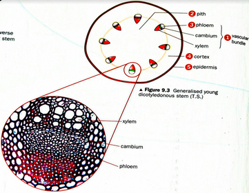
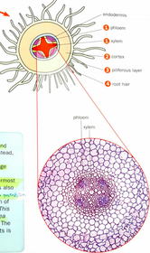
| 3. Compare and contrast the vascular tissues organised in the stems, roots and in the leaves. (i) Arrangement in stem | - Xylem & phloem are grouped tgt to form vascular bundles - The vascular bundles are arranged in a ring around the central region called the pith - Phloem lies outside & xylem lies on the inside with tissue called cambium btwn them. Cambium cells can divide & differentiate to form new xylem & phloem tissues which lead to the thickening of the stem. - Region btwn the pith & the epidermis ➞ cortex. Both cortex & pith serves to store up food substances, such as starch - The stem is covered by a layer of cells called the epidermis. The epidermal cells are protected by a waxy, waterproof cuticle that greatly reduces evaporation of water from the stem. |
| 3. (ii) Arrangement in roots | - Xylem & phloem are not bundled that. Instead, they alternate with each other. - Cortex of the root is also a storage tissue - Epidermis of the root is the outermost layer of cells. It bears root hairs & is also called the piliferous layer - Each root hair is a long & narrow protrusion of an epidermal cell. This long & narrow protrusion increases the SA:V of the root hair cell. The absorption of water & mineral salts is increased through this adaption. |
| 3. (iii) Arrangement in leaf | -The lamina has a upper epidermis made up of a single layer of closely packed cells. The upper epidermis is covered on the outside by a way & transparent cuticle layer - The mesophyll cells lies just btwn the upper & lower epidermis. It is the main site of photosynthesis. The mesophyll consists of 2 types of tissues - palisade mesophyll & spongy mesophyll (a) Palisade mesophyll ➞ consists of 1 or 2 layers of closely packed, long & cylindrical cells. Cells contain numerous chloroplasts. (b) Spongy mesophyll ➞ cells are irregular in shape. Numerous large intercellular air spaces among the cells. Cells carry out photosynthesis but contain fewer chloroplasts than the palisade mesophyll cells. Cells are covered with a thin film of moisture. Contains the transport tissue, XYLEM & PHLOEM (xylem on top, phloem below) which are grouped to form the vascular bundle. - Lower epidermis, which is coved by an outer layer of cuticle to reduce water loss through epidermal cell - Lower epidermis contain many minute openings called stomata |
| 4. Describe the process of the entry of water, in terms of water potential, from the soil to xylem. | (1) Each root hair is a fine tubular outgrowth of an epidermal cell. It grows between the soil particles, coming in close contact with the soil soln surrounding them 2) The thin film of liquid surrounding each soil particle is a dilute soln of mineral salts (3) Sap in the root hair cell is a relatively concentrated solution of sugars & various salts. thus, sap has a lower wp than the soil soln. These 2 solns are separated by the pp cell surface membrane of the root hair cell. Water enters the root hair cell by osmosis. (4) Entry of water dilutes the sap. The sap of the root hair cell now has a higher wp than that of the next cell. Hence, water passes by osmosis from root hair cell into the inner cell (5) Similarly, water passes from this cell to the next cell of the cortex. This process continues until water enters xylem vessels & move up the plant. |
| How do root hairs absorb ions or mineral salts? | When the conc of ions in the soil soln is lower than that in the root hair cell sap, ACTIVE TRANSPORT. - Root hairs absorb ions against a conc gradient by active transport - Energy for this process comes from cellular respn in the root hair cell When conc of ions is higher in soil soln than in root hair cell sap, by DIFFUSION down a conc gradient |
| 5. Describe and explain the adaptation of root hair cell for its function. | Long & narrow root hair / elongated protrusion ➞ Increases the SA:V of the cell & thus water & mineral salts can be absorbed at a faster rate. Large no of mitochondria (more mitochondria than normal cells) ➞ allows large amount of energy to be released through aerobic respn to allow for active transport of mineral salts Large central vacuole ➞ allows for large amt of mineral salts & water for storage High conc of solutes in large central vacuole ➞ decreases wp of the cell sap |
| 6. Define transpiration. | Transpiration ➞ the loss of water vapour from the aerial parts of the plant, especially through the stomata of the leaves. Water continuously moves out of mesophyll cells by osmosis to form a thin film of moisture over their surfaces down a water potential gradient. [1] Water evaporates from this thin film of moisture and move into the intercellular air spaces in the leaf. [1] The water vapour which is present in a higher concentration in the large intercellular air spaces in leaves will diffuse out of the leaves through the stomata to the drier atmosphere air at a lower concentration, down a concentration gradient. [1] |
| 7. Describe the movement of water from the leaf to the air outside. | - Water continuously moves out of the mesophyll cells to form a thin film of moisture on their surfaces - Water evaporates directly from this thin film of moisture & moves into the intercellular air spaces. Water vapour accumulates in the large air spaces near the stomata - Water vapour then diffuses through the stomata to the drier air outside the leaf ➞ transpiration - As water evaporates from the mesophyll cells , the wp of the cell sap decreases. The mesophyll cells then begin to absorb water by osmosis from cells deeper inside the leaf. These cells, in turn, remove water from the veins (from xylem vessels) - This results in transpiration pull, a suction force which pulls the whole column of water up the xylem vessels. |
| 7. [TYS 2012 9.(a)] Explain how water passes from a mesophyll cell to the atmosphere. [5] | Water is produced during the aerobic respn of the mesophyll cells. Hence, mesophyll cells has a higher water potential than the surrounding, thus, water leaves the mesophyll cell by osmosis, [1] into the thin film of moisture [1] surrounding the mesophyll cells. Water evaporates [1] and diffuses into the large intercellular air spaces [1] surrounding the spongy mesophyll cells and the vapour accumulates there. Since there is a higher concentration of water vapour in the leaf than the external atmosphere, water vapour diffuses out of the stomata, [1] down a water vapour concentration gradient, and into the atmosphere. |
| 8. Describe the movement of carbon dioxide from the leaf to the atmospheric air. | - In daylight when photosynthesis occurs, the CO2 in leaf is rapidly used up. The CO2 conc in the leaf becomes lower than that in the atmospheric air, so a diffusion gradient exists. Therefore, CO2 diffuses from the surrounding air through the stomata into the air spaces in the leaf - The surfaces of the mesophyll cells are always covered by a thin film of water so that CO2 can dissolve in it - The dissolved CO2 then diffuses into cells |
| 9. Identify and explain the factors affecting the rate of transpiration. | (i) Humidity of air (ii) Wind / air movement (iii) Temperature of the air (iv) Light |
| (i) Humidity | The intercellular air spaces are usually saturated with water vapour. There is a water vapour conc grad (WVCP) between the leaf & the atmosphere. - When the humidity of the air increases, the atmosphere will become more saturated with water vapour, thus, the WVCP between the intercellular air spaces in the leaf & the air outside the leaf becomes less steep. As a result, rate of diffusion of water vapour from the intercellular air spaces into the the surrounding air will be lower, thus resulting in a lower rate of transpiration - When the humidity decreases, the amount of water vapour in the atmospheric air decreases, thus causing the WVCP between the intercellular air spaces in the leaf & the atmospheric air to be more steep. As a result, the rate of diffusion of water vapour from the intercellular air space into the surrounding air increases, thus increasing the rate of transpiration. |
| (ii) Wind / air movement | Wind blows away the water vapour accumulated outside the stomata. This maintains the WVCG btwn the leaf & the atmosphere - When there is strong wind, there will be increased air movement and the moving air causes the water vapour molecules accumulated outside the stomata of the leaves to be blown away. This maintains the WVCG btwn the atmosphere & the intercellular air spaces in the leaves. The WVCG remains steep, thus, increasing the rate of diffusion of water vapour out of the leaves into the atmossphere & increasing the rate of transpiration. - In still air / no wind present, there will be little air movement & the water vapour molecules accumulated outside the stomata will not move away as quickly, causing the air around the stomata more humid & saturated with water vapour. This causes the WVCG to be not as steep, decreasing the rate of diffusion of water vapour out of the leaves & decreasing the rate of transpiration. |
| (iii) Temperature | An increase in the temp of air leads to an increase in the rate of transpiration. As the temp increases, the KE of the water molecules on the surface of the mesophyll cells increase & are able to escape from the thin film of moisture, leading to greater rate of evaporation of water from the thin film of moisture. Thus, the intercellular air spaces in the leaf becomes more saturated with water vapour, causing the WVCG btwn the intercellular air spaces & the atmosphere to become more steep. This increases the rate of diffusion of water vapour out of the leaf into the atmosphere & increases the rate of transpiration. |
| (iv) Light | Light affects the size of stomata on the leaf. It will therefore affect the rate of transpiration. In sunlight, the stomata open so as to allow CO2 to diffuse in photosynthesis & becomes wider. Since water vapour is more concentrated in the intercellular air spaces in the leaf than in the atmos. Water vapour diffuses out of the stomata & this increases the rate of transpiration. In darkness, the stomata closes, so less water is lost from the leaf. |
| 10. State the importance of transpiration in plants. | Although excessive transpiration is harmful to the plant, transpiration has certain uses: Transpiration pull draws water & mineral salts from the roots to the stems & leaves. Evaporation of water from the cells in the leaves remove latent heat of vaporisation, cooling the plant & prevent it from being scorched by the hot sun. Water transported to the leaves can be used in photosynthesis; to keep cells turgid & to replace water lost by the cells. Turgid cells keep the leaves spread out widely to trap sunlight for photosynthesis (increase SA:V to trap more sunlight & increase rate of photosynthesis) |
| 11. State the advantage and disadvantage of wilting. | Wilting → happens when the rate of transpiration exceeds the rate of water absorption by the roots, plant cells lose their turgor and the whole plant wilts. The turgor pressure in the leaf mesophyll cells helps to support the leaf & keep it firm & spread out widely to absorb sunlight for photosynthesis. In strong sunlight, when rate of transpiration exceeds the rate of absorption of water by roots, cells lose their turgor. Tissues become flaccid & plant wilts. |
| 11. State the advantages & disadvantages of wilting ADVANTAGES | - When the leaf folds up, the SA exposed to sunlight it reduced / lesser heat gain. This reduces rate of transpiration - Excessive loss of water causes guard cells to become plasmolysed & causes the stomata to close, decreasing rate of transpn |
| DISADVANTAGES | - Rate of photosynthesis will be reduced because water becomes a limiting factor. - As the stomata is closed, amt of CO2 entering the leaf is reduced & CO2 becomes a limiting factor, reducing the rate of photosynthesis - Leaf fold, which reduces the SA exposed to sunlight lesser SA to absorb light, thus light also becomes a limiting factor, reducing the rate of photosynthesis. |
| 12. Describe the experiments used to study rate of transpiration. (A) How can we show transpiration in a potted plant? | (1) Take a potted plant & wrap a polythene bag around the pot & the stem of the plant. Water may evaporate from the soil surface. The bag prevents such water vapour from entering the bell jar (2) Place the pot on a glass plate & cover it with a dry bell jar (3) Set up a control using similar apparatus but w/o a plant. Place the 2 bell jars side by side near an open window for 2hrs (4) Observations after 2 hrs. Test any liquid seen on the inside of the bell jar with anhydrous copper (II) sulfate. White anhydrous copper (II) sulfate turns blue when in contact with water. |
| (B) How can we show transpiration occurs mainly through the leaves? | (1) Take 2 leafy twigs about the same size from the same plant. Cut the ends of the 2 leafy twigs under water to prevent any air from entering the xylem vessels. Any air trapped in the xylem vessels would interfere with the absorption of water by the twig (2) Place each twig in a beaker of water & add a little oil to prevent evaporation (3) Strip off all leaves of 1 twig & put petroleum jelly on the ends of the petioles. Also put petroleum jelly on the stem of the leafy twig (to pervent transpiration through stem). Place each beaker under a large bell jar & allow them to stand so that they receive sunlight. (4) Observations after a few hrs. |
| (C) How can we compare the rates of transpiration of the upper & lower surfaces of a leaf? | *Exp shld be carried out on a warm dry day. If the atmos. If damp e.g. on a early morning, exp may not be successful because cobalt chloride papers turn pink the moment they come in contact with the damp air. In that case cobalt thiocynate paper may be used. Use a plant with stomata mainly on the lower surface of the leaves. Cobalt chloride paper is blue when dry & pink when in contact with water vapour. (1) Place an intact leaf btwn 2 pieces of dry cobalt chloride paper. Then sandwich the leaf btwn 2 dry glass slides help in position by rubber bands. The glass slides prevent the cobalt chloride papers from coming in contact with water vapour in the air. (2) After a short while, note colour of papers. |
| (D) How can we show that transpiration occurs largely through the stomata? | *Choose leaves in which the stomata is mainly confined to the lower surface of the leaves (refer to pg 133 to test for presence of stomata) 1. Take 3 leaves of about the same size & SA & coat the petioles with petroleum jelly to prevent loss of water by evaporation 2. Treat the 3 leaves as follows: A- Cover upper surface with petroleum jelly B- Cover lower surface with petroleum jelly C- Cover both surfaces with petroleum jelly Weigh each leaf 3. Hang the 3 leaves near window where they can get some sunlight. 4. After a few hours note condition of leaves. 5. Weigh the 3 leaves again & note the leaf that has lost the most weight. |
| E. Measuring the rate of transpiration of a shoot using a spring balance | (1) Cut a leafy shoot under water. Immerse the cut end of the shoot into a test tube of water & add a little oil to prevent evaporation. (2) Tie a string to the mouth of the test-tube & weigh the test tube on a delicate spring balance as shown in Fig 9.17. Record the mass. (3) Place the test tube in a test tube rack near the window. After 5 hours, weigh the test tube & the shoot again (loss in weight is due to transpiration) (4) Calculate the rate of transpiration as shown below: Mass of tube with shoot (initial) = a g Mass of tube with shoot (after 5hrs) = b g Rate of transpiration= (Loss in mass (g)) divide by Time taken (h) |
| F. Measuring the rate of transpiration of a potted plant under laboratory conditions | (1) Take a well-watered plant & wrap a polythene bag around the pot & stem of the plant. (2) Weigh the potted plant & place it near the window. Weigh it again after 5hrs. Loss in mass is due to loss in water vapour during transpiration (3) Record results & calculate rate of transpiration of plant under laboratory conditions. Rate of transpiration is loss of mass / time taken |
| G. Using a potometer | - A potometer is used to directly measure the rate of absorption of water by the plant & not the rate of transpiration. - The amt of water lost by the plant is less than the amount of water absorbed by the plant because some water is used in photosynthesis. - A shoot that is used in a potometer must be cut under water & the cut end is immersed in water for a few hrs before use. This is to allow the shoot to adjust to the conditions in the potometer. - Assuming the rate of transpiration is proportional to the rate of transpiration, the potometer can be used to measure the rate of transpiration in the shoot. |
| G. STEPS | (1) To measure the rate of transpiration of a shoot, prepare the shoot as follows (2) Insert the shoot through the hole in the cork in the potometer. Smear petroleum jelly around the region of the shoot which passes through the cork. This makes the apparatus airtight. Open the tap of the reservoir to fill the graduated capillary tube with water. Close the tap when the tube is full. As the shoot transpires, it absorbs water from the potometer to replace what is lost during transpiration. This causes the meniscus in the capillary tube to move from B to A. The rate of movement of the water column gives the rate of water absorption by the shoot |
| STEPS | (3) Note the reading of the water column at B. Record the time taken for the end of the water column to move from B to A. (4) Calculate the rate of transpiration. Volume of water column from B to A = a cm3 ; Time taken= b minute. Rate of transpiration = a / b (5) The end of the water column can be pushed back to B by opening the tap of the reservoir to allow a little water to flow down. In this way, the apparatus can be reset. Several readings can be taken & avg rate of transpiration is calculated. A potometer can also be used to compare the rate of transpiration under diff environmental conditions, e.g. in still air & under a strong fan. |
| 13. Define translocation. | Translocation ➞ the transport of manufactured food substances, in the form of sucrose & a.a, from leaves to all parts of the plant, bidirectionally in the phloem. (*glucose made during photosynthesis is converted into sucrose for translocation) |
| 14. Describe at least one experiment used to study translocation. A) Using aphids | (1) Using aphids in translocation studies Insects, such as aphids, feed on plant juices. The long mouthpart of each aphid penetrates the leaf or stem. (2) The aphid can be anaesthetised with CO2 while it is feeding. The body of the aphid is then cut off, leaving only the feeding stylet in the plant tissue. (3) A liquid exudes from the cut end of the proboscis. An analysis of this liquid shows that it contains sucrose & a.a (4) If the stem is sectioned & examined under the microscope, it can be seen that the feeding stylet of the aphid is inserted into the phloem sieve tube. This shows that translocation of sugars & a.a occurs in the phloem. |
| B) Using the ringing exp | (1) Cut off a complete bark including the phloem & cambium from the main stem of a woody twig. This leaves the xylem exposed. (2) Place a twig in water with the cut ring above the water level (3) Set up a control using unringed twig Observe the twigs daily. Note where roots or swelling appear. (4) Swelling at the region above the part with bark removed, which shows that the phloem carries materials from leaves to other parts. |
| 15. Identify phloem vessels and xylem vessels of root, stem and leaf for both transverse and longitudinal sections from diagrams or under the light microscope. (a) Longitudinal section of phloem | |
| (b) Stem | |
| (c) Root | |
| TYS 2010 Qn 7(bi) Describe how the potometer can be used to measure the rate of water loss from the leafy shoot. | A potometer is used to directly measure the rate of absorption of water by the plant [1] and not the rate of transpiration. The amount of water lost is less than the amount of water absorbed because some water is used for photosynthesis. Measuring the distance moved by the air bubble in a fixed amount of time will indicate the rate of water absorption. [1] We assume that the rate of water absorption is proportional to the rate of transpiration, [1] thus the distance moved by the air bubble in a fixed time is the rate of water loss in the shoot. |
| 7(bii) Explain the purpose of the waterproof seal. | It prevents the entry of air at the connection point between the shoot and the photometer because the air bubble will prevent the plant from absorbing water. [1] It prevents water loss through evaporation that would cause the air bubble to move faster than the rate of absorption of water. [1] |
| 17. State how root pressure, capillary action and transpiration pull contributes to movement of water up the xylem. TRANSPIRATION PULL | - Evaporation of water from the leaves removes water from the xylem vessels. - This results in a suction force which pulls water up the xylem vessels - Transpiration pull ➞ suction force due to transpiration - It is the main force in drawing water & mineral salts up the plant in the xylem vessels - Transpiration stream ➞ stream of water up the plant |
| TRANSPIRATION PULL PROCESS | - Water continuously moves out of the mesophyll cells to form a thin film of moisture on their surfaces - Water evaporates directly from this thin film of moisture & moves into the intercellular air spaces. Water vapour accumulates in the large air spaces near the stomata - Water vapour then diffuses through the stomata to the drier air outside the leaf ➞ transpiration - As water evaporates from the mesophyll cells , the wp of the cell sap decreases. The mesophyll cells then begin to absorb water by osmosis from cells deeper inside the leaf. These cells, in turn, remove water from the veins (from xylem vessels) - This results in transpiration pull, a suction force which pulls the whole column of water up the xylem vessels. |
| 19. Describe how a molecule of glucose from the mesophyll cell can reach the other parts of the plant. | - Glucose is synthesized in mesophyll cells during photosynthesis from water & CO2 - The glucose is converted into sucrose in the mesophyll cells & the companion cells of the phloem tissue then load sucrose from the mesophyll cells into the sieve tubes by active transport. Energy for the active transport of sucrose into the sieve tubes by the companion cells is released through aerobic respn of the companion cells. - Sucrose is then transported to other parts of the plant, such as to the roots for storage, in the sieve tubes. |
| CHAPTER 8: TRANSPORT IN HUMANS | ... |
| 1. Describe the main functions of blood | (a) Acts as a transport medium in carrying substances from 1 part of the body to another (b) Protective function: protects body from disease-carrying organisms (pathogens) (c) Clotting of blood at wounds to prevent excessive loss of blood. Clotting seals the wound & prevents entry of bacteria into the bloodstream. |
| 1. (a) Transport function What substances is transported in blood? | |
| 1. (a) Transport of O2 | As blood passes through the lungs, O2 diffuses from the alveoli into the blood in the blood capillaries Haemoglobin in the RBCs have a great affinity for O2, hence combine reversibly with O2 to form oxyhaemoglobin. The RBCs then transport oxyhaemoglobin to all tissues of the body As blood passes through tissues containing very little O2, the oxyhaemoglobin releases O2. The O2 then diffuses in solution into the tissue cells. |
| MOCK Exam Qn8.(c) Explain why ppl may experience shortness of breath at their 1st attempt at exercising at high altitudes. Suggest the advantage for athletes to exercise at high altitudes over a period of time before competing.[5] (refer to tb pg14) | At high altitudes, the air pressure & conc of O2 in the atmosphere is lower . The body cannot obtain sufficient O2 for respn & to maintain its metabolic rate. To take in more O2, one breathes deeper & faster, hence experiences shortness of breath. For athletes, their bodies begin to produce more RBCs to compensate for lower conc of O2 (acclimatisation). Increasing the proportion of RBCs also increases the haemoglobin content per unit volume of blood. This means more O2 can be transported to tissue cells per unit time. Increase in no of RBCs means greater carrying capacity for O2 of blood. |
| 1. (b&c) Protective function of blood (i) Clotting of blood | Blood exposed to air will soon clot. Clotting of blood prevents excessive loss of blood & prevents entry of foreign particles (bacteria) into bloodstream Process: - When blood vessels are damaged, damaged tissues & blood platelets release an enzyme known as thrombokinase. - Thrombokinase converts the protein prothrombin, normally present in the plasma, into thrombin. Calcium ions must be present before this can take place - Thrombin is another enzyme which catalyses the conversion of soluble protein fibrinogen into insoluble threads of fibrin. - The fibrin then entangles blood cells & the whole mass forms a blood clot / a scab over the wound & sealing it, preventing excessive loss of blood & entry of micro-organisms. |
| (i) Clotting of blood | Normally, in undamaged blood vessels, blood does not clot due to the presence of anti-clotting substance, Heparin (produced in the liver). When thrombokinase is produced, it neutralises the action of heparin so that clotting takes place. When blood clots, serum (same composition as plasma except w/o clotting factors) is left behind. |
| (ii) Phagocytosis | Phagocytosis → process of engulfing or ingesting foreign particles, such as bacteria, & digesting them by the WBCs - Phagocytes (a type of WBC) can destroy foreign particles that enter the blood, e.g. bacteria that enters a wound. - They engulf the bacteria by flowing over them & enclosing them → ingest them → ingested bacteria is digested in the phagocyte. In the process of ‘fighting’ with the bacteria at the site of the wound, some phagocytes are killed & dead phagocytes tgt with dead bacteria form pus. |
| (iii) Pdtn of antibodies | - When disease-causing organisms (pathogens) such as bacteria & viruses enter the bloodstream, they stimulate lymphocytes to produce certain chemical substances called antibodies. Antibodies protect our bodies against certain diseases by: - Destroying the bacteria e.g. attaching to them & causing their bacterial membrane to rupture - Causing bacteria to agglutinate / clump tgt so that they can be easily ingested by the phagocytes - Neutralising harmful toxins produced by the bacteria |
| (iii) Pdtn of antibodies - immune system | - Antibodies may stay in the blood long after the disease has been overcome. Thus, a person who has recovered becomes immune / resistant to that infection. - Some types of dead bacteria are sometimes injected into the bodies of certain animals, e.g. horses, to induce the formation of antibodies in the blood. Antibodies are extracted from the animal’s serum & injected into human beings to prevent them from certain diseases. - Antibody pdtn may also be directly induced in the human body by exposing the person to weakened or dead forms of pathogens → immunisation / vaccination. The dead / weakened form of pathogen stimulates the person’s immune system to produce antibodies against the pathogen. - Immune system includes WBCs & their pdts, e.g. antibodies. The immune system causes an immune response to foreign particles & help keep body free from diseases. |
| 2. Identify the components of blood and state their structure and function. - Main components of blood | (a) Red blood cells (b) White blood cells (c) Plasma (d) Platelets |
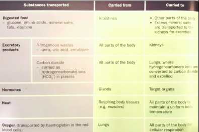
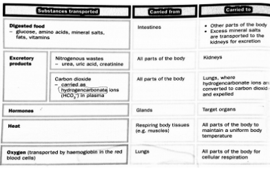
| 2. (c) Plasma | Plasma → A pale yellowish liquid Helps to transport substances, tgt with blood cells arnd the body 90% water & rest is a complex mixture of dissolved substances: - Soluble proteins (fibrinogen, prothrombin & antibodies) - Fibrinogen & Thrombin play impt role in blood clotting & these proteins are made in the liver. Antibodies help to fight diseases. - Dissolved mineral salts (hydrogencarbonates, chlorides, sulfates & phosphates of calcium, sodium & potassium) Co2 is carried as hydrogencarbonate ions in the plasma. Calcium ions are essential for blood clotting. - Food substances (a.a, glucose, fats, mineral salts, vitamins & proteins) These are transported from small intestine to arnd the body & excess mineral salts to kidney for excretion - Excretory products (urea, uric acid, creatinine) Co2 present as hydrogencarbonate ions & are transported to lungs to be removed. Urea, uric acid & creatinine transported to kidney for excretion. - Hormones e.g. Insulin Transported to target organs from endocrine glands. |
| Thematic Test 4: Transport 2.(a) Describe the role of plasma in Man’s circulatory system & discuss its features that enable it to do so. [6] | - Plasma is made up of predominantly water & water is a universal solvent -This allows substances to dissolve in the plasma & be transported in the plasma - Plasma has a high specific heat capacity which allows plasma to distribute heat effectively arnd the body w/o causing a change in the blood temp or composition - Water is non-compressible - It allows blood to be pumped by the heart for transportation |
| 2. (a) RBCs | (i) Circular, biconcave, flattened disc Increases SA:V of RBC to absorb & release O2 at a faster rate as there is a faster rate of diffusion of O2 (ii) Contains haemoglobin (iron-containing protein) Haemoglobin binds reversibly with O2 to form oxyhaemoglobin. This allows O2 to be transported arnd from lungs to all parts of body (iii) Does not hv a nucleus Can carry more haemoglobin & thus more O2 (iv) Elastic, has a flexible cell surface membrane & can turn bell-shaped Allows it to squeeze through blood vessels (capillaries) smaller than itself in diameter RBCs are produced in the bone marrow. Lifespan of RBCs: 3-4mths. When they are worn out, they are destroyed in the spleen. Haemoglobin released from destroyed RBCs is brought to liver & broken down. |
| 2.(b) White blood cells (Lymphocytes & Phagocytes) | Larger than RBCs but fewer in number Features: - Colourless & does not contain haemoglobin - Irregular in shape & contains a nucleus - Can move, change shape & squeeze through walls of the thinnest blood capillaries into spaces among tissue cells 2 main kinds: Phagocytes & Lymphocytes Play a vital role in keeping the body healthy by fighting diseases |
| 2. (b) Lymphocytes | - Lymphocytes have a large, rounded nucleus & a relatively small amt of non-granular cytoplasm Round in shape & limited movements Produce antibodies that protect the body from disease-causing microorganisms |
| 2. (b) Phagocytes | Phagocytes engulf, injest & digest foreign particles e.g. bacteria Has lobed nucleus & granular cytoplasm |
| 2. (d) Platelets | They are membrane-bound fragments of cytoplasm from certain bone marrow cells Play a part in blood clotting (by releasing the enzyme thrombokinase) |
| 3. Describe the process of blood clotting and the components involved. | - When blood vessels are damaged, the damaged blood vessels & platelets release an enzyme, thrombokinase - Thrombokinase converts the soluble protein, prothrombin, which is normally found in the plasma, into thrombin. Calcium ions must be present before this can take place. - Thrombin is another enzyme which converts soluble fibrinogen into insoluble threads of fibrin. - The threads of fibrin entangle RBCs & the whole mass forms a scab/clot over the wound, sealing it to prevent excessive loss of blood & entry of bacteria |
| 4. Identify and label the structures of the human heart. | |
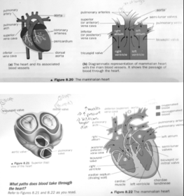
| 4. Structure of heart | Heart is surrounded by a ‘bag’ → pericardium. Pericardium is made up of 2 layers: - Inner layer is in contact with the tissues making up the heart - Btwn the 2 pericardial membranes is a fluid which helps to reduce the friction when the heart is beating Heart consists of 4 chambers: - Upper 2 chambers → right & left atria (singular: atrium) which have comparatively thinner muscles than ventricles since they only need to force blood into ventricles & do not require a high pressure - Lower 2 chambers → right & left ventricles that have comparatively thicker muscular walls esp left atrium since it has to pump blood arnd the body & requires a high pressure. Whereas, right ventricle only needs to pump blood to the lungs, which is closer - Left & Right sides of heart separated by muscular wall → median septum, which prevents the mixing of deoxygenated blood in the right side & oxygenated blood in the left side, which would reduce the amt of O2 carried to tissue cells |
| Common heart conditions: (1) 'Hole in the heart' | - The hole in the heart at the median septum might cause oxygenated blood in the left atrium & deoxygenated blood in the right atrium to mix tgt, reducing the amt of O2 in the blood. - The heart has to pump faster & harder to transport sufficient O2 to the body - Not advised to do vigorous sports because during vigorous sports, the heart has to beat faster & pump more blood to carry O2 to various parts of the body This strains the heart & causes it to fail |
| (2) 'Leaky Valves' (MOCK exam p2 Qn3. bii) Rheumatic fever is a common infection that causes heart valve disease. As a result the heart valves may become “leaky”, that is they may not be able to close completely. Suggest & explain how this affects the function of the heart. (2) | - Blood may flow back from the ventricle to atria. Thus, lower volume of blood pumped to lungs & the rest of the body. Reduced delivery of O2 & nutrients to body tissues. Heart muscles need to pump harder & faster to force more blood out. |
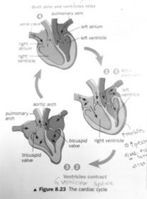
| 5. Describe the cardiac cycle in terms of what happened, muscular contraction and working of valves, during systole and diastole. | |
| Cardiac cycle | (1) The muscles of the atria contract, the atrio-ventricular valves (bicuspid & tricuspid valves open) & blood is forced into the relaxed ventricles (2) After a short pause, the muscles of the ventricles contract. The rise in pressure in the ventricles forces the atrio-ventricular valves to close to prevent the backflow of blood back into the atria, producing a loud ‘lub’ sound. (3) The semi-lunar valves open, blood flows from the right ventricle into the pulmonary arch & into the left ventricle into the aortic arch. |
| Cardiac cycle | (4) As the muscles of the ventricles contract, the atria relax. The left atrium receives blood from the pulmonary veins & the right atrium receives blood from the venae cavae (consisting of superior & inferior vena cava) . (5) The ventricles then relax. The fall in pressure of the ventricles (become lower than arches) causes the semi-valves to close to prevent backflow of blood back into the ventricle from the 2 arches. This produces a softer ‘dub’ sound. The AV valves open (pressure in ventricles lower than atria) & blood flows from the atria into the ventricles. (6) The muscles of the atria contracts again & the whole cycle repeats |
| 6. Understand the importance of a double circulation | Double circulation → how blood passes through the heart 2x in one complete circuit. It consists of the pulmonary circulation & the systemic circulation. - In systemic circulation, oxygenated blood is pumped from the left side of the body to the rest of the body & deoxygenated blood is returned to the right side of the heart by the veins & the vena cava. - In pulmonary circulation, deoxygenated blood is pumped to the lungs from the right side of the heart & oxygenated blood brought back to the left side of the heart by pulmonary veins from the lungs. |
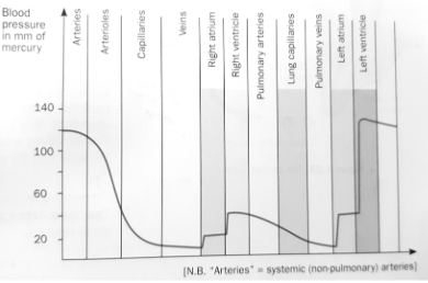
| 6. Advantages of a double circulation | - Blood entering the lungs is at a lower pressure compared to blood leaving the heart. This ensures that the blood flows more slowly through the lungs, allowing sufficient time for the blood to be well oxygenated before it is returned to the heart. - The heart pumps oxygenated blood at a higher pressure to the rest of the body, through systemic circulation, so that oxygenated blood can be distributed to the body tissues more quickly. This helps to maintain a high metabolic rate in mammals. |
| 7. Describe the difference between pulmonary and systemic circulation. | - Pulmonary circulation => circulation linking heart & lungs. From the heart, the pulmonary arteries bring deoxygenated blood to the lungs. The pulmonary veins return oxygenated blood back to the heart. - Systemic circulation => circulation of blood around the body. Oxygenated blood leave the left side of the heart through the aorta to be distributed to all parts of the body (except lungs). Veins carry blood from all parts of the body back to the right side of the heart through the venae cavae. |
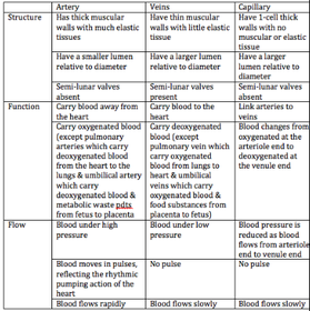
| 8. Identify the main three blood vessels | - Arteries - Capillaries - Veins |
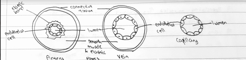
| 8. Arteries | Arteries → blood vessels carrying blood away from the heart. The large artery that leaves left side of the heart is the aorta & branches to form smaller arteries - Arteries receive blood directly from the heart. - Need to be able to withstand the immense pressure of the blood as it is forced out of the heart. Arteries have walls which are thick, muscular & elastic. The elastic layer is much thicker nearest the heart. - Thick elastic walls help to withstand the high blood pressure in the artery. Elasticity enables the artery wall to stretch & recoil or spring back. This helps to push the blood in spurts along the artery & also gives rise to the pulse. - The contraction & relaxation of muscles in the arterial wall bring about constriction & dilation of the artery. When the artery constricts, its lumen becomes narrower & less blood flow through it per unit time. On the other hand, when an artery dilates, its lumen becomes wider & more blood flows through it per unit time. - Most arteries carry oxygenated blood (except pulmonary arteries & umbilical arteries) |
| 8. Arterioles | Arterioles → arteries branch to form smaller arteries & branch again to form tiny vessels known as arterioles & ultimately their branches become very tiny blood vessels called capillaries |
| C) Using isotopes | (1) Carbon-14 is a radioactive carbon isotope. Its presence can be detected by an X-ray photographic film (2) A leaf is provided with CO2 containing the radioactive carbon (3) When photosynthesis takes place, the sugars formed will contain radioactive carbon. (4) The stem is cut & a section is exposed onto the X-ray photographic film (5) Radioactive substances are found present in phloem, since radioactive substances causes X-ray film to darken. |
| 8. Capillaries | Capillaries → microscopic blood vessels that are found between the cells of almost all the tissues. They have walls made up of only a single layer of flattened cells called the endothelium. - Endothelium is partially permeable, it enables certain substances to diffuse quickly through the capillary walls. - Capillaries branch repeatedly. The numerous branches provide a large SA for exchange of substances btwn the blood & the tissue cells. When an arteriole branches into capillaries, the total cross-sectional area increases. This lowers the blood pressure in the capillaries. The flow of blood is slowed down, giving more time for the exchange of substances. |
| 8. Venules & Veins | Venules → Before capillaries leave an organ / tissue, they unite to form small veins called venules Veins → blood vessels carrying blood towards the heart from the various parts of the body. - Venules in turn join to form bigger veins. - The blood pressure in the veins is much lower than the blood pressure in the arteries. Blood flows more slowly & smoothly in the veins. Hence the walls of veins need not be as thick & muscular as those of arteries of the same size. - Veins also contain less elastic tissues. Instead, veins have internal valves along their length to prevent the backflow of blood. Valves are folds of inner walls, shaped like half-moons. Hence, they are also called semi-lunar valves. - The movement of blood along the veins is assisted by the action of skeletal muscles on the veins. Exercising the muscles increases the pressure exerted on the veins & moves blood along more quickly. - Veins carry deoxygenated blood (except for pulmonary veins & umbilical veins) |
| 8. Heart | Blood is kept circulating throughout body by the means of a muscular pump,the heart. When the heart relaxes, it fills up w blood, when it contracts, blood is squeezed out w great force. Blood is then circulated through blood vessels which direct blood flow round the body. |
| 9. Compare and explain the differences in characteristics between the blood vessels. | |
| 9. Drawing of the 3 main blood vessels | |
| 10. State the importance of valves. | As the blood pressure in the veins are low, the semi-lunar valves help prevent the backflow of blood (flow of blood backwards) |
| THEMATIC TEST: TRANPORT Qn 2.(b) Explain how the heart functions as a pump & keeps blood flowing in 1 direction. [6] | Heart is made up of cardiac muscles that would contract to pump blood out of the heart & relax to fill up with blood to be pumped. The median septum separates the deoxygenated blood from the oxygenated blood. The heart has vales e.g.: Semi-lunar valves that prevent the backflow of blood from the aorta & pulmonary artery back into the left & right atrium respectively Bicuspid & tricuspid valves prevent backflow of blood from left & right ventricles to the left & right atrium respectively |
| 12. Identify the main blood vessels to and from the heart, lungs, liver and kidney. - Arteries | Arteries leaving the heart: - Pulmonary arteries -> carrying deoxygenated blood from right ventricle to lungs - Aorta -> carry oxygenated blood from left ventricle to rest of the body - From the aortic arch, following arteries arise: - Arteries to head, neck & arms - Aortic arch curls backwards to the left side of the heart & continues downwards as the dorsal aorta Dorsal aorta distributes oxygenated blood to areas below the heart: - Hepatic artery to liver - Renal arteries, one to each kidney - Arteries to stomach & intestines |
| 12. -Veins | Blood returned to heart by the main veins: - Pulmonary veins -> bringing oxygenated blood to left atrium from the lungs Superior vena cava returns deoxygenated blood from head, neck & arms to right atrium Inferior vena cava returns deoxygenated blood from lower regions of the body to right atrium Inferior vena cava collects blood from various veins from lower parts of the body: - Renal veins bringing blood from the kidneys - Hepatic vein bringing blood from the liver - Veins from the gut do not open directly into the inferior vena cava. They unite to form the hepatic portal vein which enters the liver & branches into numerous capillaries there. The hepatic portal vein arises from capillaries in the gut wall at one end & gives rise to capillaries in the liver at the other end. |
| 13. Describe the process of the transfer of materials from blood to tissue cells. -Tissue fluid | Tissue fluid → Medium for dissolved substances to move between the tissue cells and the blood capillaries, based on concentration gradient. Tiny spaces btwn tissue cells contain a colourless liquid, the tissue fluid. The tissue cells are bathed with tissue fluid which carries substances in solution btwn the tissue cells & the blood capillaries Dissolved food substances & oxygen diffuse from blood in the blood capillaries into the tissue fluid & then into the cells. Metabolic waste pdts diffuse from the cells into the tissue fluid, then through the blood capillary walls into the blood. The blood transports these to the excretory organs for removal. |
| RBCs change their shape as they flow through narrow blood capillaries | Since blood capillaries are narrow, the RBCs can only move through the lumen of the blood capillaries in a line, one behind another. The RBCs may become bell-shaped as they pass through the narrow blood capillaries. Advantages: - Diameter of RBCs is reduced so that it can easily pass through the lumen of the capillaries - The cell increases its SA:V to speed up the absorption of or release of O2 -Rate of blood flow is reduced, giving more time for, & thus increasing the efficiency of, exchange of materials btwn the blood & the tissue cells. |
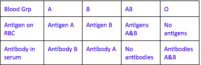
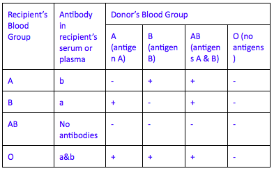
| 14. Identify the ABO blood groups and discuss the results when different blood groups are mixed together. | - Surfaces of RBCs contain special proteins called antigens. These same antigens are found on all your RBCs. - Blood plasma contain natural antibodies which are always present in your blood. These natural antibodies will not react with the antigens on RBCs on the same person, but will react with antigens on RBCs from another person, causing clumping of RBCs or agglutination. Agglutination → clumping of cells, usually as a result of reaction between specific antigens and antibodies in the blood and lymph. |
| 14. Identify the ABO blood groups and discuss the results when different blood groups are mixed together. | |
| *CONSIDER EFFECT OF RECIPIENT’S PLASMA (ANTIBODY) ON DONOR’S RBCS (ANTIGENS) | |
| 15. Describe coronary heart disease. | - Coronary arteries lie on the outside of the heart & carry blood to heart muscles - Blood supply to heart can be greatly reduced due to blockage of coronary arteries, causing a heart attack - During a heart attack, blood flow to a particular region of the heart may be completely blocked. - Due to the blocked blood flow, that part if the heart does not receive sufficient oxygen & nutrients. That part of the heart dies. - Extensive heart muscle damage is often fatal as the heart is no longer able to pump blood to various parts of the body. |
| TYS 2010 QN 1.(b) State the functions of the coronary arteries. [2] | Coronary arteries transport oxygenated blood from the heart to the heart cardiac muscles. *State what the blood vessel transports & direction it transports the substances |
| 16. Explain the cause of coronary heart disease and state the preventive measures, as well as treatment methods. CAUSE | Fatty substances may be deposited on the inner surface of the coronary arteries (atherosclerosis) This narrows the lumen of these arteries & increases blood pressure. Such an affected artery develops a rough inner surface, which increases the risk of a blood clot being trapped in the artery. A blood clot that forms in an artery is called thrombosis. If it occurs in the coronary arteries, supply of blood & oxygen to heart muscles may be completely cut off. Oxygen is needed in aerobic respn to release energy for the activities of the muscle cells. W/o O2, heart muscle cells may be damaged & heart attack occurs. |
| Factors increasing the risk of CHD, preventive measures & treatment methods | Factors increasing the risk of atherosclerosis & coronary heart disease: - Diet rich in cholesterol & saturated animal fats - Emotional stress - Smoking Preventive measures: - Proper diet, substituting animal fats with plant fats as they do not stick to inner surface of arteries. This also lowers cholesterol level in blood. - Proper stress management reduces risk of heart attack - Avoid smoking as cigarette smoke contains nicotine & CO2 which increases the risk of coronary heart disease - Regular physical exercise has LT beneficial effects on circulatory system. It strengthens heart & maintains elasticity of arteriole walls. Risk of high bp & hypertension greatly reduced. Treated by: having a coronary bypass / angioplasty |
| 17. Describe how valves open & close | The AV valves open when the blood pressure in the atrium is higher than the ventricles & close when blood pressure in ventricles higher than atria to prevent backflow of blood back into the atria. Semi-lunar valves open when blood pressure in the ventricles higher than the arches (pulmonary & aortic) & closes when the ventricles relaxes, blood pressure fall below that of the arches, to prevent backflow of blood back into the ventricles from the arches. |
| 18. State the importance of the medium septum. | The medium septum separates the right & left side of the heart. Prevents the mixing of deoxygenated in the right side & oxygenated blood in left side Mixing of deoxygenated & oxygenated blood decreases the amt of oxygen in the blood carried to the tissue cells ‘Hole in the heart’ condition: - The hole in the heart at the median septum causes deoxygenated in the right atrium & oxygenated blood in the left atrium to mix tgt, reducing the amount of O2 in the blood pumped to the tissue cells - The heart have to pump faster to transport sufficient O2 to the body - Thus, patients with atrial septal defect not advised to engage in vigorous sports. - During vigorous sports, heart hv to beat even faster & pump more blood to carry O2 to various parts of the body & the respiring tissues. This strains the heart & causes it to fail |
| 19. Define tissue rejection and describe its preventive measures. | - Tissue rejection occurs when the organ transplanted from a donor is rejected by the recipient's immune system. Any organ from another person may be treated as a foreign body by the recipient's immune system. The recipient’s lymphocytes may respond by producing antibodies to destroy the transplanted organ. Tissue rejection will not be a problem if the tissue to be transplanted is from the same person. |
| 19. Preventive measures | - A tissue match is necessary to reduce the risk of rejection. The tissues of both the donor & the recipient must be as genetically as close as possible. Siblings, parents, & close relatives of a recipient are likely to hv similar genes. - Use of immunosuppressive drugs which inhibit the responses of the recipient’s immune system. However there are problems such as: the recipient having lower resistance to many kinds of infection & having to continue to take drugs for the rest of his life |
| 20. State the pressure differences in different parts of the circulatory system. | |
| Blood Pressure | - Blood pressure in arteries is highest during ventricular systole when blood is forced into arteries. It decreases during ventricular diastole. - Blood pressure highest near the aortic arch Weaker the further the arteries are away from the heart - Blood pressure lowest at veins. It reaches almost 0mm at the venae cavae, just before the venae cavae open into the right atrium of heart |
| 21. Identify the parts of the body that transports substances to and from. | |
| 22. Differentiate between clotting and agglutination. | Clotting occurs when blood vessels or tissues are damaged to prevent entry of microorganisms & excessive loss of blood by forming a clot to seal wounds. - Beneficial to the body as it prevents entry of bacteria & excessive blood loss Agglutination is the clumping of the RBCs of the donor in the recipient’s body after a blood transfusion. Such clumps blocks up small blood vessels & obstructs blood flow. This occurs when the antigens of the RBCs of the donor reacts with the antibodies in the recipient’s body. - Not beneficial to body as it can be fatal & cause death as |
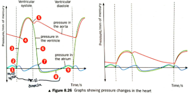
| 24. Determine, from the graph showing the changes in pressure in the left side of the heart, the length of four events: ventricular systole and diastole and atrial systole and diastole. | |
| Changes in pressure from the graph | (1) A slight increase in the ventricular pressure due to contraction of left atrium, forcing blood into relaxed ventricle (2) Ventricle begins to contract, the bicuspid valve closes & pressure increases (3) Pressure in ventricle continues to increase as it contracts (4) Pressure in left ventricle becomes higher than in the aorta. Semi-lunar valves in aorta open. (5) Ventricle begins to relax, aortic valve closes to prevent backflow of blood (6) Pressure in ventricle continues to decrease as it relaxes (7) Bicuspid valves open as pressure in ventricle becomes lower than in atrium (8) Pressure in ventricle gradually increases as blood continues to enter ventricle from atrium (9) Cycle repeats |
| 25. Determine, from the graph showing the changes in pressure in the left side of the heart, the length of four events: semilunar valves in the aorta open and close and bicuspid valve open and close. | - Bicuspid valve open when pressure in atrium higher than ventricle - Bicuspid valve close when pressure in ventricle higher than atrium to prevent backflow of blood. - Semi-lunar valves open when pressure in ventricle higher than aortic arch - Semi-lunar valves close when pressure in ventricle becomes lower than aortic arch |
| 26. Determine, from the graph showing the changes in pressure in the left side of the heart, the graph representing the pressure in the atrium, pressure in the ventricles and pressure in the aorta. |
Want to create your own Flashcards for free with GoConqr? Learn more.