6770046
Description
Flashcards by Andrew Street, updated more than 1 year ago
|
|
Created by Andrew Street
over 7 years ago
|
|
| Question | Answer |
| 3626 Describe the four phases of wound healing. | 1) Haemostasis (immediate). Collagen exposed, platelets aggregate & degranulate, releasing inflammatory mediators. Clotting & complement cascades activated. Thrombus formation & reactive vasospasm achieve haemostasis. 2) Inflammation (0–3 days). Vasodilation & ^capillary permeability allow inflammatory cells to enter & cause swelling. Neutrophils ^inflammatory response by release of cytokines, reduce infxn by bacterial killing, & debride damaged tissue. Macrophages follow & secrete cytokines, growth factors, & collagenases. They phagocytose bacteria & dead tissue & orchestrate fibroblast migration, proliferation, & collagen production. 3) Proliferation (3 days–3 weeks). Fibroblasts migrate into wound & synthesize collagen. Specialized myofibroblasts containing actin cause wound contraction. Angiogenesis stimulated by hypoxia & cytokines & granulation tissue forms. 4) Remodelling (3 weeks–1y). Reorientation & maturation of collagen fibres ^wound strength (scars 80% strength) http://oxfordmedicine.com/view/10.1093/med/9780199699476.001.0001/med-97801996994 |
| 3631 List type of wounds. | * Puncture * Bruise * Contusion (from blunt trauma) * Abrasion * Incision (inflicted with a sharp object) * Laceration (inflicted by a less sharp object like broken glass * Ulcer (break in epithelial continuity) * Avulsion ( 'an avulsion is an injury in which a body structure is forcibly detached from its normal point of insertion by either trauma or surgery.' Wiki) * Degloving ('...a type of avulsion in which an extensive section of skin is completely torn off the underlying tissue, severing its BD supply.' Wiki) * Amputation (total, near total) & subluxation From lecture: https://www.dropbox.com/home/P-Year%20Surgical%20Lectures?preview=Wound+Healing+-+Miss+J.+Odili.pptx |
| 3633 Explain healing of the skin by 1st & 2nd intention. | Skin - first intention healing: This takes place where there is close apposition of clean wound edges. • Thrombosis in cut BD vessels prevents haematoma formation. • Coagulated BD forms a surface scab which keeps the wound clean. • Fibrin precipitates to form a weak framework between the two edges. • Capillaries proliferate to bridge the gap. • Fibroblasts secrete collagen into the fibrin network. • Basal epidermal cells bridge the gap & are eventually resorbed. • The elastic network in the dermis cannot be replaced. Skin - second intention healing: This takes place in wounds where skin edges cannot be cleanly apposed. • There is phagocytosis to remove debris. • Granulation tissue to fill in defects. • Epithelial regeneration covers the surface. http://oxfordmedicine.com/view/10.1093/med/9780199699476.001.0001/med-9780199699476-chapter-3#med-9780199699476-div1-98 |
| 3636 Describe secondary bone union & what are some disadvantages. | Secondary bone union, with formation of a callus, is the natural situation eg Rx with a cast. Disadvantages are that the bone heals in the position that its left in & may lead to malunion with possible loss of function due to deformity or shortening. |
| 3636 Describe the first phase of secondary bone healing. | 1) Initial phase (lasts approx 1wk) - haematoma & inflammation: * Torn vessels at # bleed producing haematoma & clot. Haematoma may continue to expand during the first 36h. * Injured tissue & platelet activation causes inflammatory cascade via release of GF's & and cytokines. * Macrophages, fibroblasts, osteoclasts, chondroblasts migrate to #. * Fibroblasts & chondroblasts organize haematoma into collagen & granulation tissue, with angiogenesis. * Osteoclasts & macrophages remove dead bone & tissue. http://oxfordmedicine.com/view/10.1093/med/9780199699476.001.0001/med-9780199699476-chapter-15#med-9780199699476-div1-279 |
| 3636 Describe the second phase of secondary bone healing. | 2) Second phase (approx 1wk-4mths) - callus formation (soft & hard): * Osteoblast proliferation & differentiation results in callus formation. * Intramembranous/periosteal hard callus forms peripherally, with endochondral (fibrocartilagenous/bridging) soft callus forming alongside. * Soft callus is calcified by chondroblasts and subsequently resorbed by chondroclasts. * BD vessels invade into callus bringing osteoblastic type cells, resulting in ossification into woven bone. * # will have united now & be P free. 3) Third phase - remodelling: * Woven bone replaced with lamellar bone. |
| 3636 Describe the third phase of secondary bone healing. | 3) Third phase - remodelling: * Woven bone replaced with lamellar bone. * Final remodelling occurs when swelling around # decreases; trabeculae can be seen crossing the fracture site on radiographs & medullary canal is recreated. * Remodelling is most marked in children & follows the mechanical forces applied to the bone in a physiological environment. http://oxfordmedicine.com/view/10.1093/med/9780199699476.001.0001/med-9780199699476-chapter-15#med-9780199699476-div1-279 |
| 3636 Describe primary bone healing. | Primary bone healing does not produce callus. It occurs when # fragments are reduced ‘anatomically’ & ‘interfragmentary compression’ is achieved with ‘absolute stability’. There is no motion between fracture surfaces (e.g. compression plating techniques or lag screw fixation). * Inflammatory response is much reduced. * Areas of direct contact undergo some activity. * Any gaps are invaded with BD vessels & cells differentiate into osteoblasts, laying down woven and lamellar bone (gap healing). * Osteoclasts acting as ‘cutting cones’ pass directly across # site, leaving channels that are filled with BD vessels & allowing osteoblasts to fill them with lamellar bone. * No callus is formed & union takes much longer to achieve, with the strength of the healing process being borne by the mechanical properties of the fixation device. * Remodelling occurs as in secondary healing. http://oxfordmedicine.com/view/10.1093/med/9780199699476.001.0001/med-9780199699476-chapter-15#med-9780199699476-div1-279 |
| 3637 What factors may adversely affect # healing? | * Degree of local trauma (bone loss, soft tissue trauma & interposition, neurovascular injury, open #'s). * Inadequate reduction & immobilization. * Infxn. * Location of fracture. Which bone & where on bone i.e. metaphysis versus diaphysis (see below)? * Disturbances of ossification, e.g. metabolic bone disease, osteoporosis, local pathological tumour. * Age, poor nutrition, smoking, drugs (especially NSAIDs), diabetes. http://oxfordmedicine.com/view/10.1093/med/9780199699476.001.0001/med-9780199699476-chapter-15#med-9780199699476-div1-279 |
| 3638 What is delayed union & non-union of a #? | Delayed union = failure of union to occur in 1.5x the normal time for # union. Non-union = failure of union to occur within twice the normal time to # union (expect open fractures to take twice the normal time). * Hypertrophic non-union. Excess mobility or strain at # site. There is good BD supply with healing potential. Appears as large callus (elephant's foot pattern) on X-rays. Usually requires stabilization to allow callus progression. * Atrophic non-union. Due to poor BD supply resulting from initial injury or surgical intervention. There is poor healing potential. Usually require stabilization and biological augmentation to heal. http://oxfordmedicine.com/view/10.1093/med/9780199699476.001.0001/med-9780199699476-chapter-15#med-9780199699476-div1-279 |
| 3640 Describe factors affecting wound healing. | * Age * Nutritional status * Infxn * Immune status * Comorbidities - esp diabetes * Smoking * Previous irradiation * Immobility * Incontinence From lecture: https://www.dropbox.com/home/P-Year%20Surgical%20Lectures?preview=Wound+Healing+-+Miss+J.+Odili.pptx |
| 3641 Give sources of surgical infxn. | * Pt's own body flora: > Failure of correct aseptic technique. > Contaminated surgery. * Indirect contact: > Contact from hands of doctors, nursing staff, pt's, visitors. > Contaminated surfaces, e.g. door handles, cups. * Direct inoculation: > Surgeon or environmental flora through failure of aseptic technique. > Contaminated instruments or dressings. > Colonization of indwelling drains, catheters, intravenous lines. * Airborne contamination: > Skin & clothing of staff, pt's, & visitors. > Air flow in operating theatre or ward. * Haematogenous spread: > IV & intra-arterial lines. > Contaminated infusions. > Sepsis at other anatomical sites. * Food & waterborne. * Faecal-oral. http://oxfordmedicine.com/view/10.1093/med/9780199699476.001.0001/med-9780199699476-chapter-3#med-9780199699476-div1-109 |
| 3642 Describe four categories of wounds. | 1) Clean - non-traumatic wounds with no break in surgical technique, no septic focus, & no viscus opened (e.g. hernia repair). 2) Clean contaminated - non-traumatic wounds with contaminated entry into a viscus, but with minimal spillage (e.g. elective cholecystectomy). 3) Contaminated - clean, traumatic wounds or significant spillage from a viscus or acute inflammation (e.g. emergency appendectomy). 4) Dirty - includes traumatic wounds from a dirty source or when significant bacterial contamination or release of pus is encountered. http://oxfordmedicine.com/view/10.1093/med/9780199699476.001.0001/med-9780199699476-chapter-3#med-9780199699476-div1-98 |
| 3643 List common organisms causing wound/skin infxn's. | * Gram +ve: > S.aureus (may be MRSA) > S. pyogenes > C. perfringens * Gram -ve: > P. aeruginosa * Anaerobes: > Bacteriodes OHCM p420-423. 'Most wound infections are acquired from the pt's own flora. The majority are skin organisms (e.g. Staphylococcus aureus, Staphylococcus epidermidis), although the second commonest cause is contamination from opened viscera during surgery (e.g. Escherichia coli from the GI tract, Pseudomonas from the biliary tree). http://oxfordmedicine.com/view/10.1093/med/9780199699476.001.0001/med-9780199699476-chapter-2#med-9780199699476-div1-58 |
| 3644 Outline steps to reduce transmission of infxns due to surgery. | * Identify infected pt's by serology. * Identify potentially infected pt's by risk factors (e.g. IV drug users at risk from HBV carriage). * Specific procedures for the care of infected pt's (e.g. barrier nursing for C. diff). * Careful disposal of disposable items related to pt care. * Specific treatment & sterilization of non-disposable equipment. * Additional/specific precautions for theatre staff: > Make all procedures ‘safe’ procedures by having the highest standards of safety & care using instruments & sharps. > Wearing of plastic aprons in procedures with expected soiling with urine/faeces/ascites. > Wearing of two pairs of gloves to reduce the risk of skin exposure when gloves tear. > Wearing of re-enforced gloves for procedures with a high risk of penetrating injury (e.g. fragmented fractures). > Wearing of glasses, goggles, or visors for eye protection. > Handle all sharps using a transfer container: never pass them hand to hand. > Don't allow unnecessary blood or fluid spillage. http://oxfordmedicine.com/view/10.1093/med/9780199699476.001.0001/med-978019969 |
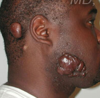
| Keloid scarring - grows beyond the boundary of the wound. | |
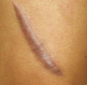
| Hypertrophic scar - remains within the original wound. |
Want to create your own Flashcards for free with GoConqr? Learn more.