7415294
Description
Flashcards by Megan Popp, updated more than 1 year ago
|
|
Created by Megan Popp
about 7 years ago
|
|
| Question | Answer |
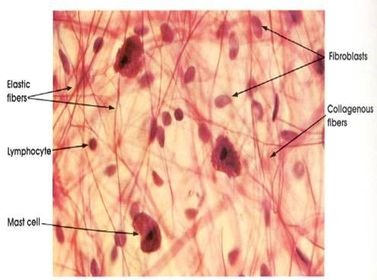
| what type of tissue prep method was used? | spread prep |
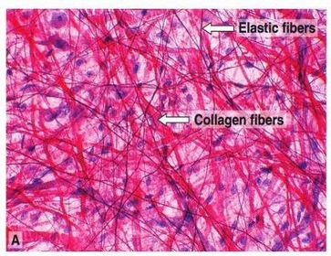
| what type of slide prep was used ? | spread prep |
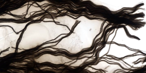
| what type of preparation was used for this specimen? | Teased prep |
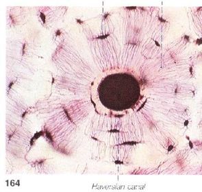
| what is this and what type of prep was used? | bone, ground prep |
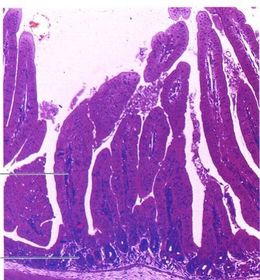
| What type microscopy and what type of stain was used | Light Microscopy H&E |
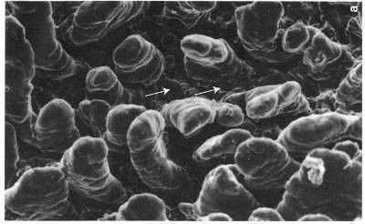
| What type of microscopy was used ? | scanning electron |
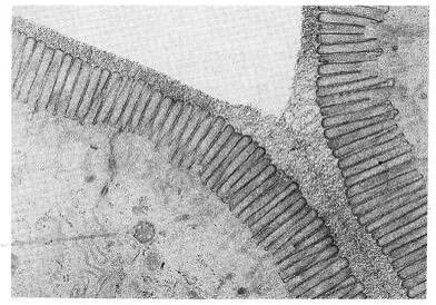
| What type of microscopy was used ? | transmission electron |
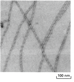
| Name this cytoskeletal element | microfilaments, made of action |
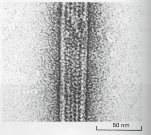
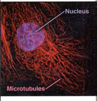
| name this cytoskeletal element | microtubule, made of tubulin |
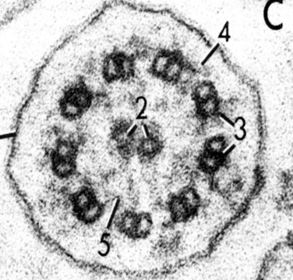
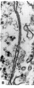
| Name this cytoskeletal element | Cilium, arranged in a 9+2 |
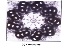
| Name this cytoskeletal element | centriole, arranged in 9*3 arrangement |
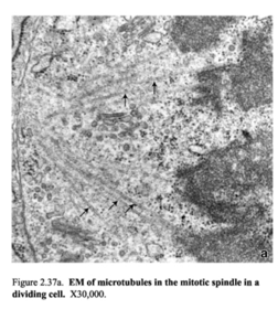
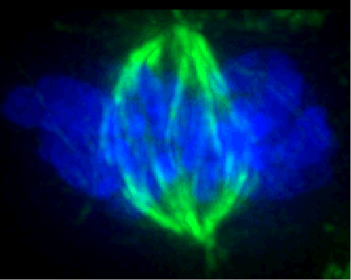
| What is this formation and what is it made of? | Mitotic spindle, made of microtubules |
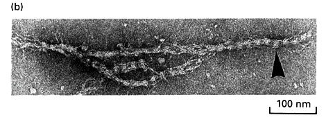
| What is this cytoskeletal element? | intermediate filament |
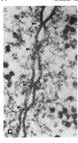
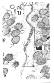
| What is the junction type located at the arrows? | Tight junction |
| What is the junction type located at "AdB"? | Adheren Junction |
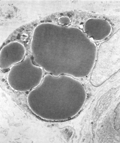
| What is a metabolic by-product found on the cytoplasm? | inclusions (Shown: lipid droplets) |
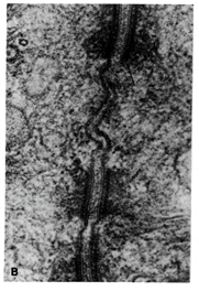
| What is located below the adheren junction and how can it be seen? | Desmosomes, connecting both cells. you can see hairy look around each side of the meeting cells |
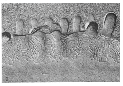
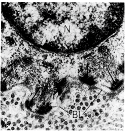
| What type of junction can be see in this and how? | This shows a hemidesmosome. you can see that the dark hairy area is only located on one side. |
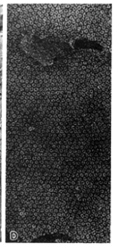
| Where will you find a gap junction? | they are usually between the adheren junction and the desmosomes. You can see that they have a beaded look. This picture shows the pores of connexons |
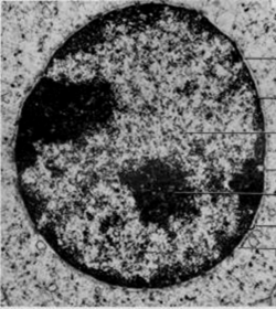
| What is the pale substance within this nucleus? Dark? | Pale: euchromatin Dark: heterochromatin (you can distinguish this from the nucleoli because the nucleoli is more dense) |
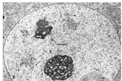
| What is the purpose of the darkest circle that the arrow is pointing to? | Nucleoli, within the nucleus it is the site where rRNA is transcribed and assembled with ribosomal proteins to make functional ribosomes |
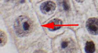
| Name the dense, dark, substance | nucleoli |
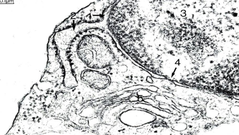
| Label 2 3 4 | 2. Lumen of RER, black dots are proteins that are spit out by the RER, usually for ejection 3. Heterochromatin 4. Nuclear pore |
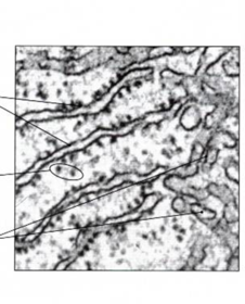
| 4 things seen in this: RER Lumen of RER Smooth ER Free Ribosomes | Labels can be seen on lecture |
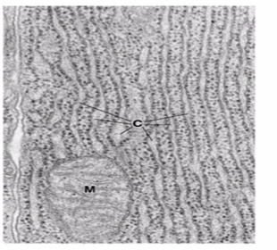
| This is an electron micrograph (EM) of?What is C labeling? | This is an EM of the RER C. cytoplasm between the RER |
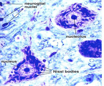
| See Nissl Bodies- stacks of RER Nucleus, Heterochromatin, nucleolus | |
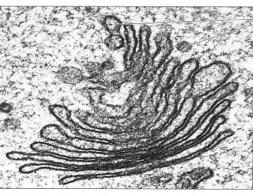
| TEM of Golgi, see, cis, medial, and trans | |
| This is showing a lysosome with ingested material | |
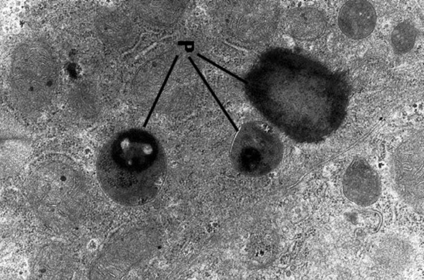
| What are the dark circles and what is their function | Peroxisomes produce H2O2 and are involve din alcohol detox and breakdown of long chain fatty acids |
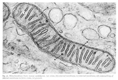
| Mitochondria | |
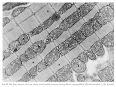
| What is this showing and why is there so many? | This is muscle tissue, mitochondria (darker) are found in between the contractile units because muscle contraction requires so much ATP |
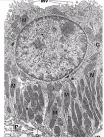
| Mitochondria are located at the _____ side of this cell. | Basil - this is because there are probably enzymes located there that need the energy. Mitochondria are located in the area where they are used (you can also see nuclear envelope, cytoplasm, heterochromatin) |
| Name this stage of mitosis in IF | Interphase |
| Name this stage of mitosis in IF | Prophase centrosomes separate, chromosomes condense |
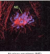
| Name this stage of mitosis in IF | Prometaphase Nuclear envelope (NE) breaks down chromosomes attach to spindle |
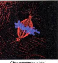
| Name this stage of mitosis in IF | Metaphase chromosomes align on spindle equator |
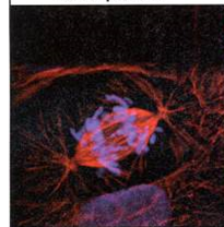
| Name this stage of mitosis in IF | Anaphase A sister chromatids separate and move to poles |
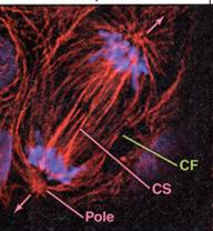
| Name this stage of mitosis in IF | Anaphase B Cleavage furrow (CF) assembles Organized central spindle assembles Poles separate |
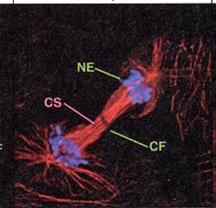
| Name this stage of mitosis in IF | Telophase Cleavage furrow constricts Nuclear envelope reassembles |
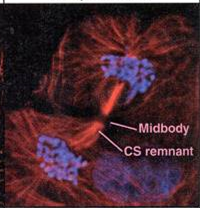
| Name this stage of mitosis in IF | Cytokinesis Chromosomes decondense Interphase microtubule network reforms Daughter cells separate |
Want to create your own Flashcards for free with GoConqr? Learn more.