
Flashcards
Histology Circulatory System

Cards in this deck (33)
- 1Circulatory System Functions? Components?Functions:
Delivery of oxygen, nutrients and other compounds, e.g. hormones
Removal of carbon dioxide and waste products
Thermoregulation
Components:
Cardiovascular
Lymphatic (water as a waste product) - 2Blood Vessels What are the three main types? What is a common term used regarding cell type of the cardiovascular system?Three main types:
Arteries
Capillaries
Veins
Common Term:
Endothelium - 3EndotheliumInternal surface of all vessels lined with single layer of squamous cells.
**Simple squamous epithelium**
Think blood, think "Squeamish!" - 4Blood Vessel Structure (3 layers) Tunica Intima? Tunica Media? Tunica Adventitia? Vasa vasorum?Tunica Intima:
Endothelium, basal lamina and internal elastic lamina
Tunica Media:
Concentric layers of smooth muscle, maybe elastic fibers and external elastic lamina
Tunica Adventitia:
Connective tissue, fibroblasts
Vasa vasorum:
Vessels of the vessel
YOU MUST KNOW resident cells, cell types, and hallmarks of all layers
*Every verse; has an endothelium! - 5Blood Vessel layers ORDER (From innermost to outermost)Tunica Intima --> Internal Elastic Lamina (part of Tunica Intima)
--> Tunica Media (smooth muscles, can have some elastin, collagen type I) --> Tunica Adventitia (mostly collagen type III made by fibroblasts & some elastin) - 6—Muscular Versus Elastic Artery
Muscular Artery: thinner tunica intima, prominent internal elastic lamina, up to 40 years layers of smooth muscle
*Function = distribute blood
Elastic Artery: thick tunica intima, elastic lamellae in tunica media
*Function = control of the peripheral resistance & systemic pressure, control flow to capillaries
- 7—Muscular Artery: thinner tunica intima, prominent internal elastic lamina, up to 40 years layers of smooth muscle
*Function = distribute blood
A = Tunica Adventitia
(contains fibroblasts that make collagen Type III)
M= Tunica Media
(contains smooth muscle layers, NO striations, collagen type I)
I = Tunica Intima
(Border is the Internal elastic lamina = "loose rubber band")

- 8—Larger Muscular Artery
-can see some elastin being made in Tunica media
(the lighter purple wavy lines)
-there is still an endothelial layer, even though it is not seen in this image
-External elastic lamina is the border or end of the Tunica media, where Tunica Adventitia begins
Vasa vasorum:
Vessels of the vessel

- 9Arteries 3 Types?Elastic, Muscular, & Arterioles
- 10Arteries ELASTIC arteries Characteristics? Function?THICK tunica intima (I)
elastic lamellae in tunica media (M)
(True layers: alternating smooth muscle and elastin, smooth muscle and elastin, etc.)
-elastin tends to be under-stained
*Function: stabilize variation in pressure
A = Tunica adventitia (fibroblasts-collagen type III)
ALL vessels have an endothelial layer!

- 11Arteries MUSCULAR arteries Characteristics? Function?THINNER tunica intima
prominent internal elastic lamina
up to 40 years layers of smooth muscle (NO striations)
*Function = distribute blood
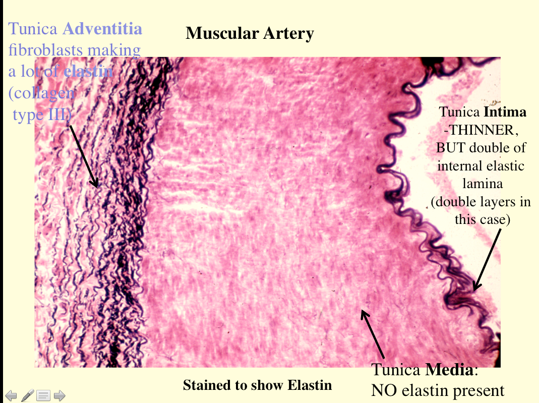
- 12Type of artery? What kind of innervation is seen at the letter A?Muscular Artery
Innervation at Tunica Adventitia:
UNmyelinated sympathetic
nerve fibers
Norepinephrine: vasoconstriction
Acetylcholine: vasodilation

- 13What is shown in the image? What are characteristics of this vessel type?Small Artery
M = Tunica media
E = endothelial cells (single layer)
Artery = >2 layers of smooth muscle in tunica media
*to count layers look at the nuclei of smooth muscle cells, SEPARATE from the single layer of endothelial cells

- 14What kind of artery is shown in the image?ARTERIOLE
1-2 layers of smooth muscle in tunica media (at most 2 layers, no striations, centrally placed nucleus)
Function: control of peripheral resistance and systemic pressure, control flow to capillaries
- 15Capillaries Characteristics?Endothelial TUBE with basal lamina
8-10 mm
Lumen of these common cells are larger than lumen of a capillary:
RBC is 7.5 microns, plasma cell is 15 microns, mast cell is 20-30 microns
**0.25 – 1 mm in length** - 16—E = endothelial layer
P = Pericyte (just outside the endothelial layer)
-a regenerative cell, can differentiate into endothelial cells to fill a clot
*Note the tube-like depiction of the capillary with RBC's inside

- 17—CAPILLARY
-BONUS: Can you identify a mast cell, plasma cell, smooth muscle cell, and the type of artery in the upper left corner?
- 18Types of Capillaries 3 Types1. Continuous
2. Fenestrated
3. Discontinuous, sinusoidal - 19What type of capillary is shown? Characteristics?1. Continuous
-Continuous endothelium, pinocytosis
-Muscle, CT, exocrine glands & nerve
-Found in respiratory system (gas exchange, small ions, water)
- 20What type of capillary is shown? Characteristics?Fenestrated
(2 Types)
-Large fenestrations (60-80 nm) in wall for rapid exchange of material
1. Kidney, intestine, endocrine glands (diaphragm)
2. Renal glomerulus (no diaphragm)
-Can be found in Pancreas
(digestive enzymes, proteins, liquids, fluids)
- 21What type of capillary is shown?Discontinuous, Sinusoids
-Enlarged capillary (30-40 microns)
-discontinuous endothelium and BM
-Spleen, Bone Marrow, Liver (a lot of material can pass through, RBC's & WBC's)

- 22What type of capillary is shown?Fenestrated!
Continuous basement membrane, but pores within the endothelial cells)
D = fenestration with density/diaphragm (acts as barrier over fenestration)
- 23Veins 2 TypesVenules & Veins
- 24What are the boxes indicating?V = VENULES
Bigger than capillaries, thin walls
Leaky, Diapedesis
A = Arteriole (shown for comparison)

- 25What is being depicted in the image?Diapedesis
(Leukocyte Migration)
- 26What is shown in the picture and what is the arrow pointing at?VEIN
THIN intima and media
Well developed adventitia
Valves
Poor IEL
maybe 1-2 layers of smooth muscle
*A TON of tunica adventitia

- 27—More adventitia and less smooth muscle for muscular VEIN


- 28—


- 29—Artery = thick tunica media and thin tunica adventitia
*the reverse is true for vein

- 30Lymphatics (mostly water) 2 Main componentsLymph
Excess fluid in the tissues
Lymphatic vessels
-Endothelium lined, thin walled vessels
*Anchoring filaments*
-Larger vessels have poorly organized intima, media and adventitia
-Valves
-No RBC, but can be WBC
-Turbid lumen - 31What type of vessel shown? What are the arrows indicating?Lymphatic Vessel
V = Valves
*Note the turbid lumen, NO RBC's, NO tunica adventitia, thin wall around the whole thing

- 32What type of vessel is shown? What are the red asterisks indicating?LARGE Lymphatic Vessel
Red asterisk = anchoring filaments
(look like tent stakes)
Note: hardly no smooth muscle, turbid lumen
- 33—


Information
- Description
- Histology (Alb) Final Exam
- Created by
- Marissa Alvarez
- Views
- 61
- Rating
More on Blood Vessel Structure and Histology