
Mind map
The Epithelial Tissue
Map content
The Epithelial Tissue
Characteristics
- Avascular tissue
- Covers the body surfaces, cavities and forms glands
- It is a barrier between the exterior and the underlying connective tissue
- Epithelial cells
- Closely apposed to each other
- Bound by cell junctions
- Tissue polarity
- Apical domain
- Lateral domain
- Basal domain
- Basement membrane
Functions
- Secretion
- Absorption
- Transportation
- Mechanical protection
- Receptor function
Classification of the epithelium
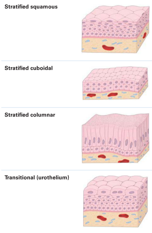
- Depending on the amount of layers
- Simple
- Monolayer
- Stratified
- Two or more layers
- Pseudostratified
- Monolayer with different heights
- Simple
- Depending on the form
- Squamous
- Cuboidal
- Columnar
- Transitional
- The last layer is the one that defines the epithelium
- Depending on the amount of layers
Apical domain
- Mircofilaments
- Microvilli (1-3 µm)
- Core of actin filaments
- Anchored to the terminal web
- Passive movement
- Contraction of the terminal web
- Localization
- Epithelial cells
- Increase absorptive surface
- Stereocilia (120 µm)
- Core of actin filaments
- Anchored to the terminal web
- Passive movement
- Fluid flow
- Vibration of endolymph
- Localization
- In sensory hais cells in the inner ear
- Microvilli (1-3 µm)
- Microtubules
- Cilia
- Motile (5-10 µm)
- Structure
- Axoneme
- Basal body
- 9+2 pattern of microtubules
- Active movement
- Localization
- Flagela
- Structure
- Primary (2-3 µm)
- Structure
- Axoneme
- Basal body
- 9+0 pattern of microtubules
- Passive movement
- Fluid flow
- Structure
- Nodal (5-6 µm)
- Structure
- Axoneme
- Basal bodie
- 9+0 pattern of microtubules
- Active movement
- Rotational movement
- During enbryonal development
- Structure
- Motile (5-10 µm)
- Cilia
- Mircofilaments
Lateral domain (cell to cell adhesions)
- Occluding junctions
- No communication between cells
- Barrier
- Near the apical domain
- Zip conformation
- No communication between cells
- Anchoring junctions
- Provide mechanical strength
- Types
- Zonula adherens
- Binding to actin filaments
- Proteins
- Cadherines
- Calcium dependent
- Nectins
- Calcium independent
- Cadherines
- Macula adherens
- Binding to intermediate filaments
- Proteins
- Desmocollin
- Desmoglein
- Proteins
- Binding to intermediate filaments
- Zonula adherens
- Communicating junctions
- There is communication between cells
- Connexins
- Stimuli regulated
- Occluding junctions
Basal domain
- Basement domain
- Dense layer of a proteinic extracelular matrix (no cells)
- Functions
- Attachment site to the undelying connective tissue
- Filters substances
- Scaffold during tissue regeneration
- Cell signalling
- Layers
- Basal lamina
- Laminin polymer (most abundant)
- Type IV collagen
- Reticular lamina (not always present)
- Type VII collagen
- Reticular fibrils (most abundant)
- Fibrillin microfibril
- Elasitc fiber
- Basal lamina
- Infoldings
- Increase the cell surface
- Facilitate interactions
- Cell to extracellular matrix junctions
- Focal adhesions
- Actin filaments
- They cross the whole basement membrane
- Hemidesmosomes
- Intermediate filaments
- To the basement membrane
- Focal adhesions
- Basement domain
Glands
- Types
- Exocrine glands
- Secretion to the lumen
- Types
- Merocrine
- Exocytosis
- No cell damage
- Pancreas
- Apocrine
- The product takes part of the cell
- Loss of cytoplasm
- Mamary glands
- Holocrine
- Programmed necrosis
- Cell death
- Cebaceous glands
- Merocrine
- Endocrine glands
- Secretion into the connective tissue
- Example
- Hormones
- Exocrine glands
- Types of signaling
- Paracrine signaling
- The signal reaches the epithelial cell that is next to it
- Mediated by receptors
- Autocrine signaling
- Own regulation
- Negative feedback
- Mediated by receptors
- Paracrine signaling
- Types
Images and files in the map
Information
- Description
- Mind Map on The Epithelial Tissue, created by Daniel Burriel Burghall on 16/02/2025.
- Created by
- Daniel Burriel Burghall
- Views
- 0
- Rating