
Mind map
RESPIRATORY SYSTEM - Ms Matthews
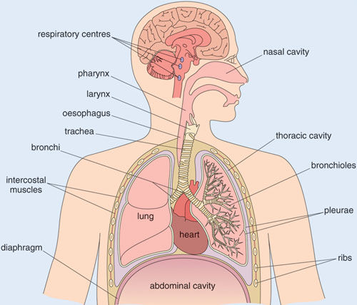
Map content
RESPIRATORY SYSTEM - Ms Matthews
LOWER RESPIRATORY SYSTEM
- BRONCHI/BRONCHIOLES
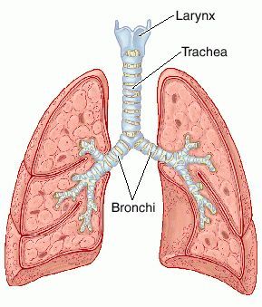
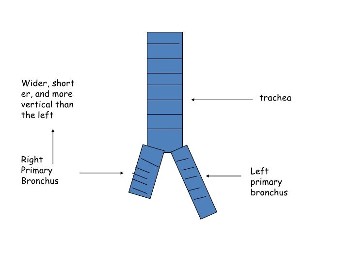
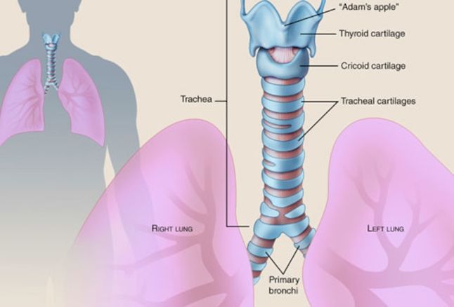
- 2 BRONCHI EMERGE at the BIFURCATION OF THE TRACHEA
- The RIGHT bronchus is WIDER, SHORTER and MORE VERTICALLY POSITIONED
- If something falls down a patients throat for example a dental file then due to the anatomic feature of the bronchi it will fall into the right lung.
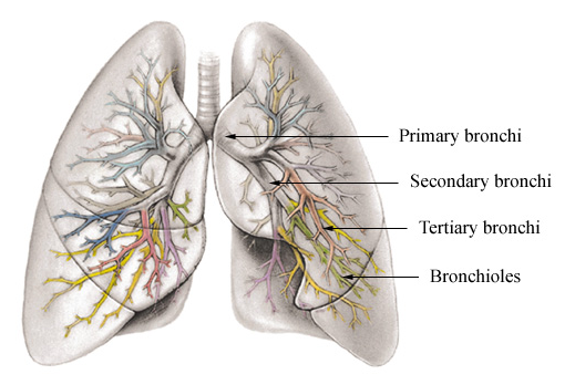
- THEY THEN DIVIDE AND SUBDIVIDE INTO SMALLER BRONCHIOLES
- It has the same tissues as the TRACHEA
- OUTER: FIBROUS TISSUE and CARTILAGE
- MIDDLE: MUSCLE
- INNER: CILIATED COLUMNAR EPITHELIUM
- MIDDLE: MUSCLE
- OUTER: FIBROUS TISSUE and CARTILAGE
- FUNCTIONS:
- CONTROL THE ENTRY OF AIR by contraction and relaxation of involuntary muscle which regulates volume of air entering lungs
- WARM + MOISTEN AIR
- SUPPORT
- COUGH REFLEX
- REMOVAL of particular matter by MACROPHAGES
- COUGH REFLEX
- SUPPORT
- WARM + MOISTEN AIR
- CONTROL THE ENTRY OF AIR by contraction and relaxation of involuntary muscle which regulates volume of air entering lungs
- The RIGHT bronchus is WIDER, SHORTER and MORE VERTICALLY POSITIONED
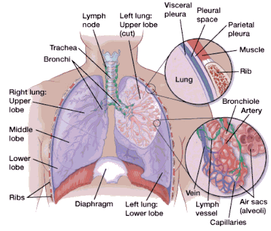
- ALVEOLI
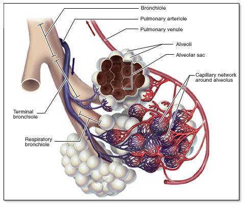
- These are found at the end of the bronchioles
- Sac-like structures (0.3mm diameter), composed of SIMPLE SQUAMOUS EPITHELIAL CELLS. (So that diffusion can occur)
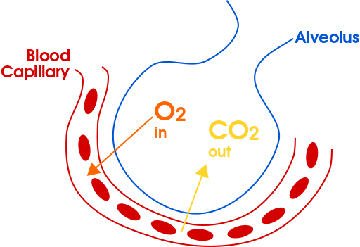
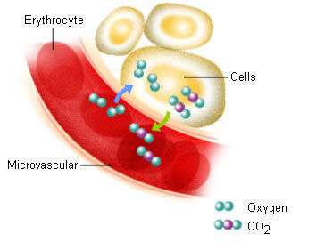
- Each one is surrounded by a dense CAPILLARY NETWORK where GASEOUS EXCHANGE OCCURS
- Sac-like structures (0.3mm diameter), composed of SIMPLE SQUAMOUS EPITHELIAL CELLS. (So that diffusion can occur)
- FUNCTION
- TRANSFER OF GASES - EXTERNAL RESPIRATION
- - TYPICAL PAIR OF HUMAN LUNGS CONTAIN 700 MILLION ALVEOLI - ELASTIC FIBRE ALLOW FOR STRETCH AS FILL WITH AIR
- DIFFUSION of CO2 AND O2 gasses between the ALVEOLI and the BLOOD
- INTERNAL: DIFFUSION of CO2 AND O2 between BLOOD CAPILLARIES and the BODY CELLS
- TRANSFER OF GASES - EXTERNAL RESPIRATION
- These are found at the end of the bronchioles
- 2 BRONCHI EMERGE at the BIFURCATION OF THE TRACHEA
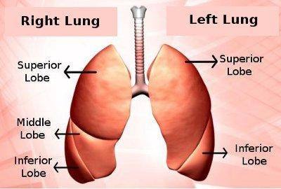
- LUNGS
- A pair of cone shaped structures
- Each lung is divided by deep fissures/lobes
- Made up of:
- Bronchioles and Alveoli
- Supported by a framework of alveolar and elastic tissue
- Permeated by blood vessels, nerves and lymphatics
- Made up of:
- Each lung is divided by deep fissures/lobes
- A pair of cone shaped structures
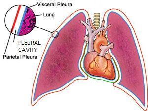
- PLEURA
- Serous membrane that surrounds the lungs
- This prevents friction between the lungs and ribs during movement
- 2 layered
- Outer PARITEL LAYER
- There is PLEURAL CAVITY in- between them. This contains SEROUS FLUID which lubricates to...
- Inner VICERAL LAYER
- Outer PARITEL LAYER
- Serous membrane that surrounds the lungs
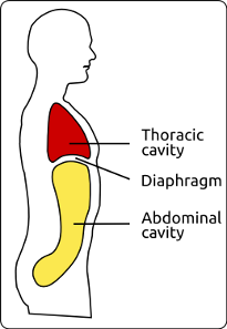
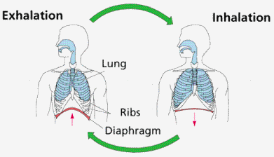
- DIAPHRAGM
- THE MUSCLES OF BREATHING
- A sheet of muscle
- It seperates the THORACIC and ABDOMINAL CAVITIES
- It consists of a CENTRAL TENDON from which muscle fibres radiate to attach to lower ribs, sternum and vertebral column
- INHALATION (AIR PULLED INTO LUNGS): LUNGS EXPAND ----- DIAPHRAGM CONTRACTS AND BECOMES FLAT
- AS IT CONTRACTS AND RELAXES BREATHING TAKES PLACE
- EXHALATION (AIR (CO2) IS PUMPED OUT OF LUNGS): LUNGS DILATE -----DIAPHRAGM RELAXES AND IS DOME SHAPED
- INHALATION (AIR PULLED INTO LUNGS): LUNGS EXPAND ----- DIAPHRAGM CONTRACTS AND BECOMES FLAT
- CONTRACT SIMULTANEOUSLY
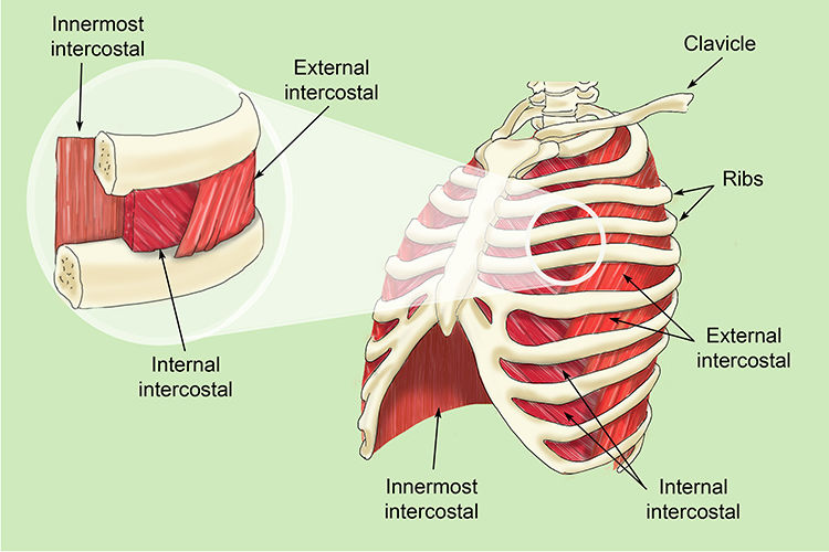
- RIBS AND INTERCOSTAL MUSCLES
- 12 PAIRS OF RIBS
- LYING BETWEEN THEM ARE 11 PAIRS OF INTERCOSTAL MUSCLES ARRANGED IN 2 LAYERS
- EXTERNAL INTERCOSTAL MUSCLES: These pass downwards and FORWARD
- INTERNAL INTERCOSTAL MUSCLES: These pass downwards and BACKWARDS
- LYING BETWEEN THEM ARE 11 PAIRS OF INTERCOSTAL MUSCLES ARRANGED IN 2 LAYERS
- 12 PAIRS OF RIBS
- BRONCHI/BRONCHIOLES
UPPER RESPIRATORY SYSTEM
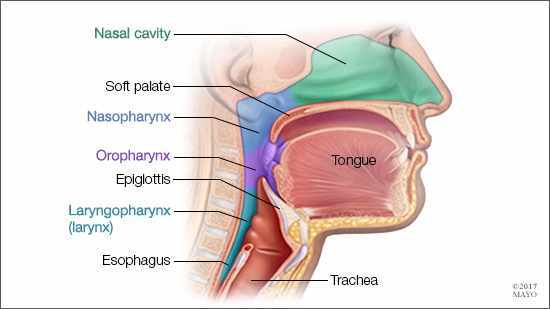
- THE NASAL CAVITY
- NOSE
- This is the FIRST PASSAGE WAY for air and is the organ for SMELL
- OUTER - CARTILAGE
- INSIDE AND OUTSIDE COVERED - SKIN
- LINED - CILIATED MUCOUS MEMBRANE
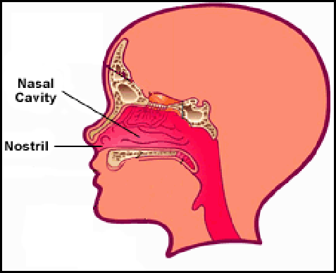
- TWO NOSTRILS that lead to the BONY NASAL CAVITY
- This is the FIRST PASSAGE WAY for air and is the organ for SMELL
- LARGE IRREGULAR CAVITY divided by a CARTILAGE SEPTUM
- The FLOOR is formed by the HARD and SOFT PALATE
- It's anterior part is formed by a hyaline cartilage (LW).
- LINED with CILIATED (to increase the surface area) MUCOUS SECRETING COLUMNAR EPITHELIUM MEMBRANE
- Cilia is a type of cell extension, they are microscopic hair like projections from the cell membrane at the free surface of a cell. They move in a rhythmic manner to propel the substances along the surface in one direction.
- The FLOOR is formed by the HARD and SOFT PALATE
- FUNCTIONS
- WARM and MOISTEN air entering the nostrils - this is to ensure that the air is the same temp as the body
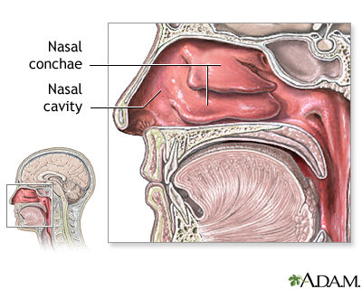
- The NASAL CONCHAE is a narrow, curled, spongy bones protruding from the nasal cavity walls providing rapid warming and humidification of air as it passes to the lungs.
- FILTER dust, bacteria and other foreign matter from air via CILIATED MUCOUS MEMBRANE, to PREVENT it getting into THE LUNGS
- WARM and MOISTEN air entering the nostrils - this is to ensure that the air is the same temp as the body
- NOSE
- THE PHARYNX
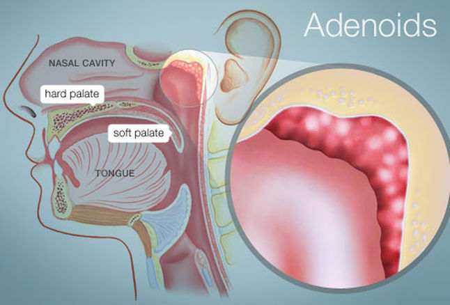
- 1. This extends from BEHIND THE NOSE ABOVE THE SOFT PALATE. You can find the ADENOIDS on it's posterior wall.
- 2. BEHIND THE ORAL CAVITY
- 3. NARROW INFERIOR PART OF THE PHARYNX continues as the Oesophagus with the Larynx (voice box) lying anterioly.
- 2. BEHIND THE ORAL CAVITY
- The EPIGLOTTIS stops food from going down the trachea. If it does you begin to COUGH.
- 1. This extends from BEHIND THE NOSE ABOVE THE SOFT PALATE. You can find the ADENOIDS on it's posterior wall.
- FUNCTIONS
- PASSAGE way for AIR (naso) & FOOD (oro/laryngo)
- WARMS and HUMIDIFYS air
- TASTE - through the OLFACTORY NERVE ENDINGS
- HEARING - EUSTACHIAN TUBE enters NASO PHARYNX for SATISFACTORY HEARING
- PROTECTION - LYMPHATIC TISSUES, TONSIL and ADENOIDS which produce ANTIBIODIES
- COMPOSITION
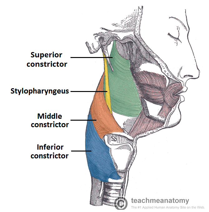
- LAYER OF INVOLUNTARY CONSTRICTOR MUSCLES - responsible for swallowing
- The naso has CILIATED COLUMNAR EPITHELIUM OF Mucous membrane lining
- In the ORO it is lined with STRATIFIED SQUAMOUS EPITHELIUM
- LW: Stratified - contain several layers, various sizes and continual cell division occurs in the basal layer this pushes cells above nearer and nearer to the surface where they are shed. The deepest layer contains columnar or cuboidal epithelial that can divide and as they grow to the surface they become squamous.
- In the ORO it is lined with STRATIFIED SQUAMOUS EPITHELIUM
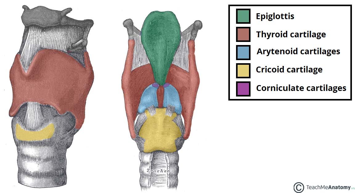
- THE LAYRNX (VOICE BOX)
- Consists of THYROID, ARYTENOID AND CRICOID CARTILAGES. They are connected by ligaments and membranes
- VOCAL BOX: The interior part of the larynx has the vocal cords, it is folds of mucous membrane and muscle
- LONGER in MALES: leads them to have a deeper voice. Men: 4.5cm Women: 3cm
- FUNCTIONS
- Produce SOUNDS OF SPEECH: PITCH, RESONANCE AND VOLUME
- PROTECTS the RESPIRATORY TRACT
- PASSAGE WAY FOR AIR: Between the pharynx and trachea
- Consists of THYROID, ARYTENOID AND CRICOID CARTILAGES. They are connected by ligaments and membranes
- THE TRACHEA
- consists of 16-20 INCOMPLETE C-SHAPED RINGS OF HYALINE CARTILAGE
- LW HYALINE CARTILAGE: It's glass like, transluent, bluish white and smooth. It has few fibred but many chondrocytes and plentiful ground susbstance. it might be fused to bone or in seperate pieces
- OUTER: FIBROUS ELASTIC TISSUE
- MIDDLE: SMOOTH MUSCLE
- INNER LINING: CILIATED COLUMNAR EPITHELIUM
- MIDDLE: SMOOTH MUSCLE
- FUNCTIONS
- SUPPORT: The CARTILAGE prevents obstruction of the airway as head and neck move, keeping the passage open.
- Incomplete cartilage: allows DISTENSION (EXPANSION) of the OESOPHAGUS during swallowing.
- WARMS, MOISTENS AND FILTERS AIR
- consists of 16-20 INCOMPLETE C-SHAPED RINGS OF HYALINE CARTILAGE
- THE NASAL CAVITY
Images and files in the map
Information
- Description
- diploma dental hygiene and therapy Mind Map on RESPIRATORY SYSTEM - Ms Matthews, created by Rachna Rai on 05/11/2017.
- Created by
- Rachna Rai
- Views
- 22
- Rating