
Mind map
Supporting structures and systems of the Brain
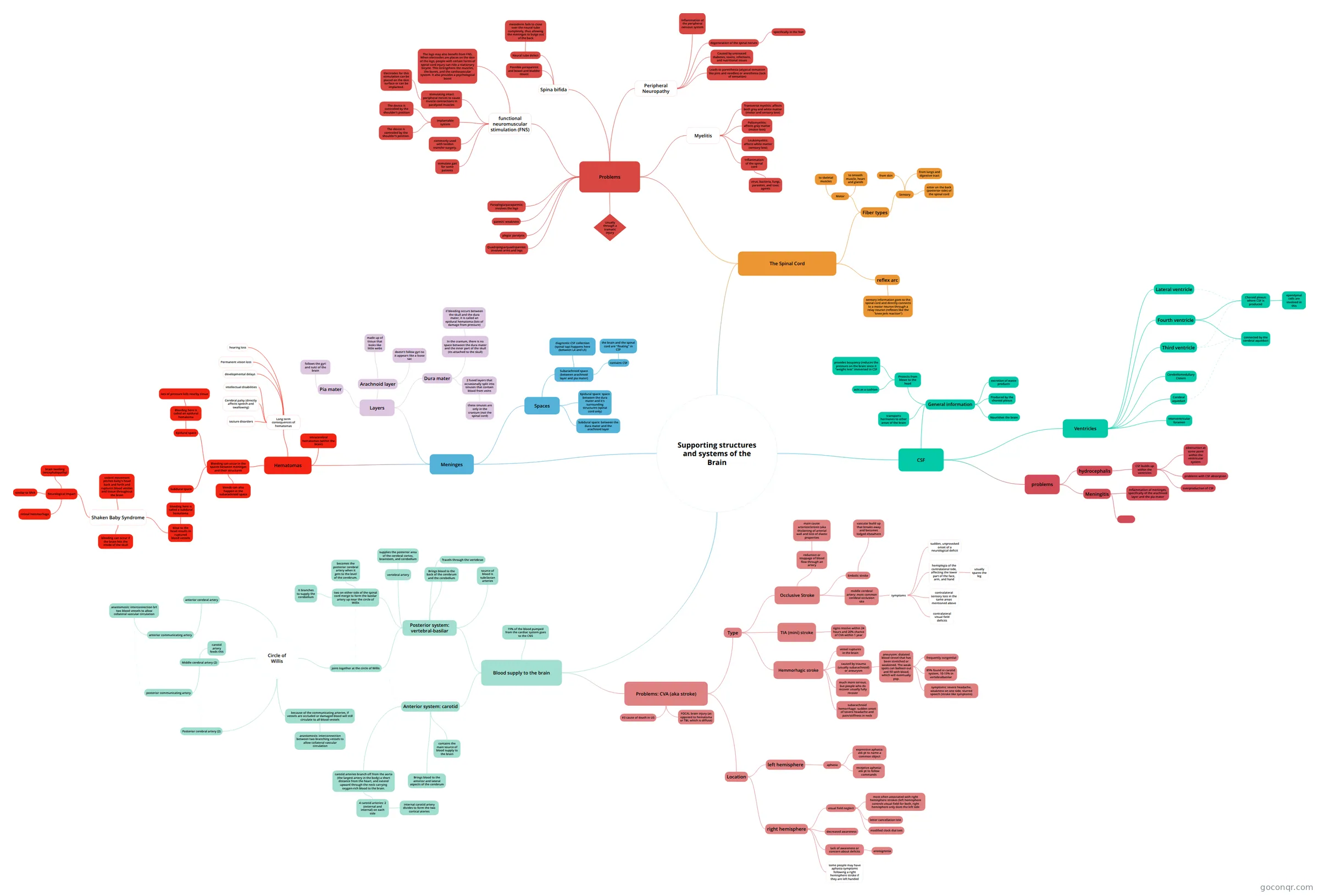
Mind map: Supporting structures and systems of the brain
Map content
Supporting structures and systems of the Brain
Meninges
- Spaces
- Epidural space: space between the dura mater and it's surrounding structures (spinal cord only)
- Subdural space: between the dura mater and the arachnoid layer
- Subarachnoid space (between arachnoid layer and pia mater)
- contains CSF
- the brain and the spinal cord are "floating" in CSF
- diagnostic CSF collection (spinal tap) happens here (between L4 and L5)
- contains CSF
- Layers
- Dura mater
- 2 fused layers that occasionally split into sinuses that contain blood from veins
- these sinuses are only in the cranium (not the spinal cord)
- In the cranium, there is no space between the dura mater and the inner part of the skull (its attached to the skull)
- if bleeding occurs between the skull and the dura mater, it is called an epidural hematoma (lots of damage from pressure)
- 2 fused layers that occasionally split into sinuses that contain blood from veins
- Arachnoid layer
- made up of tissue that looks like little webs
- doesn't follow gyri so it appears like a loose sac
- Pia mater
- follows the gyri and sulci of the brain
- Dura mater
- Hematomas
- Bleeding can occur in the spaces between meninges and their structures
- Epidural space
- Bleeding here is called an epidural hematoma
- lots of pressure kills nearby tissue
- Bleeding here is called an epidural hematoma
- Subdural space
- bleeding here is called a subdural hematoma
- blow to the head results in ruptured blood vessels
- Shaken Baby Syndrome
- violent movement pitches baby's head back and forth and ruptures blood vessles and tissue throughout the brain
- bleeding can occur if the brain hits the inside of the skull
- Neurological Impact
- brain swelling (encephalopathy)
- retinal hemmorhage
- similar to MVA
- Shaken Baby Syndrome
- blow to the head results in ruptured blood vessels
- bleeding here is called a subdural hematoma
- bleeds can also happen in the subarachnoid space
- Epidural space
- Intracerebral hematomas (within the brain)
- Long term consequences of hematomas
- Permanent vision loss
- hearing loss
- seziure disorders
- developmental delays
- intellectual disabilities
- Cerebral palsy (directly affects speech and swallowing)
- Bleeding can occur in the spaces between meninges and their structures
- Spaces
CSF
- General information
- Nourishes the brain
- Protects from blows to the head
- acts as a cushion
- provides bouyancy (reduces the pressure on the brain since it "weighs less" immersed in CSF
- Produced by the choroid plexus
- excretion of waste products
- transports hormones to other areas of the brain
- Ventricles
- Lateral ventricle
- Interventricular foramen
- Third ventricle
- Cerebral aqueduct
- Fourth ventricle
- Choroid plexus: where CSF is produced
- ependymal cells are involved in this
- connected by the cerebral aqueduct
- Choroid plexus: where CSF is produced
- Cerebellomedullary Cistern
- problems
- Meningitis
- Inflammation of meninges, specifically of the arachnoid layer and the pia mater
- hydrocephalis
- CSF builds up within the ventricles
- overproduction of CSF
- obstruction at some point within the ventricular system
- problems with CSF absorption
- CSF builds up within the ventricles
- Meningitis
- General information
The Spinal Cord
- Fiber types
- Motor
- to skeletal muscles
- to smooth muscle, heart and glands
- Sensory
- from skin
- from lungs and digestive tract
- enter on the back (posterior side) of the spinal cord
- Motor
- reflex arc
- sensory information goes to the spinal cord and directly connects to a motor neuron through a relay neuron (reflexes like the "knee jerk reaction")
- Problems
- Usually through a tramatic injury
- Paraplegia/paraparesis: involves the legs
- Quadriplegia/quadriparesis: involves arms and legs
- paresis: weakness
- plegia: paralysis
- functional neuromuscular stimulation (FNS)
- stimulating intact peripheral nerves to cause muscle contractions in paralyzed muscles
- Electrodes for this stimulation can be placed on the skin surface or can be implanted.
- implantable system
- The device is controlled by the shoulder’s position
- The device is controlled by the shoulder’s position.
- commonly used with tendon transfer surgery.
- The legs may also benefit from FNS. When electrodes are places on the skin of the legs, people with certain forms of spinal cord injury can ride a stationary bicycle. This strengthens the muscles, the bones, and the cardiovascular system. It also provides a psychological boost
- stimulate gait for some patients
- stimulating intact peripheral nerves to cause muscle contractions in paralyzed muscles
- Spina bifida
- Neural tube defect
- mesoderm fails to close over the neural tube completely, thus allowing the meninges to bulge out of the back
- Possible paraparesis and bowel and bladder issues
- Neural tube defect
- Myelitis
- Inflammation of the spinal cord
- virus, bacteria, fungi, parasites, and toxic agents
- Poliomyelitis: affects gray matter (motor loss)
- Leukomyelitis: affects white matter (sensory loss)
- Transverse myelitis: affects both gray and white matter (motor and sensory loss)
- Inflammation of the spinal cord
- Peripheral Neuropathy
- Inflammation of the peripheral nervous system
- degeneration of the spinal nerves
- specifically in the feet
- Caused by untreated diabetes, toxins, infections, and nutritional issues
- Leads to paresthesia (atypical sensation like pins and needles) or anesthesia (lack of sensation)
- Fiber types
Blood supply to the brain
- 15% of the blood pumped from the cardiac system goes to the CNS
- Posterior system: vertebral-basilar
- Brings blood to the back of the cerebrum and the cerebellum
- Travels through the vertebrae
- joins together at the circle of Willis
- Circle of Willis
- Posterior cerebral artery (2)
- Middle cerebral artery (2)
- carotid artery feeds this
- posterior communicating artery
- anterior cerebral artery
- anterior communicating artery
- anastomosis: interconnection b/t two blood vessels to allow collateral vascular circulation
- anterior communicating artery
- because of the communicating arteries, if vessels are occluded or damaged blood will still circulate to all blood vessels
- anastomosis: interconnection between two branching vessels to allow collateral vascular circulation
- Circle of Willis
- source of blood is subclavian arteries
- two on either side of the spinal cord merge to form the basilar artery up near the circle of Willis
- It branches to supply the cerebellum
- becomes the posterior cerebral artery when it gets to the level of the cerebrum.
- vertebral artery
- supplies the posterior area of the cerebral cortex, brainstem, and cerebellum
- Anterior system: carotid
- Brings blood to the anterior and lateral aspects of the cerebrum
- contains the main source of blood supply to the brain
- carotid arteries branch off from the aorta (the largest artery in the body) a short distance from the heart, and extend upward through the neck carrying oxygen-rich blood to the brain.
- 4 carotid arteries: 2 (external and internal) on each side
- internal carotid artery divides to form the two cortical ateries
- 4 carotid arteries: 2 (external and internal) on each side
- Problems: CVA (aka stroke)
- #3 cause of death in US
- FOCAL brain injury (as opposed to hematoma or TBI, which is diffuse)
- Type
- Occlusive Stroke
- reduction or stoppage of blood flow through an artery
- main cause: arteriosclerosis (aka thickening of arterial wall and loss of elastic properties
- Embolic stroke
- vascular build up that breaks away and becomes lodged elsewhere
- middle cerebral artery: most common cerebral occlusion site
- symptoms
- sudden, unprovoked onset of a neurological deficit
- hemiplegia of the contralateral side, affecting the lower part of the face, arm, and hand
- usually spares the leg
- contralateral sensory loss in the same areas mentioned above
- contralateral visual field deficits
- symptoms
- reduction or stoppage of blood flow through an artery
- Hemmorhagic stroke
- vessel ruptures in the brain
- caused by trauma (usually subarachnoid) or aneurysm
- aneurysm: dialated blood vessel that has been stretched or weakened. The weak spots can balloon out and fill with blood, which will eventually pop.
- frequently congenital
- 85% found in carotid system, 10-15% in vertebralbasilar
- symptoms: severe headache, weakness on one side, slurred speech (stroke like symptoms)
- aneurysm: dialated blood vessel that has been stretched or weakened. The weak spots can balloon out and fill with blood, which will eventually pop.
- much more serious, but people who do recover usually fully recover
- subarachnoid hemorrhage: sudden onset of severe headache and pain/stiffness in neck
- A subarachnoid hemorrhage also causes pain and stiffness of the neck. Subarchnoid blood may block CSF by plugging the arachnoid villi resulting in an acute communicating hydrocephalus with diffuse cerebral dysfunction that may appear as progressive dementia with gait abnormality and incontinence
- TIA (mini) stroke
- signs resolve within 24 hours and 20% chance of CVA within 1 year
- Occlusive Stroke
- Location
- left hemisphere
- aphasia
- expressive aphasia: ask pt to name a common object
- receptive aphasia: ask pt to follow commands
- aphasia
- right hemisphere
- some people may have aphasia symptoms following a right hemisphere stroke if they are left handed
- visual field neglect
- most often associated with right hemisphere strokes (left hemisphere controls visual field for both, right hemisphere only does the left side
- letter cancellation test
- modified clock dial test
- decreased awareness
- lack of awareness or concern about deficits
- anosognosia
- left hemisphere
Information
- Description
- Neural Bases Mind Map on Supporting structures and systems of the Brain, created by Stashia Casimir on 05/12/2017.
- Created by
- Stashia Casimir
- Views
- 4
- Rating