
Mind map
Cardiovascular Examination
Map content
Cardiovascular Examination
Exposes from WAIST UP
- Positions at 45 degrees
General Inspection around bed
- Shortness of Breath
- MI, congestive heart failure, anaemia, arrhythmia
- GTN Spray near bed
- pre-existing angina, ACS, possible MI
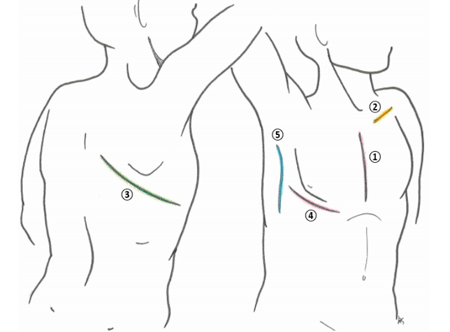
- Scars
- 1. Median sternotomy
- Heart valve replacement
- CABG
- Heart transplant
- 2. Pacemaker insertion
- Pre-existing arrythmia
- Bradycardia & heart block
- May have suffered from MI in the past
- Muscular dystrophy can cause bradycardia
- Sick sinus syndrome
- Bradycardia & heart block
- Heart failure
- Cardiac resynchronization therapy (CRT)
- Biventricular pacing
- Hypertrophic cardiomyopathy
- Pre-existing arrythmia
- 3. Posterolateral thoracotomy
- Pneumonectomy or lobectomy, chest wall resection, oesophageal surgery
- 4. Anterolateral thoracotomy
- Various pathology
- 5. Axillary thoracotomy
- Pneumonectomy & pneumothorax
- 1. Median sternotomy
- Visible apex beat
- lean patient or e.g. left ventricular aneurysm
- Listen for metallic heart valve ticking
- Oxygen
- Shortness of Breath
Examine Hands
- Nails
- Clubbing
- ABC = Atrial myxoma, bacterial infective endocarditis, congenital cyanotic heart failure
- Koilonichia (spoon shaped nails)
- Iron deficiency anaemia
- Splinter haemorrhage
- Infective endocarditis
- Quinke's Sign = Visible pulsation in nail beds
- Aortic regurgitation
- Xanthomata = Fatty deposits on tendons
- Suggests hyperlipidaemia
- Osler's nodes (tender) & Janeway lesions (haemorrhagic)
- Infective endocarditis
- Clubbing
- Temperature changes
- Capillary refil time
- Poor perfusion of extremities may be caused by hypovolaemia, hypothermia, peripheral vascular disease or just cool peripheries (with associated vasoconstriction)
- Cyanosis
- Nails
Radial Pulse
- Rate (>100bpm = Tachy, <60bpm = Brady)
- Rhythm
- Irregular
- Regularly irregular = Ectopic beats
- Variable heart block or premature ventricular excitation
- Irregular irregular = Atrial fibrillation
- Regularly irregular = Ectopic beats
- Variation of rate with breathing
- Accelerates on inspiration & slows on expiration
- Sinus arrythmia
- Slows on inspiration (pulsus paradoxus)
- Pericardial effusion, constrictive pericarditis
- Severe pneumothorax (esp. tension pneumothorax), severe asthma & COPD
- NB: Here, systolic bp falls by >10mmHg on inspiration
- Signifies severity of cardiac tamponade
- Accelerates on inspiration & slows on expiration
- Irregular
- Radio-femoral delay
- Coarctation of aorta
- More common in turner's syndrome
- Coarctation of aorta
- Radio-radial delay
- Occlusion or stenosis on one side
- Dissection of thoracic aorta
- Aortic aneurysm
- Radio-brachial delay
- Aortic stenosis
- Character
- Bounding pulse
- Waterhammer/Collapsing pulse
- Aortic regurgitation
- Hyperdynamic state e.g. pregnancy
- Fast & weak pulse = Shock
Blood Pressure
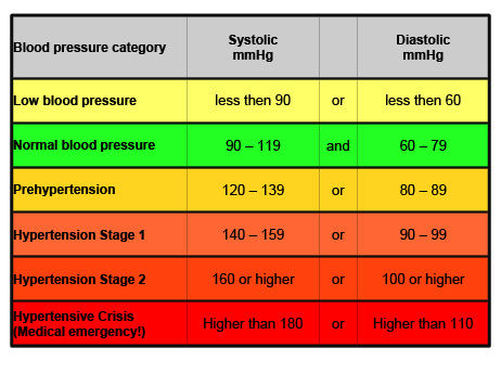
- Narrow Pulse pressure: Diastolic < 25% Systolic
- Extremely low, i.e. 25 mmHg or less = Very low stroke volume, as in Congestive Heart Failure and/or cardiogenic shock
- Aortic Stenosis
- Wide pulse pressure (consistently greater than 100 mmHg)
- Aortic regurgitation
- Wide PP & bradycardia & increased ICP = Cushing's triad (Head trauma related to intracranial hemorrhage or oedema)
- Narrow Pulse pressure: Diastolic < 25% Systolic
Face
- Eyes
- Jaundice
- Pallor
- Anaemia
- Congunctival haemorrhage
- Infective endocarditis
- Xantholasma
- Hyperlipidaema
- Mouth
- Central Cyanosis
- Poor oxygenation due to MI, heart failure, valvular disease, congenital heart disease
- Dental hygiene
- Infective endocarditis
- High arched palate
- Marfan's = Increased risk of mitral valve prolapse & aortic aneurysm
- Central Cyanosis
- Cheeks
- Mitral facies
- Severe mitral stenosis
- Rosy cheeks, whilst rest of face has bluish tinge due to cyanosis
- Mitral facies
- Eyes
Neck
- Carotid Pulse
- Normal = Smooth, relatively rapid upstroke and a smooth, more gradual downstroke
- Difference between the two = atherosclerosis
- Hyperkinetic = Higher stroke volume
- Hypokinetic = Low stroke volume
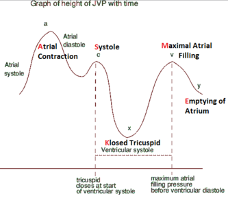
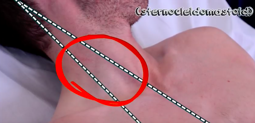
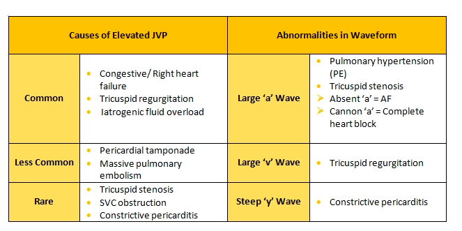
- JVP
- Indicates pressure in venous system awaiting entry to right atrium (SVC)
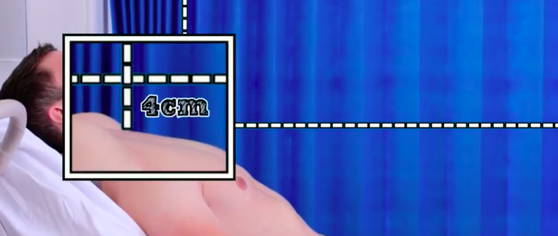
- Measurement: Vertical distance (in cm) between horizontal lines drawn from upper level of venous pulsation & sternal angle
- Normal = <4cm
- JVP + 5cm = right heart filling pressure in cm of water
- Total of 9cm = Raised
- JVP + 5cm = right heart filling pressure in cm of water
- Normal = <4cm
- Indicates pressure in venous system awaiting entry to right atrium (SVC)
- Carotid Pulse
Chest
- Scars (see above)
- Visible pulsations
- Obvious deformity
Palpation
- Apex Beat
- Normal site = 5th left intercostal space, 1cm medial to mid-clavicular line
- Parasternal heave
- Palm of hand to left of sternum with fingers lifted slightly off chest
- Right ventricular enlargement
- Right ventricular hypertrophy in chronic respiratory disease
- Thrills
- Aortic stenosis, pulmonary stenosis, mitral regurgitation
- Apex Beat
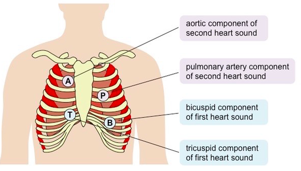
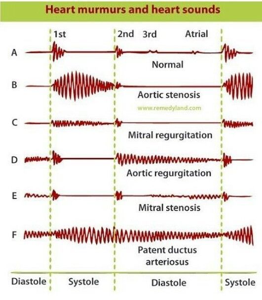
Auscultation
- S3 = "Kentucky" Loud in young people or athletes, congestive heart failure = Passive LV filling
- S4 = "Tennessee" Increase in stiffness of left ventricle due to scar tissue formation = Active LV filling
- Aortic Stenosis = "Potato"
- Mitral Regurgitation = Potaaaato
- Mitral Valve Prolapse = "Oliver" with hiccup
Peripheral checks
- Sacral oedema = Right sided heart failure
- Pulmonary oedema = Left sided heart failure
"To complete"
- Fundoscopy
- Peripheral pulses
- ECG, Echo etc.
Images and files in the map
Information
- Description
- Phase 1 Medicine Mind Map on Cardiovascular Examination, created by Flora Bradshaw on 02/01/2018.
- Created by
- Flora Bradshaw · Jan 2018
- Updated
- Jan 3, 2018
- Views
- 0
- Rating