
Mind map
Pathologies of growth
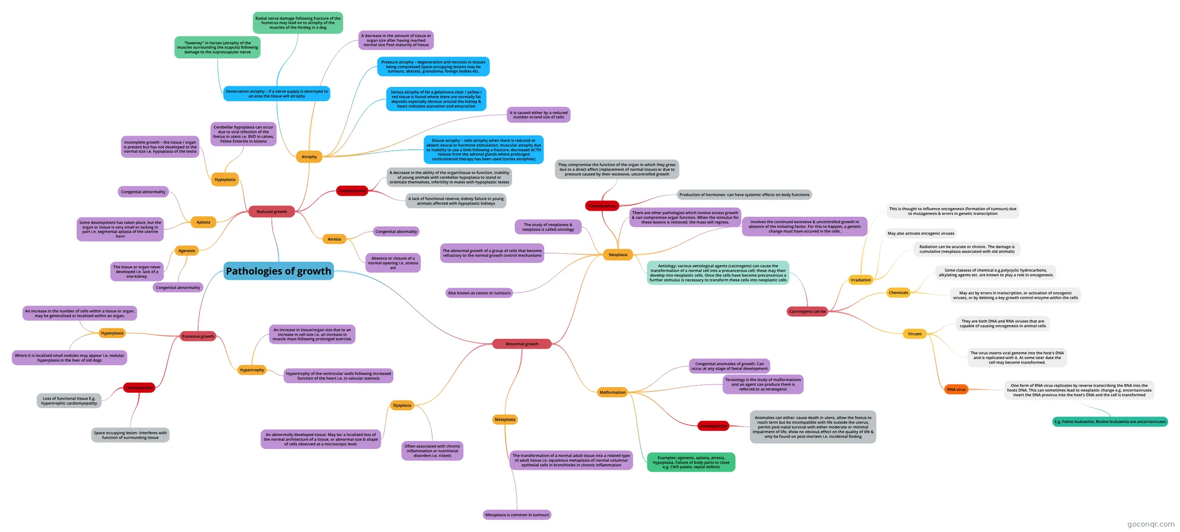
Mind map: Pathologies of growth
Map content
Pathologies of growth
Reduced growth
- Aplasia
- Some development has taken place, but the organ or tissue is very small or lacking in part i.e. segmental aplasia of the uterine horn
- Congential abnormality
- Hypoplasia
- Incomplete growth – the tissue / organ is present but has not developed to the normal size i.e. hypoplasia of the testis
- Cerebellar hypoplasia can occur due to viral infection of the foetus in utero i.e. BVD in calves, Feline Enteritis in kittens
- Atrophy
- A decrease in the amount of tissue or organ size after having reached normal size Post-maturity of tissue
- It is caused either by a reduced number or/and size of cells
- Denervation atrophy – if a nerve supply is destroyed to an area the tissue will atrophy
- “Sweeney” in horses (atrophy of the muscles surrounding the scapula) following damage to the suprascapular nerve
- Radial nerve damage following fracture of the humerus may lead on to atrophy of the muscles of the foreleg in a dog
- Pressure atrophy – degeneration and necrosis in tissues being compressed Space-occupying lesions may be tumours, abscess, granuloma, foreign bodies etc.
- Serous atrophy of fat a gelatinous clear / yellow / red tissue is found where there are normally fat deposits especially obvious around the kidney & heart indicates starvation and emaciation
- Disuse atrophy – cells atrophy when there is reduced or absent neural or hormone stimulation, muscular atrophy due to inability to use a limb following a fracture, decreased ACTH release from the adrenal glands where prolonged corticosteroid therapy has been used (cortex atrophies)
- Agenesis
- The tissue or organ never developed i.e. lack of a one kidney.
- Congenital abnormality
- Atresia
- Congenital abormality
- Absence or closure of a normal opening i.e. atresia ani
- Consequences
- A decrease in the ability of the organ/tissue to function, inability of young animals with cerebellar hypoplasia to stand or orientate themselves, infertility in males with hypoplastic testes
- A lack of functional reserve, kidney failure in young animals affected with hypoplastic kidneys
- Aplasia
Excessive growth
- Hyperplasia
- An increase in the number of cells within a tissue or organ. may be generalised or localised within an organ.
- Where it is localised small nodules may appear i.e. nodular hyperplasia in the liver of old dogs
- Hypertrophy
- An increase in tissue/organ size due to an increase in cell size i.e. an increase in muscle mass following prolonged exercise.
- Hypertrophy of the ventricular walls following increased function of the heart i.e. in valvular stenosis
- Consequences
- Loss of functional tissue E.g. hypertrophic cardiomyopathy
- Space occupying lesion- Interferes with function of surrounding tissue
- Hyperplasia
Abnormal growth
- Dysplasia
- An abnormally developed tissue. May be: a localised loss of the normal architecture of a tissue, or abnormal size & shape of cells observed at a microscopic level.
- Often associated with chronic inflammation or nutritional disorders i.e. rickets
- Metaplasia
- The transformation of a normal adult tissue into a related type of adult tissue i.e. squamous metaplasia of normal columnar epithelial cells in bronchioles in chronic inflammation
- Metaplasia is common in tumours
- Neoplasia
- The abnormal growth of a group of cells that become refractory to the normal growth control mechanisms
- Also known as cancer or tumours
- The study of neoplasms & neoplasia is called oncology
- Consequences
- They compromise the function of the organ in which they grow: due to a direct effect (replacement of normal tissue) or due to pressure caused by their excessive, uncontrolled growth
- Production of hormones- can have systemic effects on body functions
- There are other pathologies which involve excess growth & can compromise organ function. When the stimulus for these lesions is removed, the mass will regress.
- Involves the continued excessive & uncontrolled growth in absence of the initiating factor. For this to happen, a genetic change must have occured in the cells.
- Aetiology: various aetiological agents (cacinogens) can cause the transformation of a normal cell into a precancerous cell; these may then develop into neoplastic cells. Once the cells have become precancerous a further stimulus is necessary to transform these cells into neoplastic cells
- Carcinogens can be
- Irradiation
- This is thought to influence oncogenesis (formation of tumours) due to mutagenesis & errors in genetic transcription
- May also activate oncogenic viruses
- Radiation can be acurate or chronic. The damage is cumulative (neoplasia associated with old animals)
- Chemicals
- Some classess of chemical e.g.polycyclic hydrocarbons, alkylating agents etc. are known to play a role in oncogenesis
- May act by errors in transcription, or activation of oncogenic viruses, or by deleting a key growth control enzyme within the cells
- Viruses
- They are both DNA and RNA viruses that are capable of causing oncogenesis in animal cells
- The virus inserts viral genome into the host's DNA and is replicated with it. At some later date the cell may become transformed.
- RNA virus
- One form of RNA virus replicates by reverse transcribing the RNA into the hosts DNA. This can sometimes lead to neoplastic change e.g. oncornaviruses insert the DNA provirus into the host's DNA and the cell is transformed
- E.g. Feline leukaemia, Bovine leukaemia are ancornaviruses
- One form of RNA virus replicates by reverse transcribing the RNA into the hosts DNA. This can sometimes lead to neoplastic change e.g. oncornaviruses insert the DNA provirus into the host's DNA and the cell is transformed
- Irradiation
- Carcinogens can be
- Malformation
- Congenital anomalies of growth. Can occur at any stage of foetal development
- Teratology is the study of malformations and an agent can produce them is referred to as teratogenic
- Consequences
- Anomalies can either: cause death in utero, allow the foetus to reach term but be incompatible with life outside the uterus, permit post-natal survival with either moderate or minimal impairment of life, show no obvious effect on the quality of life & only be found on post-mortem i.e. incidental finding
- Examples: agenesis, aplasia, atresia, hypoplasia. Failure of body parts to close e.g. Cleft palate, septal defects
- Dysplasia
Information
- Description
- Veterinary Nursing Mind Map on Pathologies of growth, created by Rachael Hancox on 02/03/2018.
- Created by
- Rachael Hancox
- Views
- 1
- Rating