
Mind map
Digestive System
Map content
Digestive System
- very little of the gut requires the brain, the 2 gut brains are the submucosal plexus and myenteric plexus
- the enteric nervous system influences all digestive processes, the myenteric plexus exerts control over GI motility and the submucosal plexus senses the environment within the lumen, regulates GI blood flow and controls epithelial cell function
- passive absorption involves diffusion, facilitated transport is slightly faster and involves a membrane carrier and finally active transport absorption uses energy and a membrane carrier and is controllable e.g. fructose is absorbed by facilitated transport, glucose enters the cell with sodium
Components of the digestive system (alimentary canal)
- oral cavity
- mastication and chemical digestion which begins when the salivary glands secretes salvia into the mouth
- saliva consists of mainly water with small amounts of digestive enzymes, mucus, antibodies and electrolytes (salts)
- pH 6-7
- it functions to soften and lubricate food so that it can be swallowed and begins the chemical digestion of food to ensure that food can be tasted and to cleanse the teeth and mouth which keeps the bacterial population under control
- saliva is produced by either intrinsic (minor) salivary glands or extrinsic (major) salivary glands
- lingual, palatpglossal, palatal and buccal glands secrete saliva directly into the mouth and are intrinsic
- extrinsic glands: parotid, submandibular and sublingual glands and they are all paired exocrine glands and secrete saliva into the mouth via a series of ducts
- salivary amylase is produced by the acini cells, pH range is pH 4-11 with optimum pH as pH 6.9
- amylase breaks down alpha 1,4 glucosidic bonds which causes starch or glycogen to break down into maltose, maltotriose and alpha limit dextrins
- a 1,4 glucoside bonds = 1 glucose-glucose 4 bonds
- amylase breaks down alpha 1,4 glucosidic bonds which causes starch or glycogen to break down into maltose, maltotriose and alpha limit dextrins
- saliva consists of mainly water with small amounts of digestive enzymes, mucus, antibodies and electrolytes (salts)
- mastication and chemical digestion which begins when the salivary glands secretes salvia into the mouth
- pharynx
- a U-shaped fibromuscular structure that connects the oral and nasal cavities with the oesophagus
- the walls of the pharynx are formed the pharyngeal constrictor muscles which push the food into the oesophagus
- the inner circular muscles of the pharynx are split into the ... superior pharyngeal constrictor (in the oropharynx) - constricts upper pharynxmiddle pharyngeal constrictor (in laryngopharynx)- performs general sphincteric and peristaltic action in swallowing (deglutition)inferior pharyngeal constrictor (in laryngopharynx)- acts as a sphincter at the junction of the laryngopharynx and the oesophagus and provides a propulsive force for swallowing - it is further split into the superior thyropharyngeus and the inferior circopharyngeus
- the walls of the pharynx are formed the pharyngeal constrictor muscles which push the food into the oesophagus
- a U-shaped fibromuscular structure that connects the oral and nasal cavities with the oesophagus
- oesophagus
- continuous with the pharynx at the level C6, is ~25cm in length and empties into the stomach
- posterior to the trachea and aortic arch, along the right side of the descending aorta
- passes through the diaphragm at the level T10 to end at the cardiac orifice of the stomach
- upper oesophageal sphincter is made by a portion of the inferior constrictor muscle and allows the food bolus to enter the oesophagus
- voluntary control
- lower oesophageal sphincter is formed by a portion of the diaphragm, it constricts the oesophagus during respiration to prevent the contents of the stomach regurgitating
- involuntary smooth muscle
- mucosa
- lamina propria (areolar connective tissue)
- muscular mucosae (smooth muscle)
- non keratinising stratified squamous epithelia
- temperature, chemical and abrasive protection
- submucosal glands
- muscularis propria
- CILO (circular inner, longitudinal outer)
- muscle types
- upper 1/3
- skeletal
- middle 1/3
- mixed
- lower 1/3
- smooth
- upper 1/3
- muscle types
- CILO (circular inner, longitudinal outer)
- adventitia
- upper oesophageal sphincter is made by a portion of the inferior constrictor muscle and allows the food bolus to enter the oesophagus
- continuous with the pharynx at the level C6, is ~25cm in length and empties into the stomach
- stomach
- the stomach is a large, distensible muscular sac, which varies in size and shape depending on the individual
- small intestine
- large intestine
- oral cavity
Functions of the digestive system
- ingestion (the beginning of the digestive process)
- mastication begins the mechanical breakdown of food
- digestion (the mechanical and chemical breakdown of food and drink)
- secretion of enzymes, acids and buffers
- absorption of nutrients
- uptake of water and the condensing/compaction of indigestible components
- defication (elimination of waste from the body)
Phases of swallowing
- buccal phase
- phase lasts from food entering the mouth until it is propelled back towards the oropharynx
- pharyngeal phase
- phase lasts until the pharyngeal muscles contract to push the bolus through the open upper oesophageal sphincter into the upper oesophagus
- the upper oesophageal sphincter is a bundle of muscles at the top of the oesophagus
- phase lasts until the pharyngeal muscles contract to push the bolus through the open upper oesophageal sphincter into the upper oesophagus
- oesophageal phase
- peristaltic waves, breathing resumes once the bolus enters the upper oesophagus, bolus reaches the lower oesophageal sphincter which relaxes to allow the bolus to enter the stomach
- the lower oesophageal sphincter is a ring of muscle that opens to allow food and drink to pass into the stomach from the mouth
- proton pumps are given to people with acid reflex and this minimises the amount of acid in the stomach
- the lower oesophageal sphincter is a ring of muscle that opens to allow food and drink to pass into the stomach from the mouth
- peristaltic waves, breathing resumes once the bolus enters the upper oesophagus, bolus reaches the lower oesophageal sphincter which relaxes to allow the bolus to enter the stomach
- buccal phase
Stomach
- in the left hypochondrium/epigastric region (2,3)
- internal surface contains a series of ridges called the rugae which allows for distension and contraction of the muscle
- pH 1-3
- longitudinal, circular and inner oblique muscle layers
- functions
- acts as a mixing compartment, braking up food and mixing it into a semi-fluid pulp called chyme
- storage organ (can hold up to 1500ml of chyme which remains in the stomach for 2-4 hours)
- secretes gastric juice which contains water, mucous, hydrochloric acid, intrinsic factor and pepsinogen
- pH 1-3.5
- stomach ulcers can be causes by Helicobacter pylori
- when food enters the stomach through the oesophageal sphincter, peristaltic movements called mixing waves help breakdown the bolus mechanically before it mixes with gastric juice to become chyme
- the pyloric sphincter controls the movement of chyme from the pyloric antrum and pyloric canal to the duodenum
- peristaltic contractions squeeze out a small volume of chyme into the duodenum which is gastric emptying
- the remaining chyme is forced back into the body of the stomach by retropulsion
- the mixing continues
- the remaining chyme is forced back into the body of the stomach by retropulsion
- peristaltic contractions squeeze out a small volume of chyme into the duodenum which is gastric emptying
- food can cause the stomach pH to buffer and raise up to about pH4
- the pyloric sphincter controls the movement of chyme from the pyloric antrum and pyloric canal to the duodenum
- the acid secretion breaks down connective tissue and muscle fibre, activates pepsin, solubises calcium and iron and acts as a barrier to microbes
- oesophagus
- cardia
- fundus
- body
- pylorus
- the pylorus is a constriction separating the pyloric canal and the first duodenum, the pyloric sphincter guards it
- the pyloric canal is a narrow section of stomach that lies between the pyloric antrum and pylorus
- the pyloric antrum is a funnel-shaped section of the stomach that lies between the body and the pyloric canal
- some babies can get pylorus stenosis
- pyloric sphincter
- pylorus
- cardia
- mucosa
- epithelium
- simple columnar epithelium that secretes thick alkaline mucin which produces mucus when combined with water
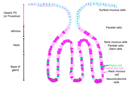
- gastric pits
- at the base of each gastric pit are a number of gastric glands, containing several types of specialised epithelial cells
- these cells release their secretions into the gastric pits which flow into the lumen of the stomach
- parietal cells
- produce hydrochloric acid and intrinsic factor
- chief cells
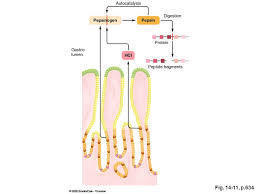
- release pepsinogen which is activated into the digestive enzyme pepsin when it comes into contact with acid produced by parietal cells
- pepsin breaks down proteins and is an endopeptidase, "endo-" means it breaks specific bonds throughout the molecule
- release pepsinogen which is activated into the digestive enzyme pepsin when it comes into contact with acid produced by parietal cells
- gastric juice is a mixture of parietal, chief and neck mucous cell secretions
- gastric enteroendocrine cells are found at the base of gastric glands
- neuroendocrine cells secrete serotonin and other hormones
- G cells secrete gastrin
- parietal cells
- stem cells are located in the neck region of the gastric gland, they have short microvilli on their apical surface
- these cells release their secretions into the gastric pits which flow into the lumen of the stomach
- foveolar cells or surface mucus cells are mucus producing cells that cover the inside of the stomach
- mucous neck cells are found at the neck of the gastric pit
- intrinsic factor is essential for vitamin B12 absorption
- at the base of each gastric pit are a number of gastric glands, containing several types of specialised epithelial cells
- mucus protects the underlying mucosa from self-abrasion that could be causes by the acidity of stomach secretions and its contents
- gastric pits
- simple columnar epithelium that secretes thick alkaline mucin which produces mucus when combined with water
- lamina propria
- loose connective tissue that is rich in capillaries, lymphatic vessel and nerves and the gastric glands protrude into it
- muscularis mucosae
- smooth muscle that compresses the gastric glands when it contracts and helps to push out their contents into the lumen of the stomach
- epithelium
- submucosa
- connective tissue and vascular plexus that supplies the mucosa and allows the mucosa to move freely over the deeper structures
- muscularis externa/propria
- oblique, circular and longitudinal muscular layers
- peritoneum
- adventitia
- serosa that covers the outer surface of the stomach and is continuous superiorly with the lesser omentum and inferiorly with the greater omentum
Pancreas
- functions to produce an enzyme-rich pancreatic juice which helps to breakdown proteins, starch and fats
- lobes of the pancreas: tail, body, neck, head (head proper and uncinate process)
- each lobe of the pancreas drains into the centrally running main pancreatic duct
- this duct joins with the common bile duct in the head of the pancreas which opens into the duodenum
- hepatopancreatic ampulla
- there may be accessory ducts that open into the duodenum independently
- the exocrine region of the pancreas (99%) is composed of clusters of secretory epithelial cells called acini
- the cells of the acini secrete a mixture of digestive enzymes, bicarbonate ions and fluid (pancreatic juice) which is used to neutralise stomach acid and aid in digestion
- amylase, trypsinogen, phospholipase and deoxyribonuclease, most have proteins as their substrates apart from phospholipase which has phospholipids as its substrate
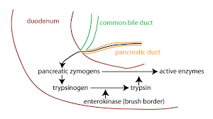
- pancreatic juice is made up of sodium bicarbonate, pancreatic amylase, trypsin (secreted as trypsinogen), elastase (secreted as proelastase), carboxypeptidase, chymotrypsin, ribonuclease and deoxyribonuclease
- pH 8-8.3
- zymogens are the inactive form
- the pancreatic juice accumulates in the intralobar ducts and finally enters the main pancreatic duct
- pancreatic juice secretion is active which means it is under control and responds and changes in composition
- the bicarbonate ions makes the pancreatic juice alkali and it helps to buffer the secretions of the stomach, it also protects the mucosa and provides the right pH for pancreatic enzymes
- non proteolytic pancreatic enzymes are released in their active form
- e.g. amylase, lipase (substrate - triglycerides), ribonuclease and deoxyribonuclease
- pancreatic amylase is slightly different to salivary amylase (they are structurally different and are coded for by different genes)
- both are digested by a 1,4 glucose-glucose bonds
- it digests starch and glycogen to maltose, maltotriose and dextrins
- pancreatic lipase acts on water insoluble triglycerides
- colipase inhibits absorption onto the lipid:water interphase which would denature lipase
- Islets of Langerhans make up the endocrine glands of the pancreas (1-2% of the overall pancreas mass), the cells secrete hormones like insulin, glucagon and somatostatin
- the cells of the acini secrete a mixture of digestive enzymes, bicarbonate ions and fluid (pancreatic juice) which is used to neutralise stomach acid and aid in digestion
- this duct joins with the common bile duct in the head of the pancreas which opens into the duodenum
- each lobe of the pancreas drains into the centrally running main pancreatic duct
- duel purpose gland
Liver
- peritoneum covers the liver but is reflected away from it in several places to from coronary ligaments which help suspend the liver in the abdomen
- the liver secretes bile acid and alkaline fluid, degrades and conjugates waste metabolic products, detoxifies and regulates nutrient metabolism
- produces bile, heparin, urea, vitamin A, plasma proteins and antibodies
- acts as a storage for fats, vitamin A, D and B12, iron and glycogen
- stores iron for at least 6 months
- right, left, caudate and quadrate lobes
- bile canaliculi are tiny channels located between the hepatocytes and transport bile to the bile ducts
- bile is an alkaline liquid
- bile is secreted by hepatocytes and contains cholesterol, bile acid, bile salts, neutral fats, electrolytes and phospholipids
- sodium, potassium and calcium electrolytes
- bile is synthesised in the liver and stored in the gallbladder
- gallbladder: fundus, body, neck, cystic duct (+hepatic duct), common bile duct
- pH 7.8
- the gallbladder is found under the inferior surface of the right lobe of the liver
- a person can survive without a gallbladder
- the principle pigment in bile is bilirubin
- the common bile duct releases bile into the duodenum where it emulsifies fats into little droplets called micelle which makes their digestion by lipases easier
- increases the surface area in which the enzyme can work on
- after emulsifying fats in the small intestine, bile can be recycled back into the gallbladder, through the ileum via hepatic portal blood
- when intestinal cells come into contact with acid chyme, they release secretin which promotes the bile ducts to secrete more bile
- bile also helps in the absorption of fats by forming complexes called micelles
- the common bile duct releases bile into the duodenum where it emulsifies fats into little droplets called micelle which makes their digestion by lipases easier
- daily output is 700-1200ml
- bile is secreted by hepatocytes and contains cholesterol, bile acid, bile salts, neutral fats, electrolytes and phospholipids
- bile is an alkaline liquid
- 1.5kg
- essential for the production of clotting factors
Intestines
- small intestine
- pH 7.5-7.8
- the main digestion and absorption site for water, proteins, carbohydrates and lipids
- duodenum
- the major duodenal papilla is an opening of the pancreatic duct into the duodenum
- jejunum
- ileum
- nutrient absorption
- completes break down and nutrient absorption
- ileum
- mixing secretions from the pancreas, liver and its own with food causes the neutralisation of acid and further digestion and absorption
- mucosa: epithelium, lamina propria, muscularis mucosae
- columnar epithelia lamina propria forms the core of each villus and consists of loose areolar tissue, a lacteal and a network of capillaries, small intestine also contains absorptive cells, goblet cells, granular cells/cells of Paneth (enzymes) and endocrine cells the muscularis mucosae is a layer of smooth muscle located at the base of the villi that aids the propulsion of food along the gastrointestinal tract
- villi -> microvilli -> brush border -> brush border enzymes
- enterokinase is found on the brush border and it activates trypsinogen into trypsin and trypsin can then go on to activate chymotrypsinogen, procarboxypeptidase, proelastase, prophospholipase and procolipase
- small intestine enterocytes of the mucosa are present especially on the microvilli
- brush border enzymes are an integral part of the membrane and not free in the lumen, peptidases, lactase, sucrase and maltase are all brush border enzymes
- submucosa
- contain lymphatic vessels, venues, arterioles, mucus secreting glands (Brunner's glands) found in the duodenum and they produce an alkaline mucus that neutralises acid chyme
- Brunner's gland secretions -> pH 7.5-8.9
- muscularis propria/externa
- inner circular and outer longitudinal muscles with the myenteric plexus between them
- serosa
- made of simple squamous epithelium and froms a visceral peritoneum covering it
- adventitia
- cells
- absorptive cells
- most numerous type of epithelial cell present on the surface of the villi
- goblet cells
- synthesise an alkaline mucus that provides lubrication and prevents digestive enzymes eroding the cell walls
- enteroendocrine cells
- often located at the base of the intestinal glands of the duodenum and the fundus of the stomach
- paneth cells
- deep within intestinal glands
- intestinal glands are in the grooves between intestinal villi and duodenal glands as deep as the muscularis mucosae, they contain hormone secreting cells, paneth cells and constantly dividing epithelial cells
- deep within intestinal glands
- stem cells
- located in the lower part of the intestinal tract and have short microvilli on their apical surface
- gives rise to all cell types in the mucosa and travel upwards to replace absorptive and goblet cells
- located in the lower part of the intestinal tract and have short microvilli on their apical surface
- absorptive cells
- 12 finger lengths long
- large intestine
- no villi
- main functions: - absorption of water and electrolytes from chyme to form solid faeces - vitamin K absorption - colonic bacteria activity production of short chain fatty acids and production of vitamins - storage of faecal matter - transport
- carbohydrates and proteins not digested in the small intestine are fermented by colonic microflora to short chain ftty acids and gases, most gut microflora can only exist in area where there is no oxygen, they are capable of any reactions, some bacteria products are useful but other are harmful toxins and carcinogens
- large muscular tube, composed of the same layer as the small intestine
- mucosa
- epithelia
- columnar epithelia: mainly goblet and absorptive cells
- there are lots of goblet cells in the large intestine
- large intestinal mucus -> pH 7.5-7.8
- lamina propria
- lacteal and network of capillaries and lymphatic nodules
- muscularis mucosae
- smooth muscle
- crypts are small tubular glands, pits or recesses
- epithelia
- submucosa
- muscularis propria/externa
- inner circular and outer longitudinal muscle
- the taenia coli is the incomplete layers of longitudinal muscle in the large intestine
- the haustrau/haustrations are ring-like circular muscles in the large intestine
- serosa
- simple squamous epithelia
- adventitia
- mucosa
- absorbs water and salts and eliminates waste
- microvilli are responsible for the absorption of mainly water and salts
- the material that enters the large intestine is very fluid but once the water has been absorbed, only undigested food and bacteria remain
- caecum
- connects to the ileum through the ileocecal valve
- ascending colon
- right colic flexure/ hepatic flexure
- transverse colon
- left colic flexure (splenic flexure)
- descending colon
- when it reaches the inner pelvis, it curves to enter the centre of the pelvis at the sigmoid colon
- sigmoid colon
- curves to enter the lesser pelvis and continue as the rectum
- rectum
- lies in front of the sacrum and passes through the pelvic diaphragm to continue as the anal canal
- anal canal
- internal and external sphincters surround it, functioning to keep the anus closed, the external anal sphincter can be voluntarily contracted
- the appendix is attached to the caecum and is made of chronic inflammatory tissue
- small intestine
Peritoneum
- covers the stomach, liver, jejunum and ileum
- a thin serous coating which continues as the meszocolon to anchor the intestines to the abdominal wall
- parietal peritoneum covers the abdominal wall
- visceral peritoneum continues from the folds to cover the abdominal organs
- fluid-filled peritoneal cavity
- visceral peritoneum continues from the folds to cover the abdominal organs
Sections of the abdomen
- 1 - right hypochondrium
- 2 - epigastric
- 3 - left hypochondrium
- 4 - right lumbar
- 5 - umbilical
- 6 - left lumbar
- 7 - right iliac fossa
- 8 - suprapubic (hypogastrium)
- 9 - left iliac fossa
- 8 - suprapubic (hypogastrium)
- 7 - right iliac fossa
- 6 - left lumbar
- 5 - umbilical
- 4 - right lumbar
- 3 - left hypochondrium
- 2 - epigastric
- 1 - right hypochondrium
Abdominal aorta
- the 3 main vessels arising from the abdominal aorta are the coeliac trunk, the superior mesenteric artery and the interior mesenteric artery
Hepatic vein
- external iliac vein (right) and common iliac vein (left)
Images and files in the map
Information
- Description
- Year 1 Mind Map on Digestive System, created by Rana Al-Khayat on 17/04/2018.
- Created by
- Rana Al-Khayat
- Views
- 0
- Rating