
Mind map
Neuroanatomy 1
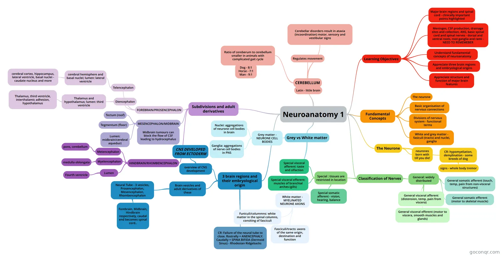
Mind map: Neuroanatomy 1
Map content
Neuroanatomy 1
Learning Objectives:
- Major brain regions and spinal cord - clinically important points highlighted
- Meninges, CSF production, drainage sites and collection, ANS, basic spinal cord and spinal nerves - dorsal and ventral roots, root ganglia and rami - NEED TO REMEMEBER
- Understand fundamental concepts of neuroanatomy
- Appreciate three brain regions and embryological origins
- Appreciate structure and function of major brain features
Fundamental Concepts
- The neurone
- Basic organisation of nervous connections
- Divisions of nervous system - functional terms
- White and grey matter - fasiculi (tracts) and nuclei, ganglia
The Neurone
- neurones born with till you die!
- CR: hypomyeliation, demylination - some breeds of dog
- signs - whole body tremor
Classification of Nerves
- General: widely distributed
- General somatic afferent (touch, temp, pain from non-visceral structures)
- General somatic efferent (motor to skeletal muscle)
- General visceral afferent (distension, temp, pain from viscera)
- General visceral efferent (motor to viscera, smooth muscles and glands)
- Special : tissues are restricted in location
- Special somatic afferent - vision, hearing, balance
- Special visceral afferent: taste and olfaction
- Special visceral efferent: muscles of branchial arches (gills)
- General: widely distributed
Grey vs White matter
- Grey matter - NEURONE CELL BODIES
- Nuclei: aggregations of neurone cell bodies in brain
- Ganglia: aggregations of nerve cell bodies in PNS
- White matter - MYELINATED NEURONE AXONS
- Funiculi/colummns: white matter in the spinal columns, consiting of fasciculi
- Fasciculi/tracts: axons of the same origin, destination and function
- Grey matter - NEURONE CELL BODIES
3 brain regions and their embryological origin
- overview of CNS development
- CNS DEVELOPED FROM ECTODERM
- Brain vesicles and adult derivatives of these
- Neural Tube - 3 vesicles, Prosencephalon, Mesencephalon, Rhombencephalon
- Forebrain, Midbrain, Hindbrain respectively, caudal end becomes spinal cord..
- Neural Tube - 3 vesicles, Prosencephalon, Mesencephalon, Rhombencephalon
- CR: Failure of the neural tube to close. Rostrally = ANENCEPHALY. Caudally = SPINA BIFIDA (Dermoid Sinus) - Rhodesian Ridgebacks
- overview of CNS development
Subdivisions and adult derivatives
- FOREBRAIN/PROSENCEPHALON
- Telencephalon
- cerebral hemisphere and basal nuclei; lumen: lateral ventricle
- cerebral cortex, hippocampus, lateral ventricle, basal nuclei - caudate nucleus and more
- cerebral hemisphere and basal nuclei; lumen: lateral ventricle
- Diencephalon
- Thalamus and hypothalamus; lumen: third ventricle
- Thalamus, third ventricle, interthalamic adhesion, hypothalamus
- Thalamus and hypothalamus; lumen: third ventricle
- Telencephalon
- MESENCEPHALON/MIDBRAIN
- Tectum (roof)
- Tegmentum (floor)
- Lumen: midbrain/cerebral aqueduct
- Midbrain tumours can block the flow of CSF leading to hydrocephalus
- HINDBRAIN/RHOMBENCEPHALON
- Metencephalon
- pons, cerebellum
- Myelencephalon
- medulla oblongata
- Lumen
- Fourth ventricle
- Metencephalon
- FOREBRAIN/PROSENCEPHALON
CEREBELLUM
- Latin - little brain
- Ratio of cereberum to cerebellum smaller in animals with complicated gait cycle
- Dog - 8:1 Horse - 7:1 Man - 9:1
- Regulates movement
- Cerebellar disorders result in ataxia (incoordination) motor, sensory and vestibular signs