
Mind map
Cardiac Alterations
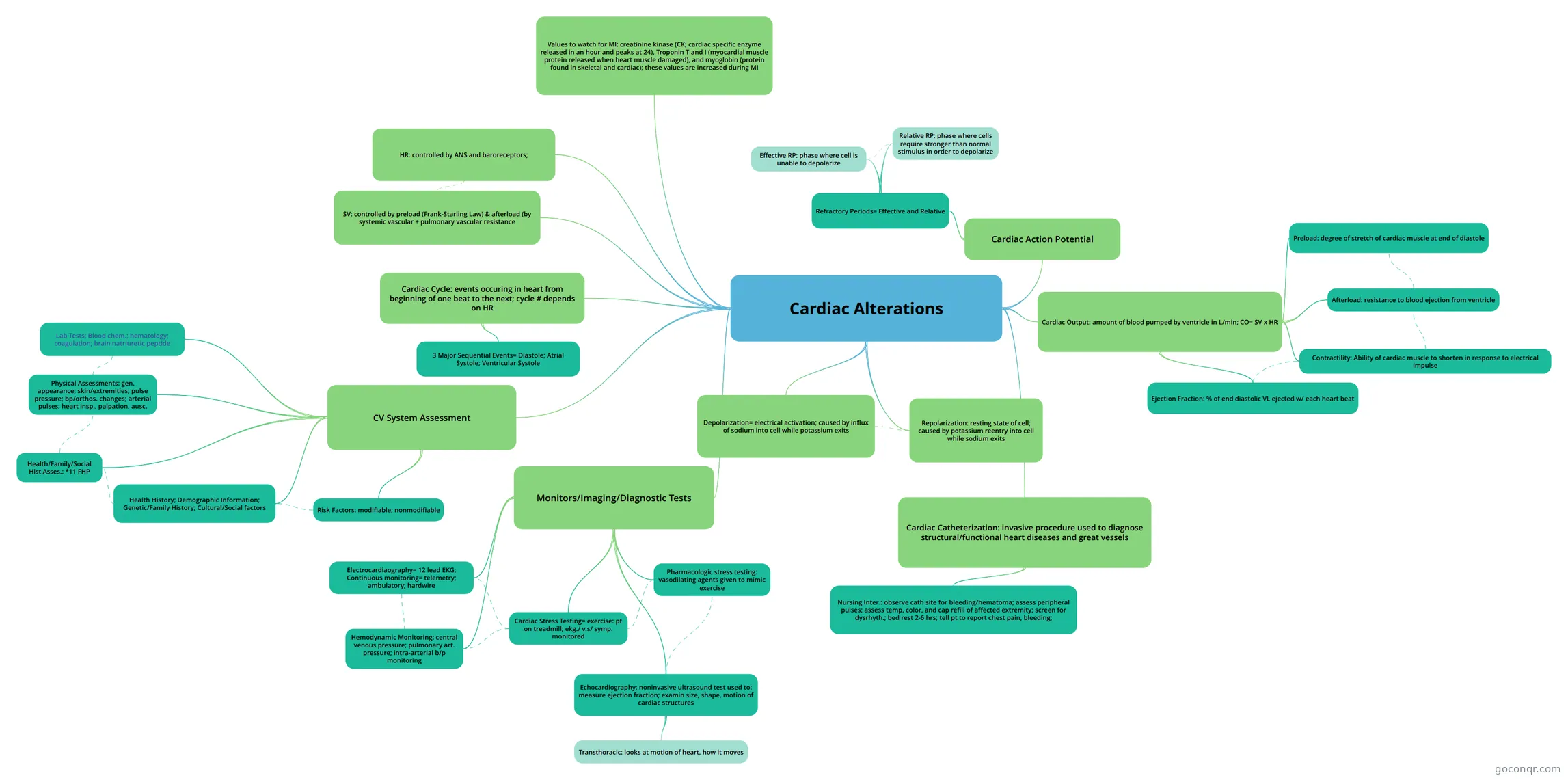
Mind map: cardiac alterations
Map content
Cardiac Alterations
Depolarization= electrical activation; caused by influx of sodium into cell while potassium exits
Repolarization: resting state of cell; caused by potassium reentry into cell while sodium exits
Cardiac Action Potential
- Refractory Periods= Effective and Relative
- Relative RP: phase where cells require stronger than normal stimulus in order to depolarize
- Effective RP: phase where cell is unable to depolarize
- Refractory Periods= Effective and Relative
Cardiac Cycle: events occuring in heart from beginning of one beat to the next; cycle # depends on HR
- 3 Major Sequential Events= Diastole; Atrial Systole; Ventricular Systole
Cardiac Output: amount of blood pumped by ventricle in L/min; CO= SV x HR
- Contractility: Ability of cardiac muscle to shorten in response to electrical impulse
- Afterload: resistance to blood ejection from ventricle
- Preload: degree of stretch of cardiac muscle at end of diastole
- Ejection Fraction: % of end diastolic VL ejected w/ each heart beat
HR: controlled by ANS and baroreceptors;
SV: controlled by preload (Frank-Starling Law) & afterload (by systemic vascular + pulmonary vascular resistance
CV System Assessment
- Health History; Demographic Information; Genetic/Family History; Cultural/Social factors
- Risk Factors: modifiable; nonmodifiable
- Physical Assessments: gen. appearance; skin/extremities; pulse pressure; bp/orthos. changes; arterial pulses; heart insp., palpation, ausc.
- Health/Family/Social Hist Asses.: *11 FHP
- Lab Tests: Blood chem.; hematology; coagulation; brain natriuretic peptide
Monitors/Imaging/Diagnostic Tests
- Electrocardiaography= 12 lead EKG; Continuous monitoring= telemetry; ambulatory; hardwire
- Cardiac Stress Testing= exercise: pt on treadmill; ekg./ v.s/ symp. monitored
- Pharmacologic stress testing: vasodilating agents given to mimic exercise
- Echocardiography: noninvasive ultrasound test used to: measure ejection fraction; examin size, shape, motion of cardiac structures
- Transthoracic: looks at motion of heart, how it moves
- Hemodynamic Monitoring: central venous pressure; pulmonary art. pressure; intra-arterial b/p monitoring
Cardiac Catheterization: invasive procedure used to diagnose structural/functional heart diseases and great vessels
- Nursing Inter.: observe cath site for bleeding/hematoma; assess peripheral pulses; assess temp, color, and cap refill of affected extremity; screen for dysrhyth.; bed rest 2-6 hrs; tell pt to report chest pain, bleeding;
Values to watch for MI: creatinine kinase (CK; cardiac specific enzyme released in an hour and peaks at 24), Troponin T and I (myocardial muscle protein released when heart muscle damaged), and myoglobin (protein found in skeletal and cardiac); these values are increased during MI
Information
- Description
- cardiac alteration information
- Created by
- Kadijah Sannoh
- Views
- 6
- Rating
More on Heart Anatomy and Function