
Mind map
Cardiovascular Anatomy
Map content
Cardiovascular Anatomy
Anatomy of the Heart
- Mediastinum; medial space between the two lateral pleural cavities. Boundaries: superior thoracic aperture (thoracic inlet), the diaphragm , the sternum and costal cartilages and the bodies of the thoracic vertebrae. Middle mediastinum contains the pericardium, the heart, origins of great vessels, nerves and other vessels.
- Pericardium: 2 layers Fibrous pericardium – tough external layer continuous with the central tendon of the diaphragm and the adventitia of great vessels Serous pericardium composed of i) Parietal layer of serous pericardium – shiny layer reflected on the heart at the great vessels Pericardial cavity - potential space that normally has a small amount fluid that acts as a lubricant ii) Visceral layer of serous pericardium (epicardium)
- Transverse pericardial sinus – passageway separating the aorta plus the pulmonary trunk from the superior vena cava (used in cardiac surgery to isolate the great vessels to begin cardiopulmonary bypass) Oblique pericardial sinus – blind-ended passageway separating the inferior vena cava plus the right pulmonary veins from the left pulmonary veins
- Excess fluid accumulation in the pericardial cavity pushes against the relatively rigid parietal layer of serous pericardium and fibrous pericardium and so the pressure is transmitted to the heart Pericardiocentesis – drainage of this fluid either through (A) Bare area of pericardium (B) Infrasternal angle between xiphoid and costal cartilage
- Arteries: coronary arteries Veins: cardiac veins that drain into the coronary sinus which opens into the right atrium
- Valves ensure unidirectional flow
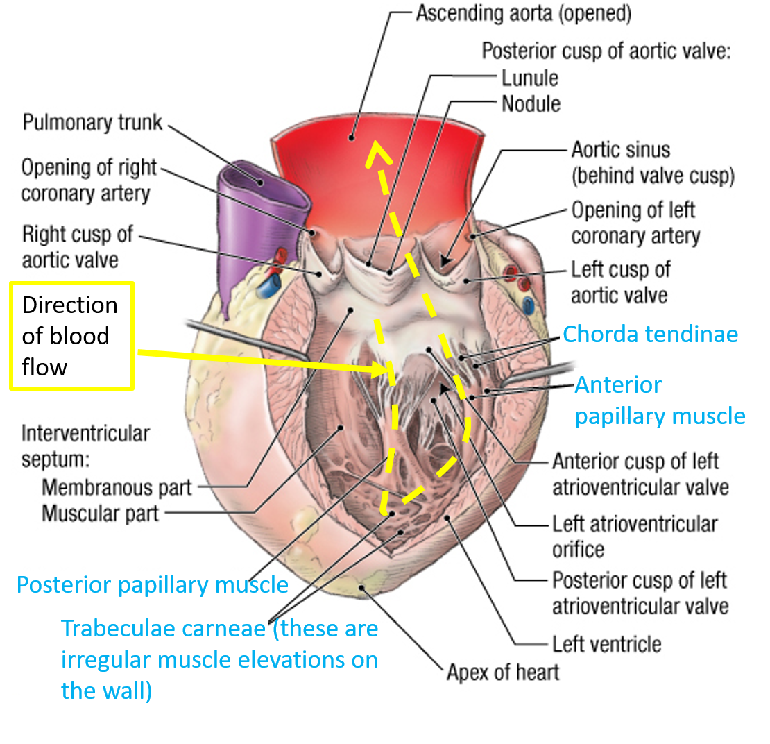
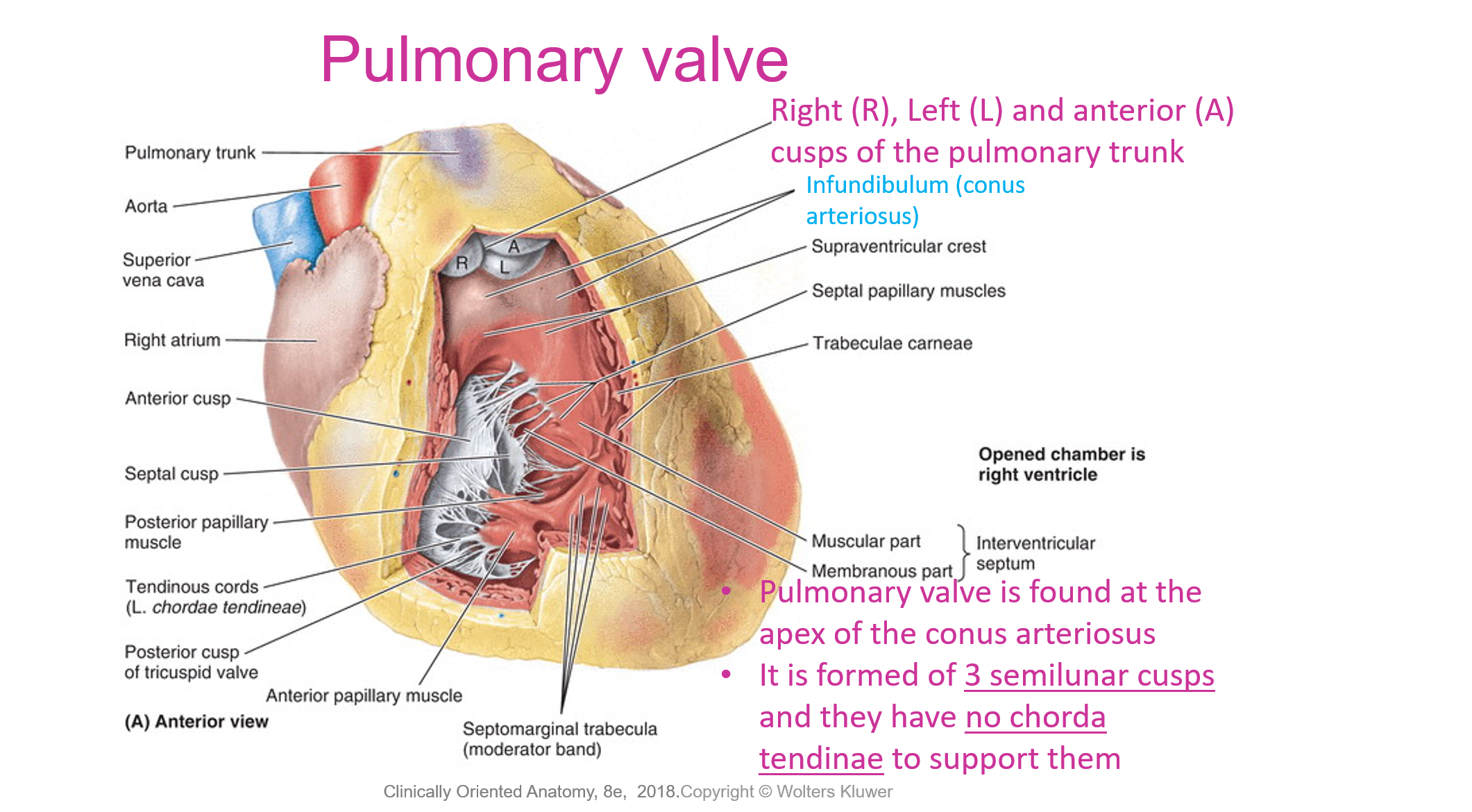
- Aortic valve is found between the left ventricle and the ascending aorta It is formed of 3 semilunar cusps and they have no chorda tendinae to support them
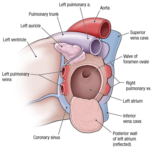
- Smooth walled left atrium Pectinate muscles (muscles that appear parallel in arrangement) are found in the left auricle
- Mitral valve is found in the left atrioventricular orifice
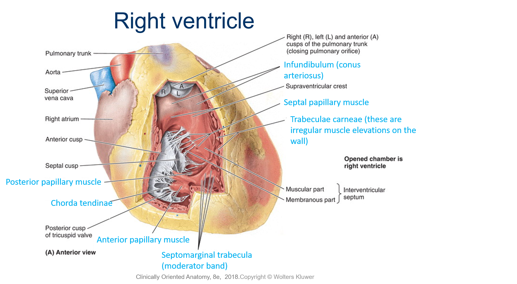
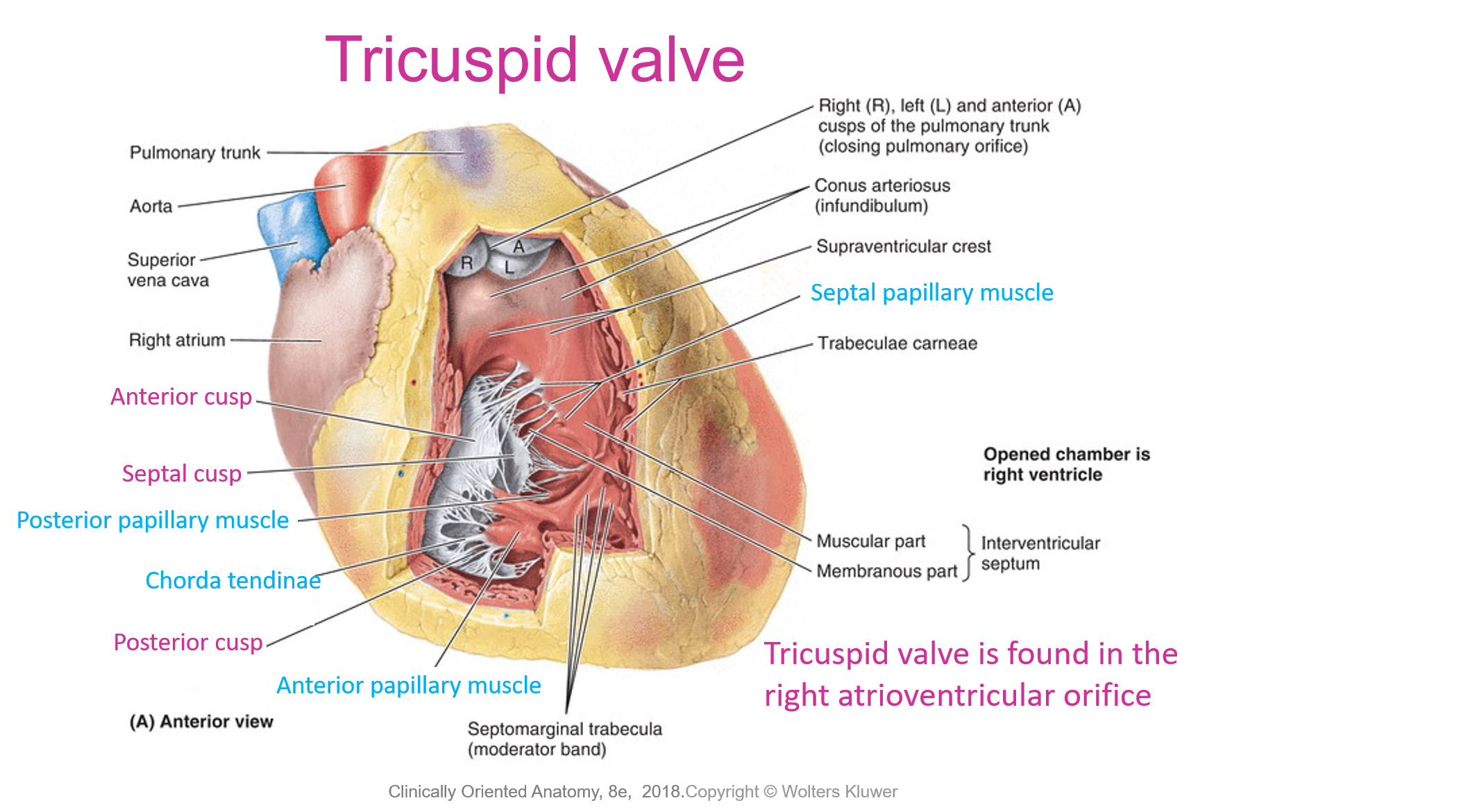
- Papillary muscles and chorda tendinae support the atrioventricular valve
- Mitral valve is found in the left atrioventricular orifice
- Left ventricle= thick Papillary muscles contract to tighten the chorda tendinae thereby pulling the cusps together They support the atrioventricular valve to resist the pressure during contraction of the ventricle
- Heart valves and their auscultation areas Two heart sounds are heard with a stethoscope: a lub (1st) sound as the blood is transferred from the atria into the ventricles (closure of mitral and tricuspid valves), and a dub (2nd) sound as the ventricles expel blood from the heart (closure of semilunar valves)
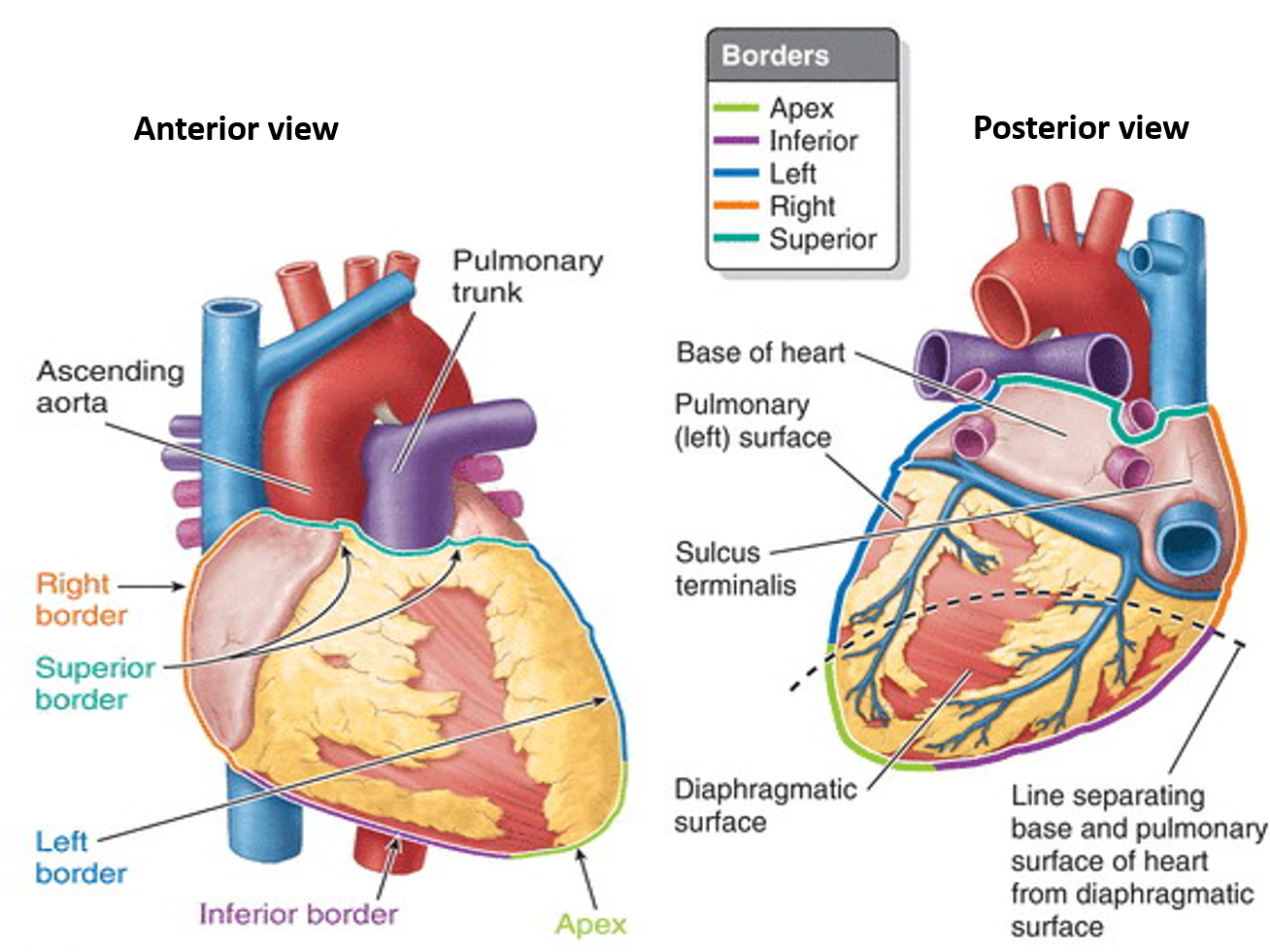
- Boundaries between chambers of the heart: posterior interventricular groove ,anterior interventricular groove, Interatrial septum, Interventricular septum. Congenital anomalies: Large atrial septal defects (ASD) allow oxygenated blood from the left atrium to be shunted to the right atrium overloading the pulmonary circulation; ventricular septal defects (VSD) cause left to right shunting also overloading the pulmonary circulation ASD and VSD loosely referred to as ‘hole in heart’
- The skeleton of the heart is a complex framework of dense collagen that forms rings surrounding the orifices of valves and the membranous parts of the interatrial and interventricular septa. The skeleton: Keeps the orifices of the atrioventricular and semilunar valves patent and prevents their over distension Provides attachment for the valves and the myocardium Insulates the atria from the ventricles so that they contract independently
- Palpating arterial pulses can help assess cardiac performance, cardiac rate and rhythm, establish the integrity of the peripheral arterial blood supply or localise a peripheral lesion
Coronary Artiries and Cardiac Veins
- Arteries: coronary arteries Veins: cardiac veins that drain into the coronary sinus which opens into the right atrium
- Coronary sulcus (atrioventricular groove) and interventricular groove
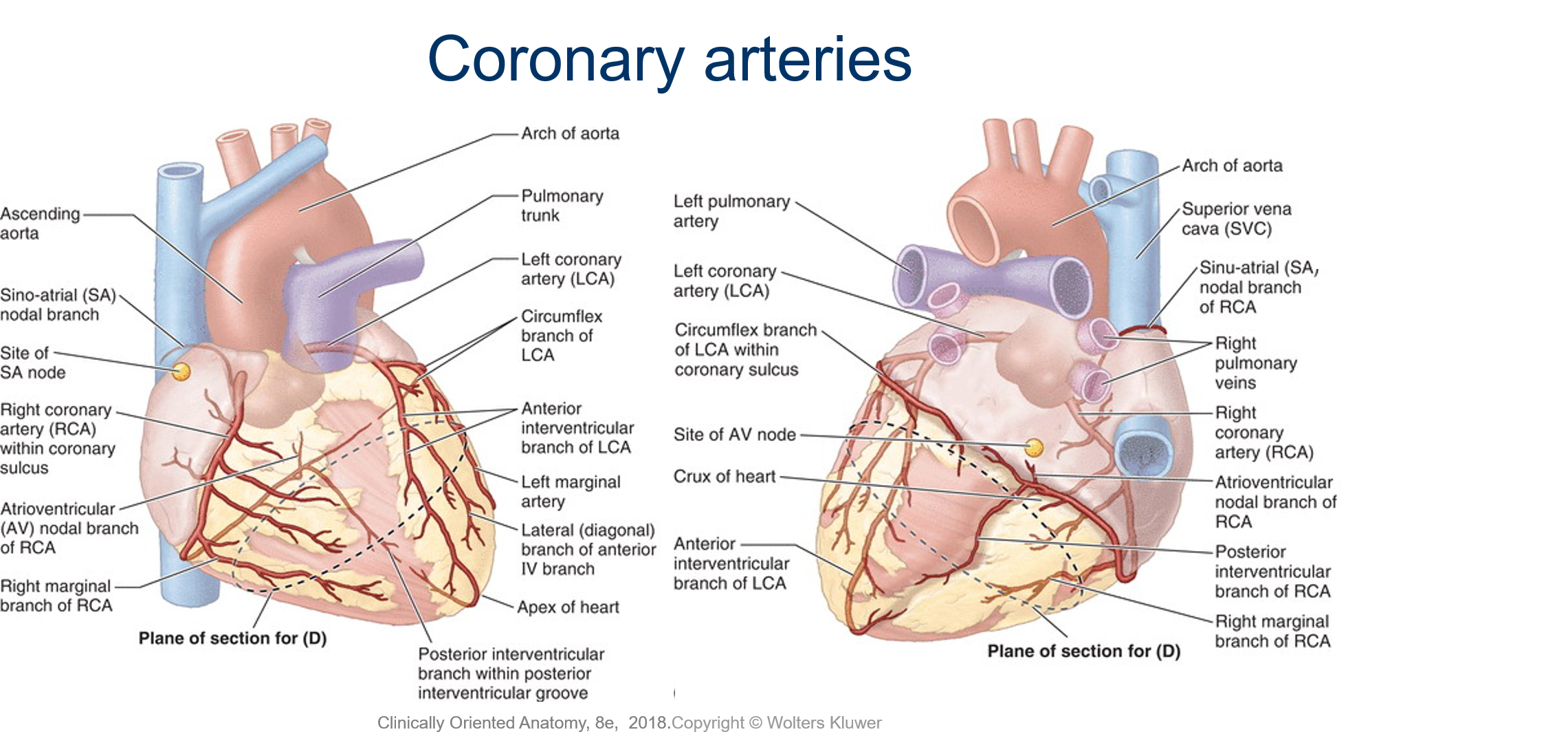
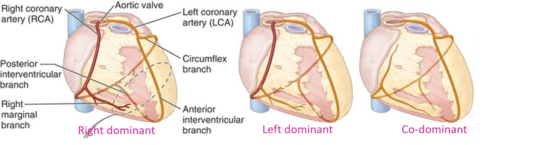
- Coronary arteries are the first branches of the aorta (ascending aorta) The left coronary artery arises from the left aortic sinus The right coronary artery arises from the right aortic sinus
- Right coronary artery branchesi) Sino atrial nodeii) Right marginal) Atrio-ventricular node) Posterior interventricular
- Right atrium, right ventricle, sinoatrial node, atrioventricular node, interatrial septum, some of the left atrium, posterior 1/3 of interventricular septum, some of the posterior of left ventricle
- Left coronary artery branches The left coronary artery has two terminal branches: circumflex and anterior interventricular. Circumflex artery branches: (A) left marginal and posterior left ventricular (not shown in diagram)
- Most of the left atrium, most of the left ventricle, most of the interventricular septum including the atrioventricular (AV) bundle and branches
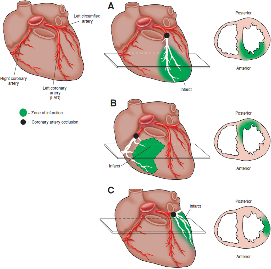
- Coronary arteries are considered functional end arteries as they do not interconnect; the borders between viable and infarcted myocardium are very distinct when a coronary artery is occluded
- Dominance is defined by the artery that gives rise to the posterior interventricular artery (posterior descending artery) The majority of individuals have dominance of the right coronary artery (RCA) i.e. the posterior interventricular artery is a branch of RCA
- Coronary artery bypass grafting uses a saphenous vein or left internal thoracic (mammary) artery to redirect blood around a blockage
- Right coronary artery branchesi) Sino atrial nodeii) Right marginal) Atrio-ventricular node) Posterior interventricular
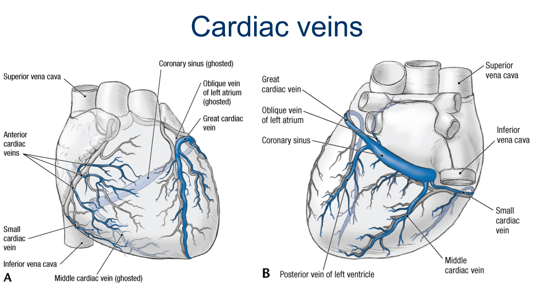
- Great cardiac vein – travels with LAD artery then circumflex artery and empties into coronary sinus Middle cardiac vein – travels with posterior interventricular artery and empties into coronary sinus Small cardiac vein – travels with right coronary artery in the lower anterior section of the coronary sulcus and empties into coronary sinus
- They are normally embedded in fat and travel just deep to the epicardium
- Lymphatic vessels follow the coronary arteries They drain into brachiocephalic nodes and tracheobronchial nodes
- Arteries: coronary arteries Veins: cardiac veins that drain into the coronary sinus which opens into the right atrium
Conduction system of the heart
- The conducting system generates and transmits electrical impulses that cause the coordinated contractions of the cardiac cycle It is formed of nodal tissue and conducting fibres
- The rate of depolarisation of cardiac muscle varies; the fastest is in the atria, the slowest in the ventricles. The contraction cycle of the heart is initiated in the atria, pushing blood into the ventricles. A wave of contraction in the ventricles then begins at the apex of the heart, pushing blood from the heart into the aorta and pulmonary trunk
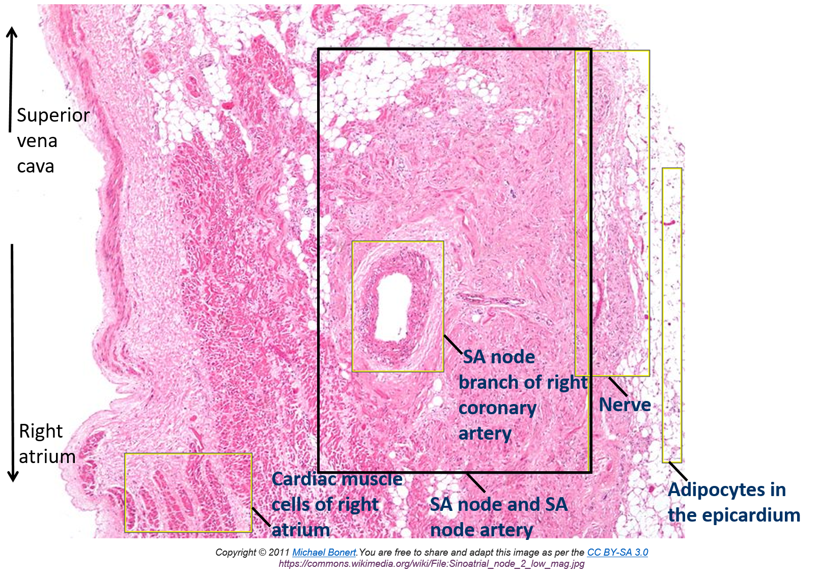
- SA node (sinus node) is at the junction of the superior vena cava and right atrium, where crista terminalis ends, deep to the epicardium Electrical impulses are generated here and spread through the cardiac muscles of the atria It has the fastest intrinsic depolarisation rate (~60-100 beats per minute) and is therefore called the pacemaker of the heart It is supplied by the sinoatrial nodal branch of the right coronary artery
- Small structure SA node artery in the centre Paler cells Poorly striated cells scattered in fibrous connective tissue Lots of nerves (sympathetics accelerate the heart rate, parasympathetics reduce the rate back to basal rate)
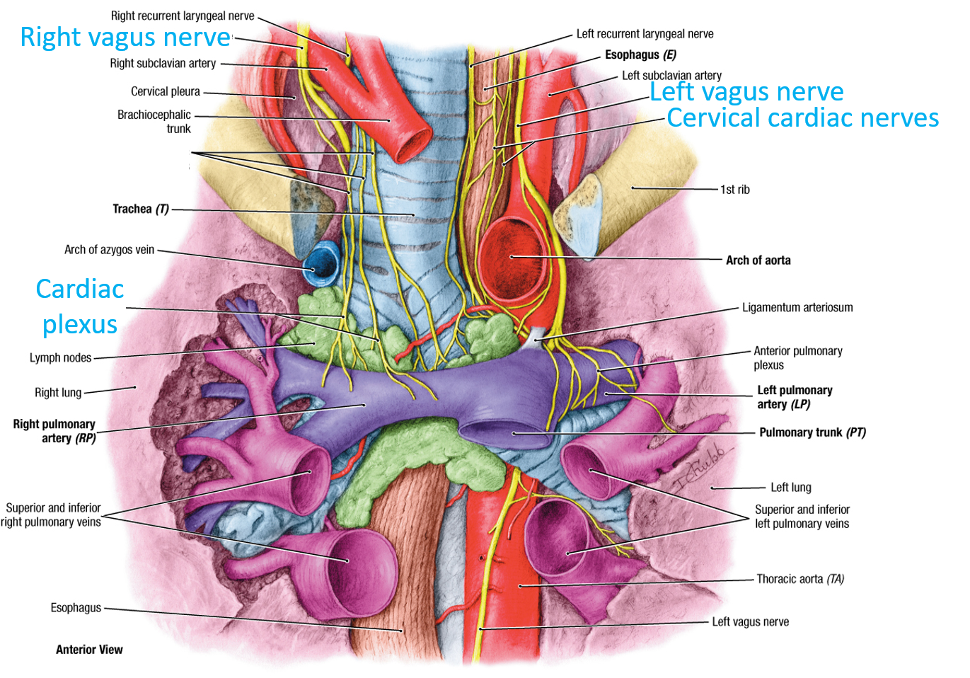
- Cardiac plexus The heart is supplied by autonomic nerve fibres from the cardiac plexus which is found posterior to the ascending aorta (anterior to the trachea) The cardiac plexus has sympathetic and parasympathetic fibres (to the heart) and visceral afferent fibres (from the heart) Fibres to the heart go to the coronary vessels and to the conducting system Sympathetics (arising from T1-T6 and synapsing in cervical and superior thoracic sympathetic ganglia) increase heart rate, parasympathetics (carried by the Vagus nerve) decrease heart rate
- SA node’s impulse passes through the muscle wall of the atrium and is then picked up at the atrioventricular (AV) node The AV node is located in the interatrial septum near the opening of the coronary sinus and is supplied by the AV nodal artery, a branch of the right coronary artery Its intrinsic rate of depolarisation is ~50 beats per minute (i.e. it is slower than the SA node) If the SA node fails to function e.g. due to poor blood supply, the area with the next fastest intrinsic rate of depolarisation i.e. the AV node, will take over driving the heart’s contractions at ~50 beats per minute The AV node distributes the signal to the ventricles via the AV bundle Sympathetics speed up conduction, parasympathetics slow down conduction
- The AV node distributes the signal to the ventricles via the AV bundle The AV bundle carries the impulse across the fibrous skeleton and along the membranous part of the interventricular septum At the junction of the membranous and muscular part of the interventricular septum, the AV bundle divides into right and left bundles and continue down the septum
- The AV bundle is found in the centre of the interventricular septum The anterior interventricular artery (left anterior descending artery) supplies most of the interventricular septum and therefore the AV bundle
- The AV node distributes the signal to the ventricles via the AV bundle The AV bundle carries the impulse across the fibrous skeleton and along the membranous part of the interventricular septum At the junction of the membranous and muscular part of the interventricular septum, the AV bundle divides into right and left bundles and continue down the septum
- SA node’s impulse passes through the muscle wall of the atrium and is then picked up at the atrioventricular (AV) node The AV node is located in the interatrial septum near the opening of the coronary sinus and is supplied by the AV nodal artery, a branch of the right coronary artery Its intrinsic rate of depolarisation is ~50 beats per minute (i.e. it is slower than the SA node) If the SA node fails to function e.g. due to poor blood supply, the area with the next fastest intrinsic rate of depolarisation i.e. the AV node, will take over driving the heart’s contractions at ~50 beats per minute The AV node distributes the signal to the ventricles via the AV bundle Sympathetics speed up conduction, parasympathetics slow down conduction
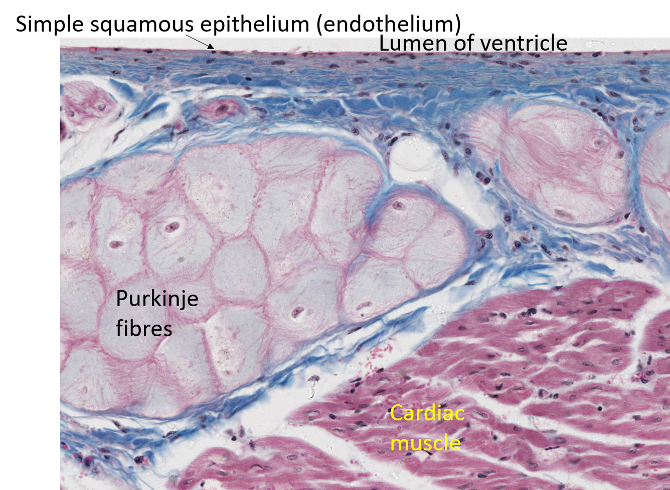
- The right and left bundles divide into fibres called Purkinje fibres that extend into the walls of their respective ventricles The Purkinje fibres on the right side stimulate the muscle of the interventricular septum (IVS) and the wall of the right ventricle, including the anterior papillary muscle through the moderator band The Purkinje fibres on the left side stimulate the IVS and the wall of the left ventricle, including the papillary muscles In complete heart block, when conduction of electrical impulses to the ventricles is interrupted, the ventricles will beat at the depolarisation rate of the Purkinje fibres (~30-40 beats per minute)
- artificial cardiac pacemaker
- Modern implantable artificial pacemakers consist of an implantable pulse generator (a computer with an integral pulse generator and battery) and leads (wires with electrodes at their tips) About the size of a matchbox and weigh <50g They sense intrinsic heart rhythm and transmit electrical impulses, if indicated, replacing defective natural pacemaker There are different types of artificial cardiac pacemakers
- The most common site to place the pocket is the pectoral region (although the axilla is used in children or the abdomen for other systems of pacemakers) The pocket can be either subcutaneous at the level of pre-pectoral fascia or for those with less adipose tissue (to prevent erosion) it is placed submuscular (below pectoralis major or even below pectoralis minor and above the ribcage)
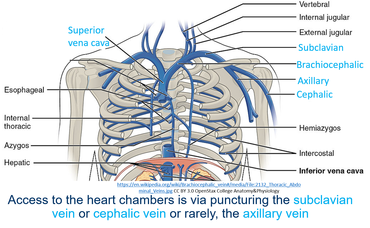
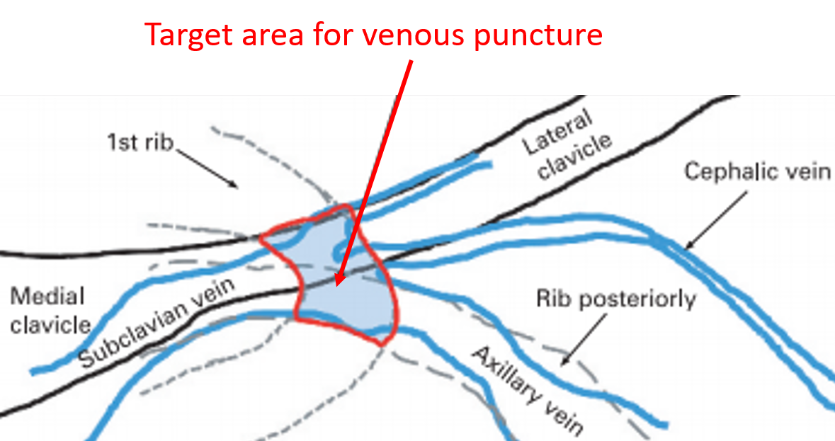
- Leads are inserted and advanced under fluoroscopy to the heart chamber and attached to the endocardium Most permanent pacemakers are implanted on the left side as this is easier for right handed surgeon and it is easier to place the leads from the left side
- Leads are inserted and advanced under fluoroscopy to the heart chamber and attached to the endocardium Most permanent pacemakers are implanted on the left side as this is easier for right handed surgeon and it is easier to place the leads from the left side
- Skin -> Subcutaneous tissue Breast -> Pectoral fascia (part of deep fascia) -> Branches of intercostal nerves -> Pectoralis major
Superior mediastinum
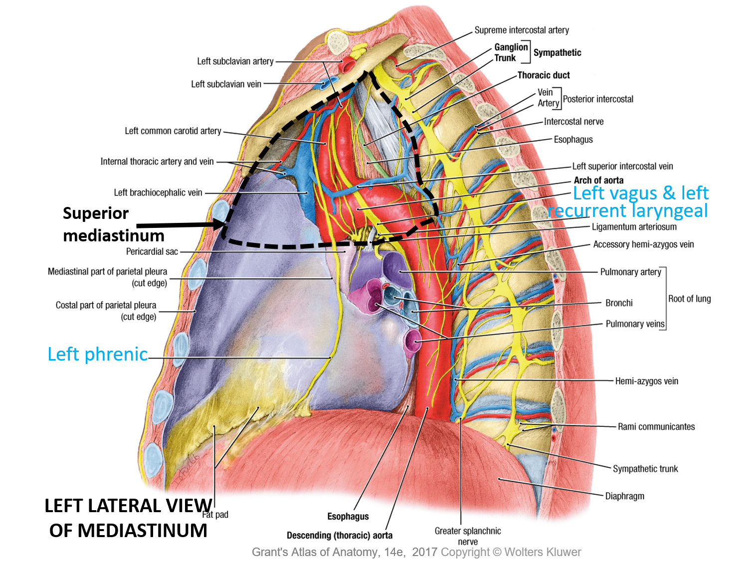
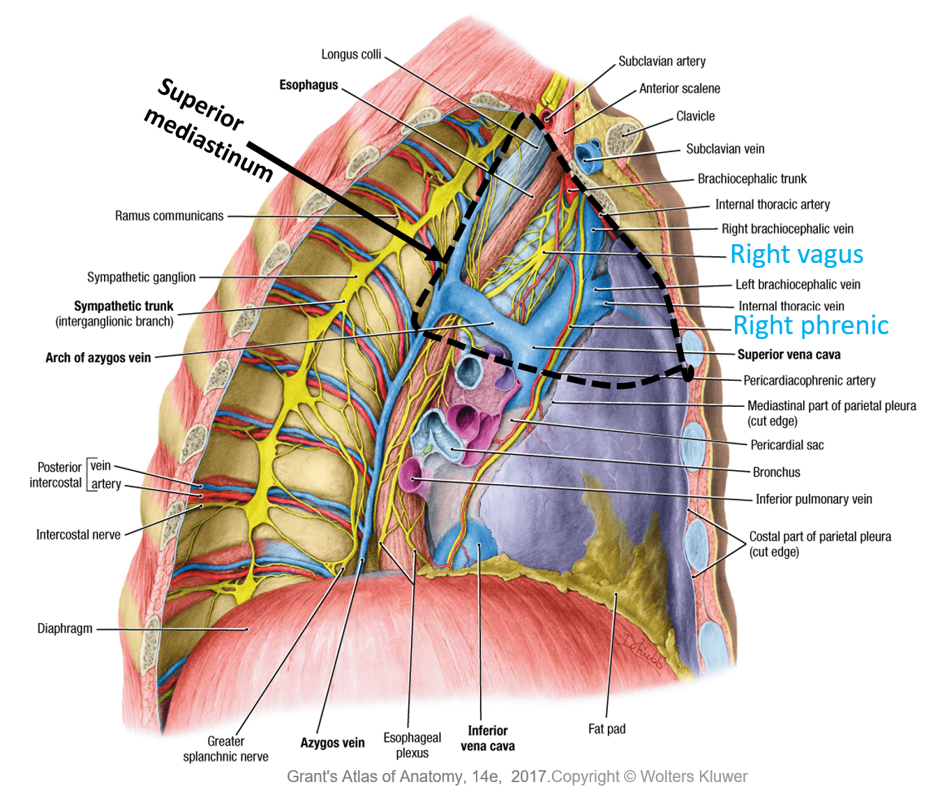
- For organisational purposes, the mediastinum is subdivided into smaller regions: The superior mediastinum: from superior thoracic aperture to the transverse thoracic plane (horizontal plane from sternal angle to the intervertebral disc of T4 and T5) and Inferior mediastinum: from transverse thoracic plane to the diaphragm The inferior mediastinum is subdivided by the pericardium into: anterior mediastinum, middle mediastinum and posterior mediastinum There will be structures that are present in more than one part of the mediastinum e.g. oesophagus
- Boundaries: Superior: superior thoracic aperture (thoracic inlet) Inferior: transverse thoracic plane Anterior: manubrium of the sternum and Posterior: bodies of T1-T4 Lateral: mediastinal pleurae The superior mediastinum is continuous with the neck superiorly and the inferior mediastinum inferiorly
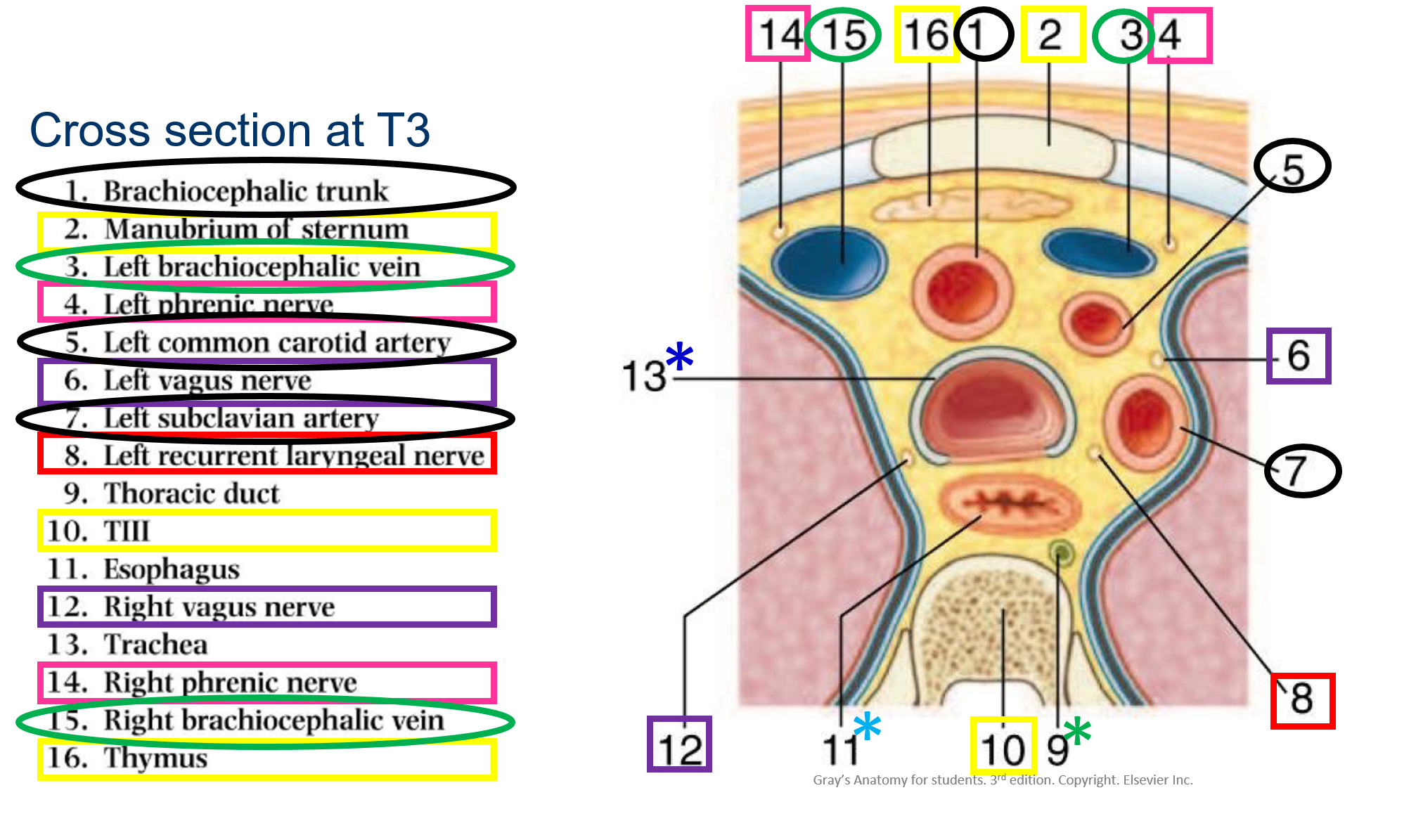
- Structure of superior mediastinum: Thymus or its remnants Nerves Arteries Ligamentum arteriosum Veins Trachea Oesophagus Terminal part of the thoracic duct Lymph nodes
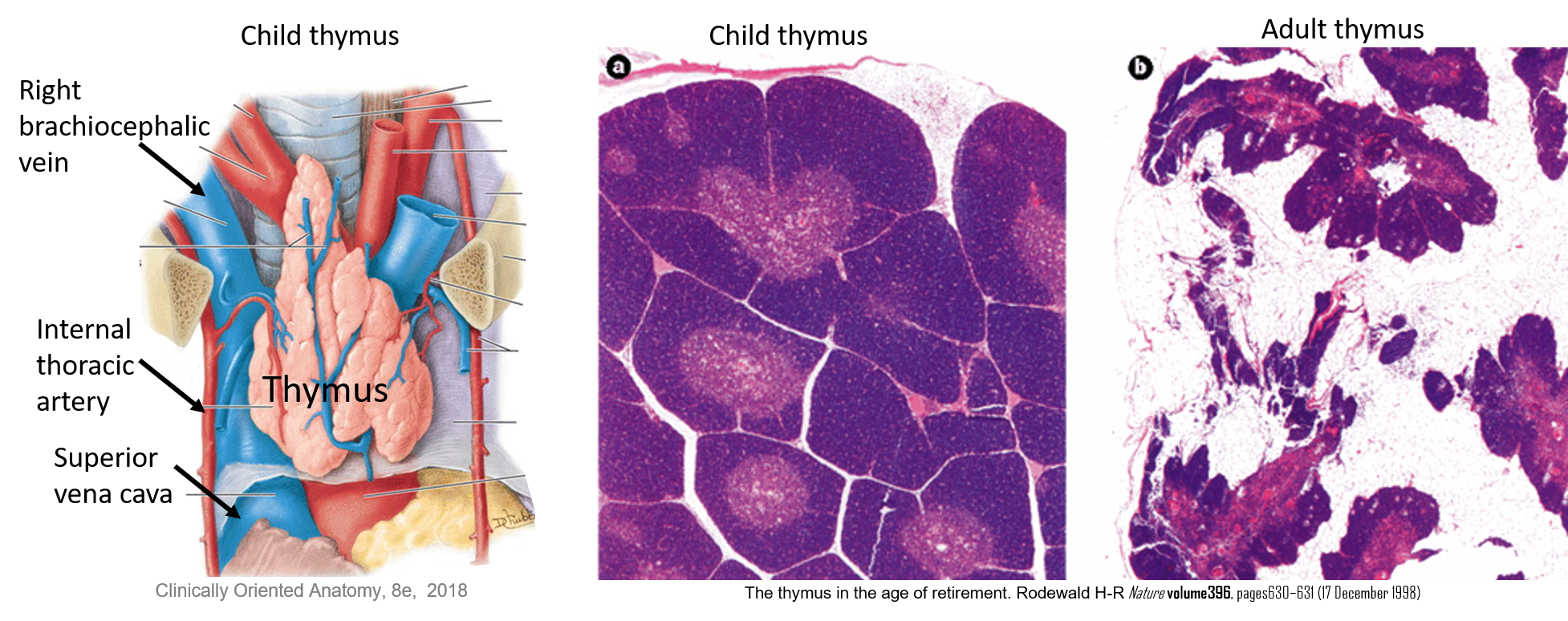
- Thymus Primary lymphoid organ (T lymphocytes mature here) It is largest and most active during neonatal and pre-puberty periods: after puberty, it becomes smaller and smaller, being replaced by fat (thymic involution) Found posterior to the manubrium
- Right and left vagus nerves and their branches to the oesophagus, cardiac plexus and pulmonary plexus Sympathetic nerves The cardiac plexus and pulmonary plexus have sympathetic and parasympathetic fibres (to the viscera i.e.heart/lungs) and visceral afferent fibres (from the viscera) The pulmonary plexus’ sympathetics regulate bronchodilation and parasympathetics regulate bronchoconstriction) Left recurrent laryngeal Right and left phrenic nerves
- Arteries: Arch of the aorta and its 3 branches: Brachiocephalic trunk Left common carotid Left subclavian Veins: Right and left brachiocephalic Superior vena cava.
- The trachea is palpated in the jugular notch as it enters the superior mediastinum: it is inclined a little to the right of the median plane The trachea’s posterior surface is flat Trachea ends at the level of sternal angle by branching into the two bronchi The oesophagus lies posterior to the trachea and anterior to T1-T4 The oesophagus is usually flat anterior-posteriorly The oesophagus continues into the inferior mediastinum
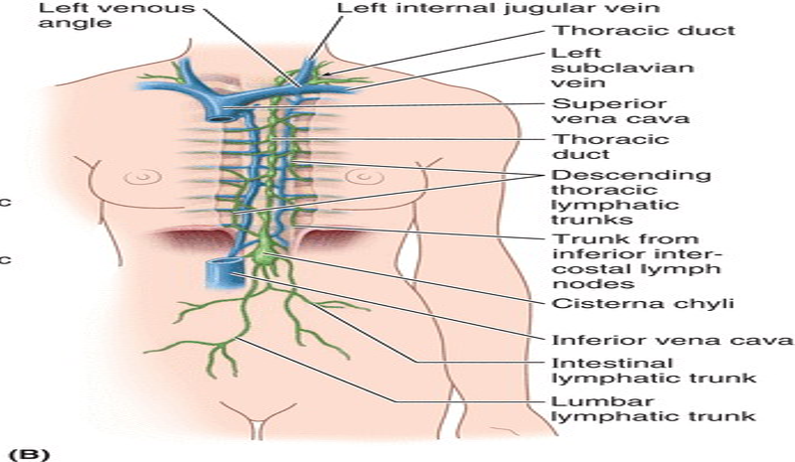
- It begins at the level of L1/L2 where the main lymphatic ducts of the abdomen unite This union of abdominal lymphatic ducts may be large enough in some individuals to form a sac known as cisterna chyli The thoracic duct ascends through the aortic hiatus in the diaphragm into the posterior mediastinum, and collects parietal and visceral drainage, particularly from the left upper quadrant of the body The thoracic duct terminates by entering the venous system at the junction of the left subclavian and internal jugular veins (the left venous angle)
- It is thin walled, dull white and appears beaded because of its valves It ascends in the posterior mediastinum among the thoracic aorta on its left, the azygos vein on its right, the esophagus anteriorly, and the vertebral bodies posteriorly At the level of the T4, T5, or T6 vertebra, it crosses to the left and ascends into the superior mediastinum. It receives branches from: the middle and superior intercostal spaces of both sides through several collecting trunks posterior mediastinal structures Near its termination, the thoracic duct receives the jugular, subclavian, and bronchomediastinal lymphatic trunks (although any or all these vessels may terminate independently)
- At the T4/T5 vertebral level Sternal angle (manubriosternal joint) Costal cartilage of 2nd rib Junction between the superior and inferior mediastinum Arch of the aorta begins, ascending aorta ends Trachea bifurcates
- Palpating arterial pulses can help assess cardiac performance, cardiac rate and rhythm, establish the integrity of the peripheral arterial blood supply or localise a peripheral lesion
- The external jugular vein is occluded at the clavicle and distends as it fills from above (indicated by the arrows). Internal jugular indicated by dotted line
- For organisational purposes, the mediastinum is subdivided into smaller regions: The superior mediastinum: from superior thoracic aperture to the transverse thoracic plane (horizontal plane from sternal angle to the intervertebral disc of T4 and T5) and Inferior mediastinum: from transverse thoracic plane to the diaphragm The inferior mediastinum is subdivided by the pericardium into: anterior mediastinum, middle mediastinum and posterior mediastinum There will be structures that are present in more than one part of the mediastinum e.g. oesophagus
Inferior mediastinum
- Middle mediastinum- It is the broadest part of the inferior mediastinum
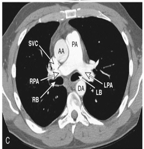
- Middle: It contains the: Pericardium Phrenic nerves Heart Ascending aorta Superior vena cava (the part that is intrapericardial) Pulmonary trunk Tracheal bifurcation and main bronchi Right and left pulmonary arteries and veins Deep part of cardiac plexus Tracheobronchial lymph nodes Inferior vena cava
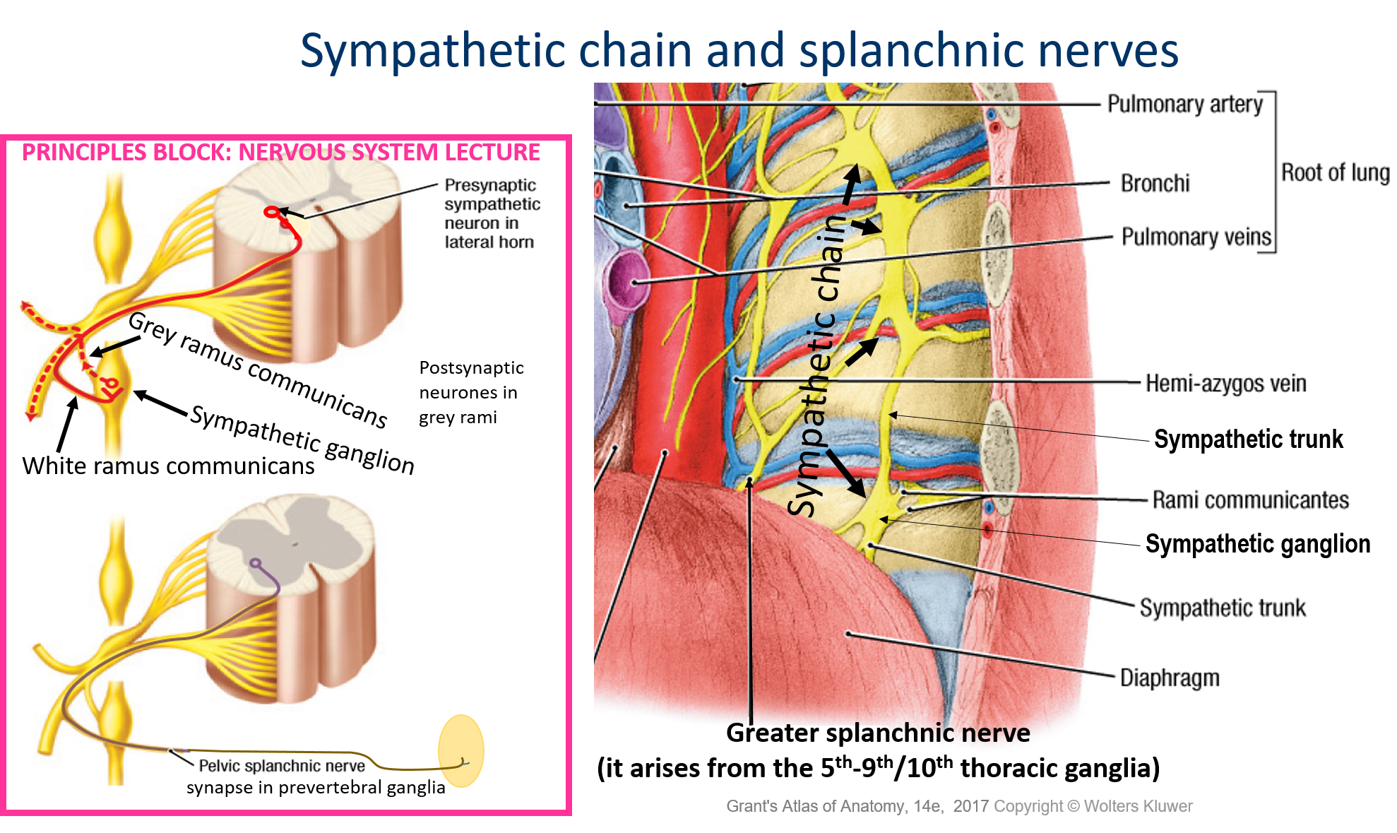
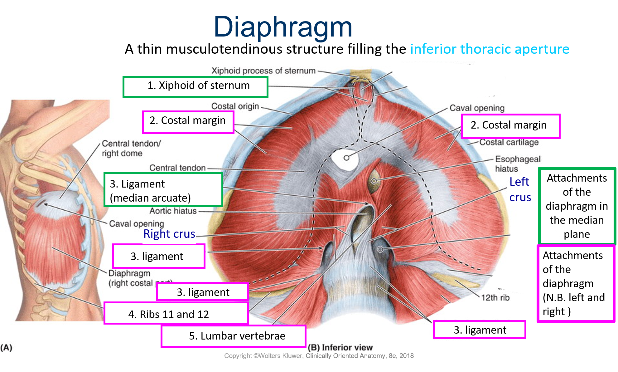
- Posterior mediastinum contains: Descending aorta Oesophagus Azygos, hemi-azygos and accessory hemi-azygos veins Thoracic duct Lymph nodes Vagal trunks (Sympathetic chains)* and (Splanchnic nerves)* There are communications to the abdomen for the: oesophagus (oesophageal hiatus of diaphragm), aorta (aortic hiatus of diaphragm), splanchnic nerves, sympathetic chains * Strictly not part of the posterior mediastinum
- Splanchnic nerves are branches from the sympathetic ganglia There are 3 large branches from the lower sympathetic ganglia of the thorax and they contain mainly preganglionic sympathetic neurones going to abdominal (the gut) and pelvic viscera i.e. visceral efferents, but also have visceral afferents They are: 1) Greater splanchnic nerve – ends in coeliac ganglion; 2) Lesser splanchnic nerve – from 9th and 10th ganglia and 3) Least (lowest) splanchnic nerve – from the 12th ganglion
- Axial CTScan AA, ascending aorta; DA, descending aorta (thoracic aorta); LB, left main bronchus; RB, right main bronchus; PA, main pulmonary artery segment i.e. pulmonary trunk; LPA, left pulmonary artery; RPA, right pulmonary artery; SVC, superior vena cava
- Azygos system and the azygos vein
- Azygos system includes the azygos vein, hemi-azygos vein, accessory hemi-azygos vein and their tributaries Azygos system drains blood from the body wall returning it back to the superior vena cava (SVC) The azygos system shows a lot of variation Azygos vein begins at L1/L2 and enters the thorax via the aortic hiatus (though there is variation in this) and ascends until T4 where it arches and enters the SVC Azygos vein forms a collateral pathway between the SVC and the inferior vena cava As the azygos vein ascends, it has tributaries: 5th- 11th right intercostal veins Hemi-azygos Accessory hemi-azygos Veins from oesophagus, mediastinum, pericardium and the right bronchial veins Right superior intercostal vein
- Variation in the azygos system Left superior intercostals, accessory hemi-azygos and hemi-azygos may be continuous or discontinuous
- Accessory hemi-azygos (superior hemi-azygos) Descends from T4 to about T8 and crosses vertebral column and enters the azygos vein Connected to left superior intercostal vein Tributaries: 4thor 5th - 8th left posterior intercostal veins Left bronchial veins Hemi-azygos (inferior hemi-azygos) Ascends from the abdomen At about T9 crosses the vertebral column and enters azygos vein Tributaries are: the lowest 4/5 left posterior intercostal veins Veins from oesophagus and mediastinum
- Posterior mediastinum contains: Descending aorta Oesophagus Azygos, hemi-azygos and accessory hemi-azygos veins Thoracic duct Lymph nodes Vagal trunks (Sympathetic chains)* and (Splanchnic nerves)* There are communications to the abdomen for the: oesophagus (oesophageal hiatus of diaphragm), aorta (aortic hiatus of diaphragm), splanchnic nerves, sympathetic chains * Strictly not part of the posterior mediastinum
- Oesophagus Connects pharynx to the stomach Begins at the inferior border of the cricoid cartilage (C6) Midline structure but as it approaches the diaphragm, it travels anterior and to the left crossing the thoracic aorta It has four constrictions: Pharyngeal-oesophageal junction Crossed by arch of aorta Crossed by left main bronchus Diaphragm Arterial supply: oesophageal branches from the thoracic aorta, bronchial arteries, left gastric artery Venous drainage: Azygos, hemiazygos and left gastric vein Lymphatic drainage: posterior mediastinal and left gastric lymph nodes Innervation: vagus and sympathetic nerves
- 1) Left vagus and 2) right vagus are anterior to the hilum of the lung; 3) they then divide and spread over the oesophagus (oesophageal plexus) which then, close to the diaphragm, form two trunks i.e. 4) anterior vagal trunk (most of its fibres are from the left vagus) and 5) posterior vagal trunk (most of its fibres are from the right vagus) which travel to the abdomen
- Thoracic aorta Begins at the lower end of T4 and ends at lower end of T12, passing through the aortic hiatus of diaphragm Branches (think of structures of the inferior mediastinum): Pericardial Bronchial Oesophageal Mediastinal Posterior intercostals i.e. 9 pairs to the lower 9 intercostal spaces Subcostals – a pair of arteries found inferior to the 12th rib Superior phrenic arteries
- 1) Left vagus and 2) right vagus are anterior to the hilum of the lung; 3) they then divide and spread over the oesophagus (oesophageal plexus) which then, close to the diaphragm, form two trunks i.e. 4) anterior vagal trunk (most of its fibres are from the left vagus) and 5) posterior vagal trunk (most of its fibres are from the right vagus) which travel to the abdomen
- Middle: It contains the: Pericardium Phrenic nerves Heart Ascending aorta Superior vena cava (the part that is intrapericardial) Pulmonary trunk Tracheal bifurcation and main bronchi Right and left pulmonary arteries and veins Deep part of cardiac plexus Tracheobronchial lymph nodes Inferior vena cava
- Middle mediastinum- It is the broadest part of the inferior mediastinum
Images and files in the map
Information
- Description
- Mind Map on Cardiovascular Anatomy, created by A C on 23/03/2019.
- Created by
- A C
- Views
- 0
- History
-
- Created by A C · Mar 23, 2019
- Copied by A C · Mar 23, 2019
- Rating