
Mind map
Cardiogenic Shock
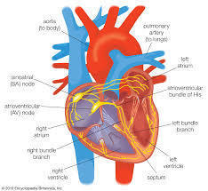
Map content
Cardiogenic Shock
Treatment/ Management (Lewis & Bucher, 2018; Morozowich & Ramakrishna, 2015)
- Medications
- Sympathomimetic drugs- Mimic SNS fight-or-flight response, Selectively activate alpha- & beta-adrenergic receptors. Two Primary Effects: Vasopressor Effects and Inotropic Effects, Some drugs may exert a mixture of both vasopressor and inotropic effects
- Epinephrine - Considered as both a vasopressor and an inotrope. Effect: Induces vasoconstriction & increases cardiac output · Mechanism: Activates both α1 & β1 adrenergic receptors
- Norepinephrine · Considered mainly as a vasopressor · Can trigger both α1 & β1 adrenergic effects · Predominantly activates α1 receptors. Arterial & venous vasoconstriction ↑, ↑ Preload, ↑MAP, ↑SVR. β1 Inotropic effects are offset by afterload from α1 receptor activation
- Vasopressin -Similar to the anti-diuretic hormone. Constricts vascular smooth muscle & enhances the vascular response to catecholamines
- Dopamine - Considered mainly as a vasopressor. Stimulates α1 & β1 receptors. Also stimulates dopaminergic receptors, DA1 & DA2 o DA1 stimulation results in renal & visceral vasodilation o DA2 stimulation inhibits norepinephrine reuptake
- Dobutamine -Ionotropic effects. Mainly stimulates β1 and β2 adrenergic receptors, with limited A-adrenergic effects · Results in β1 induced positive inotropic effects and β2 mediated vasodilation o ↑ CO, ↓SVR, ↑ Stroke volume · Preferred drug of choice to treat cardiogenic shock
- Phenylephrine -Activates α1 receptors, leading to an increase in systemic vascular resistance o ↑ SVR, ↑MAP
- Adrenergic Receptors- a class of G protein-coupled receptors that are targets of many catecholamines
- α1 Adrenergic Receptors - Induce vasoconstriction & ↑ mean arterial pressure
- α2 Adrenergic Receptors – Activation reduces norepinephrine release. Reduce blood pressure. Slows rate of cardiac electric conduction (Negative dromotropic effect)
- β1 Adrenergic Receptor- Positive chrono, dromo, inotropic effects. Increases the rate of cardiac conduction, accelerates heart rate, & strengthens cardiac contraction
- β2 Adrenergic Receptor - Induces vasodilation, & decreases mean arterial pressure
- Nitroglycerin · Dilates coronary and systemic blood vessels. ↓SVR, ↓ Preload, ↓ Afterload, ↓BP · Used for cardiogenic shock w/severe vasoconstriction
- Sympathomimetic drugs- Mimic SNS fight-or-flight response, Selectively activate alpha- & beta-adrenergic receptors. Two Primary Effects: Vasopressor Effects and Inotropic Effects, Some drugs may exert a mixture of both vasopressor and inotropic effects
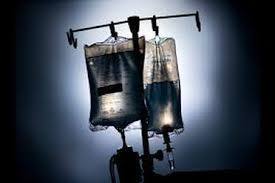
- Fluid Resuscitation. (Agnihotri & Agnihotri, 2014; Frazee & Kashani, 2016; Reddy, Weinberg, & Young, 2016; Silva, Gonçalves, & Sousa, 2018; Lewis & Bucher, 2018)
- Must be started prior to giving sympathomimetic medications - Fluid therapies are given to optimize blood flow & restore organ perfusion With Cardiogenic Shock, fluid therapies must be given with caution
- Crystalloid therapies - Fluid of choice w/shock patients- Contains ions affecting the tonicity of the fluid. It is an Isotonic solution. Must be warmed prior to administration to prevent hypothermia.
- Lactated Ringer’s Solution - Isotonic solution. Designed to better mimic plasma. A more ‘balanced’ salt solution compared to NS. Contains Lactate with several other ions & anions
- 0.9% NaCl ,most commonly given crystalloid, Contains an equal concentration of Na & Cl. Increases intravascular volume by remaining mostly in the intravascular spa
- Colloids -Usually not recommended for cardiogenic shock. Provide plasma expansion. Consists of proteins that increase the osmotic pressure of the intravascular space o Retain and attract fluids into intravascular space. Compared to crystalloids, lower volumes are required to maintain hemodynamic function · Includes albumin, starch & dextran
- Blood Transfusion · Indications: All types of shock unresponsive to crystalloids o Hemoglobin level <120g/L Packed RBCs must not contain clotting factors but should be subsequently replaced
- Crystalloid therapies - Fluid of choice w/shock patients- Contains ions affecting the tonicity of the fluid. It is an Isotonic solution. Must be warmed prior to administration to prevent hypothermia.
- Must be started prior to giving sympathomimetic medications - Fluid therapies are given to optimize blood flow & restore organ perfusion With Cardiogenic Shock, fluid therapies must be given with caution
- Medications
Main causes (Khaira, R. 2017)
- Cardiogenic shock is caused by impaired function of the heart. This type may be caused by conditions that interfere with the function of the heart as a pump. Systolic or diastolic dysfunction Compromised/decreased cardiac output (CO) When affecting the right side, pulmonary circulation is compromised
- Other health conditions such as: MI Cardiomyopathies, Severe systemic/pulmonary, HTN Blunt cardiac injury, Severe myocardial depression from sepsis, Diastolic dysfunction, Impaired ability of either ventricle to fill during diastole, and Decreased stroke volume
- Cardiogenic shock is caused by impaired function of the heart. This type may be caused by conditions that interfere with the function of the heart as a pump. Systolic or diastolic dysfunction Compromised/decreased cardiac output (CO) When affecting the right side, pulmonary circulation is compromised
Manifestations (Khaira, R. 2017)
- Signs
- Tachycardia
- < 100 beats per min
- Tachypnea
- < 25 breaths per min
- Hypotension
- >90/60
- Narrowing pulse pressure
- A drop in left ventricular stroke volume
- Increased myocardial O2 consumption
- Increased amount of oxygen that the heart requires to maintain optimal function, and myocardial oxygen supply is the amount of oxygen provided to the heart by the blood
- Tachycardia
- Symptoms
- Anxiety
- Cool Extremities
- Crackles/ pulmonary congestion
- Signs
Stages (Lewis & Bucher, 2018)
- Progressive Stage
- Cellular perfusion leads to ↑ capillary permeability. Vascular fluid & protein leak into extracellular space. ↓ Organ perfusion & ↑ systemic edema. It affects Resp, cardio, kindey, gastrointestinal, liver, hemotological systems all differently.
- Gastrointestinal System- ↓ GI perfusion , GI ulcers, bacterial translocation, & nutrient malabsorption
- Kidney Function- ↓ Kidney perfusion, Increased BUN, creatinine, urine output, AKI, metabolic acidosis
- Hematological System -↑ Risk for disseminated intravascular coagulation, Abnormal increase in blood clots, using up platelets, & clotting factors - Results in substantial bleeding
- Pulmonary System- 1st to display clinical manifestations. Pulmonary arterioles constrict, decreasing perfusion of pulmonary capillaries Ventilation-perfusion mismatch,↑ permeability of pulmonary capillaries Pulmonary edema, & bronchoconstriction, decreased gas exchange. Increased Tachypnea & crackles
- Cardiovascular System- ↓ Cardiac output- Hypotension o ↓ cerebral, coronary, & peripheral perfusion, CNS changes, cold, clammy skin, weakened peripheral pulses, MIs
- Liver Function- Buildup of ammonia, lactate, bilirubin, Jaundice, metabolic acidosis, altered drug metabolism . Kupffer cells fail to clear bacteria from the GI tract. Increased risk of Bacteremia, ↑ ALT and AST
- Cellular perfusion leads to ↑ capillary permeability. Vascular fluid & protein leak into extracellular space. ↓ Organ perfusion & ↑ systemic edema. It affects Resp, cardio, kindey, gastrointestinal, liver, hemotological systems all differently.
- Initial Stage
- Occurs at a cellular level. No clinical signs or symptoms. Cellular metabolism switches to anaerobic metabolism. Causes excess build-up of lactic acid.Lactic acid can’t be removed by the liver due to oxygen hypo-perfusion
- Refractory Stage
- Continued peripheral vasoconstriction further increases anaerobic metabolism. Lactic acid accumulation further increases capillary permeability. Venules constrict, & arterioles dilate: Blood pools in capillary bed and leaks into extravascular space . Ultimately results in myocardial & cerebral ischemia
- Compensatory Stage
- Cardiac output leads to hypotension. Baroceptors activate the SNS, releasing epinephrine & norepinephrine. Blood flow to the heart/brain is maintained while system circulation is shunted. Myocardium increases contractility and heart rate in response to SNS stimulation.
- Progressive Stage
Diagnosing (Khaira, R. 2017)
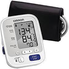
- Blood pressure measurement
- Low blood pressure entails shock
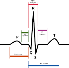
- Electrocardiogram
- This test records the electrical activity of your heart via electrodes attached to your skin. If you have damaged heart muscle, electric problems or fluid buildup around your heart, it won't conduct electrical impulses normally.
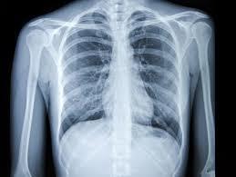
- Chest-X ray
- to check the size and shape of your heart and its blood vessels and whether there's fluid in your lungs
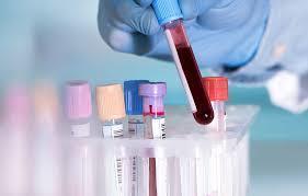
- Blood tests
- to check for organ damage, infection and heart attack, also to check arterial blood gas for level of O2 in blood
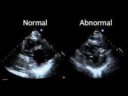
- Echocardiogram
- Used to measure contraction, which is measured as the degree of myocardial fiber shortening that occurs during systole. The most common cause of cardiogenic shock results from marked reduction in left ventricular contraction
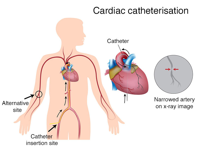
- Cardiac Catheterization (angiogram)
- A liquid dye is injected into the arteries of your heart through a long, catheter that's inserted through an artery, usually in your leg. The dye makes your arteries visible on X-ray, revealing areas of blockage or narrowing.
- Blood pressure measurement
Nursing Interventions (Lewis & Bucher, 2018; Warise, 2015)
- Neurological Status - Assess orientation to person, place and time q1. Observe for s/s of neurological deterioration r/t impaired gas exchange- Apprehension, impatience, restlessness, blurred vision, confusion, and paresthesias . Provide a calm, quiet environment to avoid sensory overload
- Cardiovascular Status - Assess for clinical manifestations of left ventricular failure- Dyspnea, bilateral crackles, s3/s4 heart sounds, ↑PAOP, ↓BP, Left ventricular failure can develop secondary to left ventricular failure o Peripheral edema, ↑CVP . Monitor the HR, CVP (2-6 mm Hg), PAP (4-12 mmHg) q15 · PAOP (6-12 mmHg) should be assed q1-2 · ECG should be monitored continuously
- Respiratory Status - Administer supplemental oxygen as ordered to maintain spO2 >90% · Continuous pulse oximetry may provide an inaccurate reading in advanced stages of shock, switch to nose, ear or forehead. Monitor respiratory rate q15-30. Tachypnea could be a sign of metabolic acidosis. Assess breath sounds q1. Crackles could indicate fluid overload from therapy or increased secretions. Monitor for accessory muscle use, nasal flaring, and neurological changes · Position p/t in semi-Fowler position to increase oxygen and ventilation. Assess ABGS as appropriate
- Renal Status- Record input/output q1 · Assess BUN and creatinine for kidney function
- Gastrointestinal Status- Auscultate bowel bounds q4. Assess for abdominal distention. Check for bloody stools
- Integumentary Status - Assess for cyanosis, pallor, and decreased capillary refill
- Other Considerations - Perform ROM exercises q3-4 to maintain normal function. Turn patient q2
- Renal Status- Record input/output q1 · Assess BUN and creatinine for kidney function
- Respiratory Status - Administer supplemental oxygen as ordered to maintain spO2 >90% · Continuous pulse oximetry may provide an inaccurate reading in advanced stages of shock, switch to nose, ear or forehead. Monitor respiratory rate q15-30. Tachypnea could be a sign of metabolic acidosis. Assess breath sounds q1. Crackles could indicate fluid overload from therapy or increased secretions. Monitor for accessory muscle use, nasal flaring, and neurological changes · Position p/t in semi-Fowler position to increase oxygen and ventilation. Assess ABGS as appropriate
- Cardiovascular Status - Assess for clinical manifestations of left ventricular failure- Dyspnea, bilateral crackles, s3/s4 heart sounds, ↑PAOP, ↓BP, Left ventricular failure can develop secondary to left ventricular failure o Peripheral edema, ↑CVP . Monitor the HR, CVP (2-6 mm Hg), PAP (4-12 mmHg) q15 · PAOP (6-12 mmHg) should be assed q1-2 · ECG should be monitored continuously
- Neurological Status - Assess orientation to person, place and time q1. Observe for s/s of neurological deterioration r/t impaired gas exchange- Apprehension, impatience, restlessness, blurred vision, confusion, and paresthesias . Provide a calm, quiet environment to avoid sensory overload
Images and files in the map
Information
- Description
- done by: Savannah Wolf-Hays, Doug Lau, Brandon Gardner
- Created by
- savvy hays
- Views
- 51
- Rating