
Mind map
Radiation 1
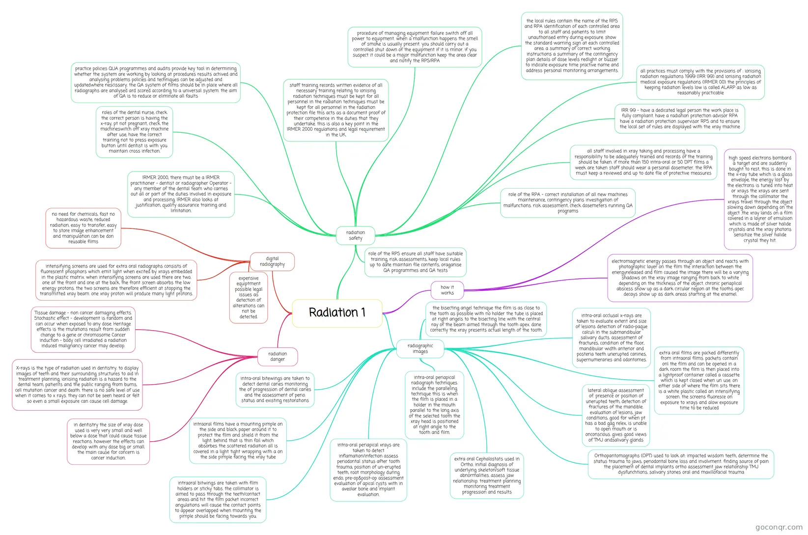
Mind map: Radiation in dentistry
Map content
Radiation 1
radiation danger
- X-rays is the type of radiation used in dentistry. to display images of teeth and their surrounding structures to aid in treatment planning. ionising radiation is a hazard to the dental team, patients and the public ranging from burns, cell mutation cancer and death. there is no safe level of use when it comes to x rays. they can not be seen heard or felt so even a small exposure can cause cell damage.
- Tissue damage - non cancer damaging effects. Stochastic effect - development is random and can occur when exposed to any dose. Heritage effects is the mutations result from sudden change to a gene or chromosome Cancer induction - body cell irradiated a radiation induced malignancy cancer may develop.
- in dentistry the size of xray dose used is very very small and well below a dose that could cause tissue reactions. however the effects can develop with any dose big or small. the main cause for concern is cancer induction.
radiation safety
- all practices must comply with the provisions of : ionising radiation regulations 1999 (IRR 99) and ionising radiation medical exposure regulations (IRMER 00) the principles of keeping radiation levels low is called ALARP as low as reasonably practicable
- IRR 99 - have a dedicated legal person the work place is fully compliant. have a radiation protection advisor RPA have a radiation protection supervisor RPS and to ensure the local set of rules are displayed with the xray machine
- IRMER 2000, there must be a IRMER practitioner - dentist or radiographer Operator - any member of the dental team who carries out all or part of the duties involved in exposure and processing. IRMER also looks at justification, quality assurance training and limitation.
- roles of the dental nurse, check the correct person is having the x-ray, pt not pregnant, check the machineswitch off xray machine after use, have the correct training not to press exposure button until dentist is with you maintain cross infection.
- procedure of managing equipment failure switch off all power to equipment. when a malfunction happens the smell of smoke is usually present. you should carry out a controlled shut down of the equipment if it is minor. if you suspect it could be a major malfunction keep the area clear and notify the RPS/RPA
- practice policies QUA programmes and audits provide key tool in determining whether the system are working by looking at procedures results achived and analysing problems policies and techniques can be adjusted and updatedwhere nescissary. the QA system of films should be in place where all radiographs are analysed ard scored according to a universal system. the aim of QA is to reduce or eliminate all faults
- role of the RPS ensure all staff have suitable training, risk assessments, keep local rules up to date maintain file contents, oraganise QA programmes and QA tests
- staff training records written evidence of all necessary training relating to ionising radiation techniques must be kept for all personnel in the radiation techniques must be kept for all personnel in the radiation protection file this acts as a document proof of their competence in the duties that they undertake. this is also a key point in the IRMER 2000 regulations and legal requirement in the UK.
- all staff involved in xray taking and processing have a responsibility to be adequately trained and records of the training should be taken. if more than 150 intra-oral or 50 DPT films a week are taken staff should wear a personal dosemeter. the RPA must keep a reviewed and up to date file of protective measures
- the local rules contain the name of the RPS and RPA identification of each controlled area to all staff and patients to limit unauthorised entry during exposure. show the standard warning sign at each controlled area. a summary of correct working instructions a summary of the contingency plan details of dose levels redlight or buzzer to indicate exposure time practive name and address personal monitoring arrangements.
- role of the RPA - correct installation of all new machines maintenance, contingency plans investigation of malfunctions. risk assessment, check dosemeters running QA programs
how it works
- high speed electrons bombard a target and are suddenly bought to rest, this is done in the x-ray tube which is a glass envelope, the energy lost by the electrons is tuned into heat or xrays the xrays are sent through the collimator the xrays travel through the object slowing down depending on the object .the xray lands on a film covered in a layrer of emulsion which is made of silver halide crystals and the xray photons sensitize the silver halide crystal they hit.
- electromagnetic energy passes through an object and reacts with photographic layer on the film the interaction between the energyreleased and film caused the image there will be a varying shadows on the xray image ranging from back to white depending on the thickness of the object. chronic periaplical abscess show up as a dark circular region at the tooths apec decays show up as dark areas starting at the enamel.
radiographic images
- the bisecting angel technique the film is as close to the tooth as possible with no holder the tube is placed at right angels to the bisecting line with the central ray of the beam aimed through the tooth apex. done correctly the xray presents actual length of the tooth.
- intra-oral periapical xrays are taken to detect inflamation/infection assess periodontal status after tooth trauma, position of un-erupted teeth, root morphology during endo, pre-op&post-op assessment evaluation of apical cysts with in aveolar bone and implant evaluation.
- Orthopantomographs (OPT) used to look at: impacted wisdom teeth, determine the status trauma to jaws, periodontal bone loss and involvment. finding source of pain the placement of dental implants ortho assessment jaw relationship TMJ dysfunchtions, salivary stones oral and maxillofacial trauma
- lateral oblique assessment of: presence or position of unerupted teeth, detection of fractures of the mandible. evaluation of lesions, jaw conditions, good for when pt has a bad gag relex, is unable to open mouth or is onconscious. gives good views of TMJ andsalivary glands
- extra oral films are packed differently from intraoral films. packets contain onl the film and can be opened in a dark room the film is then placed into a lightproof container called a cassette which is kept closed when un use. on either side of where the film sits there is a white plastic called an intensifying screen. the screens fluoresce on exposure to xrays and alow exposure time to be reduced
- intraoral films have a mounting pimple on the side and black paper around it to protect the film and shield it from the light. behind that is thin foil which absorbes the scattered radiation all is covered in a light tight wrapping with a on the side pimple facing the xray tube
- intraoral bitwings are taken with film holders or sticky tabs. the collimator is aimed to pass through the teeth/contact areas and hit the film packet incorrect angulations will cause the contact points to appear overlapped when mounting the pimple should be facing towards you.
- intra-oral bitewings are taken to detect dental caries monitoring the of progression of dental caries and the assessment of perio. status and existing restorations
- intra-oral occlusal x-rays are taken to evaluate extent and size of lesions detection of radio-paque calculi in the submandibular salivary ducts. assessment of fractures, condition of the floor, mandibular width anterior and posteria teeth unerupted canines, supernumeraries and odontomes.
- extra oral Cephalostats used in Ortho. initial diagnosis of underlying skeleton/soft tissue abnormalities. assess jaw relationship. treatment planning monitoring treatment progression and results
- intra-oral periapical radiograph techniques. include the paralleling technique this is when the film is placed in a holder in the mouth parallel to the long axis of the selected tooth the xray head is positioned at right angle to the tooth and film.
digital radiography
- expensive equiptment possible legal issues as detection of alterations can not be detected.
- intensifying screens are used for extra oral radiographs consists of fluorescent phosphors which emit light when excited by xrays embedded in the plastic matrix. when intensifying screens are used there are two, one at the front and one at the back. the front screen absorbs the low energy protons. the two screens are therefore efficient at stopping the transmitted xray beam. one xray proton will produce many light protons.
- no need for chemicals, fast no hazardous waste, reduced radiation, easy to transfer, easy to store image enhancement and manipulation can be don reusable films
Information
- Description
- Mind Map on Radiation 1, created by zoelynch on 02/03/2015.
- Created by
- zoelynch
- Views
- 6
- Rating
More on Radiation Safety and Regulations