
Mind map
The Foot
Map content
The Foot
Bones
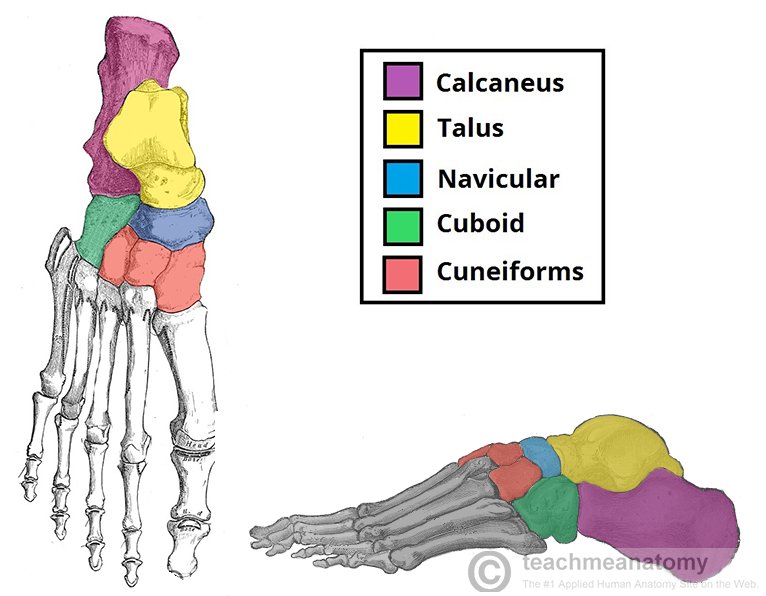
- Tarsals
- 7 irregular shaped bones
- Proximal: talus & calcaneus
- Intermediate: Navicular
- Distal: Cuboid & 3 cuneiforms
- 7 irregular shaped bones
- Metatarsals
- Located between the tarsals& phalanges
- Numbered 1-5
- They have 3 or 4 articulations
- Proximally - Tarsometatarsal joint
- Laterally - intermetatarsal joint
- Distally - Metatarsophalangeal joint
- They have 3 or 4 articulations
- Numbered 1-5
- Located between the tarsals& phalanges
- Phalanges
- Bones of toes
- 3 phalanges : proximal, intermediate & distal
- Great toe only has Prox' & Distal phalanges
- Bones of toes
- Tarsals
Muscles
- Extrinsic
- Arise from the anterior and posterior & lateral compartments of the leg
- Responsible for: eversion, inversion, plantar flexion & dorisfelxion
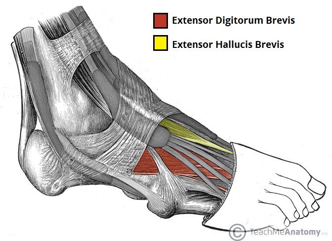
- Extensor Digitorum Brevis
- Lies deep to the tendon of extensor digitorum longus
- It originates from the calcaneus, the interosseus talocalcaneal ligament and the inferior extensor retinaculum
- It inserts into the long extensor tendons of the four medial digits.
- Aids in extension of medial 4 toes
- Lies deep to the tendon of extensor digitorum longus
- Extensor Hallucis Brevis
- Medial to extensor digitorum longus
- Originates from the calcaneus, the interosseus talocalcaneal ligament and the inferior extensor retinaculum
- Inserts into base of proximal phalanx of the great toe
- Originates from the calcaneus, the interosseus talocalcaneal ligament and the inferior extensor retinaculum
- Extends the great toe
- Medial to extensor digitorum longus
- Arise from the anterior and posterior & lateral compartments of the leg
- Intrinsic
- Can be split into 4 layers (all 4 covered in another map)
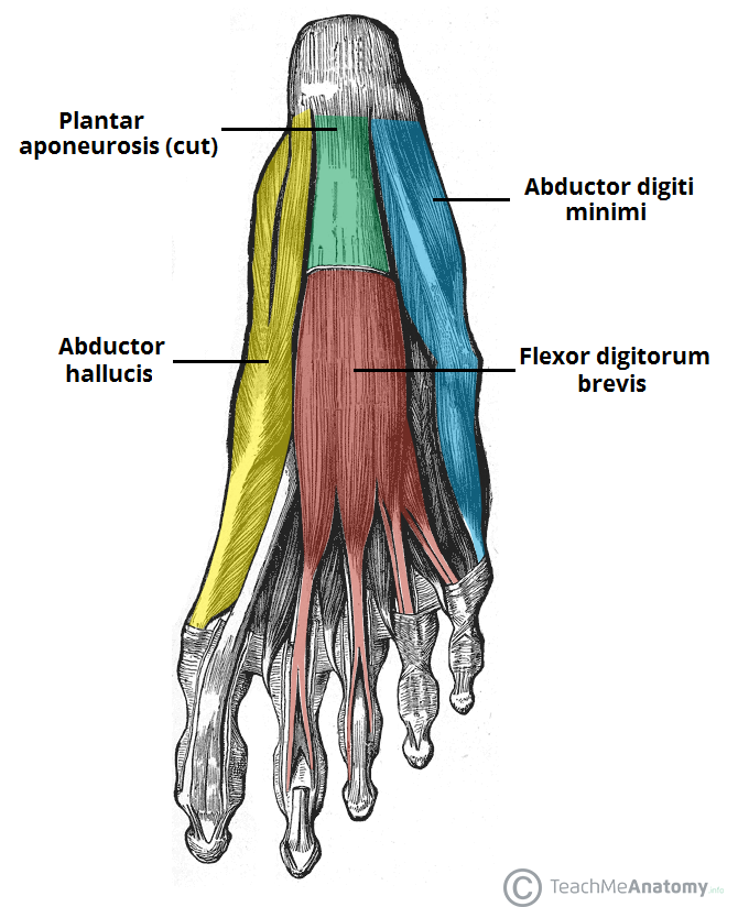
- 1st Layer
- Abductor Hallucis Longus
- Originates from medial tubercle of calcaneus, lexor retinaculum and the plantar aponeurosis
- Inserts into medial base of prox' phalanx of great toe
- Abducts and flexes great toe
- Flexor Digitorum Brevis
- Originates from medial tubercle of the calcaneus and the plantar aponeurosis
- Inserts into middle phalanges of the lateral four digits.
- Flexes lateral 4 digits
- Abductor Digiti Minimi
- Originates from medial & lateral tubercles of the calcaneus & the plantar aponeurosis
- Inserts into base of the proximal phalanx of the 5th digit.
- Abducts & flexes 5th digit
- Inserts into base of the proximal phalanx of the 5th digit.
- Originates from medial & lateral tubercles of the calcaneus & the plantar aponeurosis
- Abductor Hallucis Longus
- 1st Layer
- Can be split into 4 layers (all 4 covered in another map)
- Extrinsic
Ligament Support
- Medial Ligament
- Also known as the deltoid ligament
- Consists of 4 separate ligaments
- Which fan out from the malleolus, attaching to the talus, calcaneus and navicular bones.
- Primary action of the medial ligament is to resist over-eversion of the foot.
- Also known as the deltoid ligament
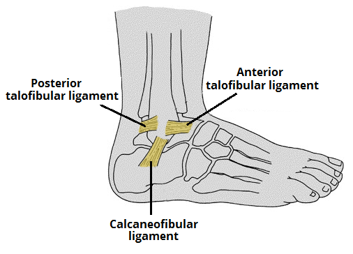
- Lateral Ligaments
- Anterior Talofibuar
- Spans between the lateral malleolus and lateral aspect of the talus.
- Posterior Talfibular
- Spans between the lateral malleolus and the posterior aspect of the talus.
- Calcaneofibular
- Spans between the lateral malleolus and the calcaneus.
- Anterior Talofibuar
- Medial Ligament
Nerve Supply
- 8 nerves innervate the foot
- Tibial Nerve
- Common Peraneal (deep & Superificial)
- Medial Plantar
- Lateral Plantar
- Plantar Digital nerves
- Calcaneal branches of tib & sural nerve
- Medial Plantar
- Sural
- Saphenous
- 8 nerves innervate the foot
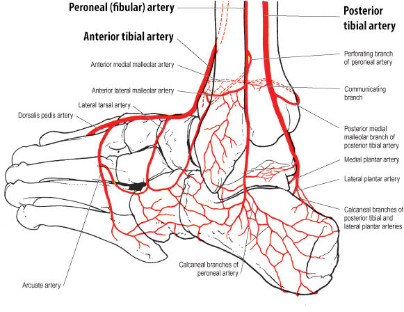
Vascular Supply
- Blood comes from 3 primary sources
- Peroneal (fibular) Artery
- Prosterior Tibial Artery
- Anterior Tibial Artery
- Blood comes from 3 primary sources
Images and files in the map
Information
- Description
- Mind Map on The Foot, created by katewarrior4 on 01/05/2015.
- Created by
- katewarrior4
- Views
- 0
- Rating