
Mind map
Protein misfolding during biosynthesis
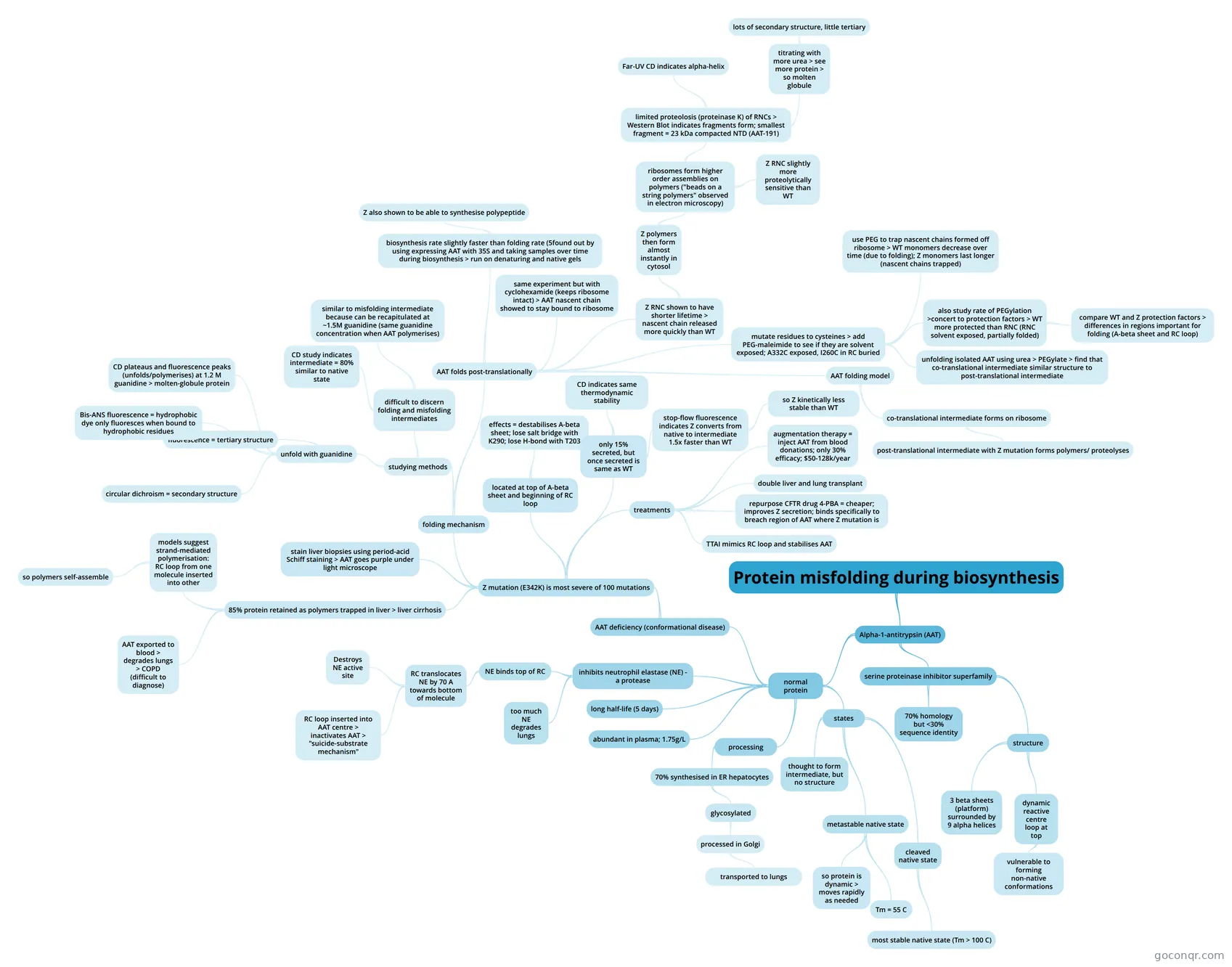
Mind map: Protein misfolding during biosynthesis
Map content
Protein misfolding during biosynthesis
Alpha-1-antitrypsin (AAT)
- normal protein
- abundant in plasma; 1.75g/L
- processing
- 70% synthesised in ER hepatocytes
- glycosylated
- processed in Golgi
- transported to lungs
- processed in Golgi
- glycosylated
- 70% synthesised in ER hepatocytes
- inhibits neutrophil elastase (NE) - a protease
- too much NE degrades lungs
- NE binds top of RC
- RC translocates NE by 70 A towards bottom of molecule
- RC loop inserted into AAT centre > inactivates AAT > "suicide-substrate mechanism"
- Destroys NE active site
- RC translocates NE by 70 A towards bottom of molecule
- long half-life (5 days)
- states
- metastable native state
- Tm = 55 C
- so protein is dynamic > moves rapidly as needed
- cleaved native state
- most stable native state (Tm > 100 C)
- thought to form intermediate, but no structure
- metastable native state
- AAT deficiency (conformational disease)
- Z mutation (E342K) is most severe of 100 mutations
- 85% protein retained as polymers trapped in liver > liver cirrhosis
- AAT exported to blood > degrades lungs > COPD (difficult to diagnose)
- models suggest strand-mediated polymerisation: RC loop from one molecule inserted into other
- so polymers self-assemble
- stain liver biopsies using period-acid Schiff staining > AAT goes purple under light microscope
- folding mechanism
- studying methods
- unfold with guanidine
- fluorescence = tertiary structure
- circular dichroism = secondary structure
- Bis-ANS fluorescence = hydrophobic dye only fluoresces when bound to hydrophobic residues
- CD plateaus and fluorescence peaks (unfolds/polymerises) at 1.2 M guanidine > molten-globule protein
- difficult to discern folding and misfolding intermediates
- CD study indicates intermediate = 80% similar to native state
- similar to misfolding intermediate because can be recapitulated at ~1.5M guanidine (same guanidine concentration when AAT polymerises)
- unfold with guanidine
- AAT folds post-translationally
- biosynthesis rate slightly faster than folding rate (5found out by using expressing AAT with 35S and taking samples over time during biosynthesis > run on denaturing and native gels
- Z also shown to be able to synthesise polypeptide
- same experiment but with cyclohexamide (keeps ribosome intact) > AAT nascent chain showed to stay bound to ribosome
- Z RNC shown to have shorter lifetime > nascent chain released more quickly than WT
- Z polymers then form almost instantly in cytosol
- ribosomes form higher order assemblies on polymers ("beads on a string polymers" observed in electron microscopy)
- limited proteolosis (proteinase K) of RNCs > Western Blot indicates fragments form; smallest fragment = 23 kDa compacted NTD (AAT-191)
- Far-UV CD indicates alpha-helix
- titrating with more urea > see more protein > so molten globule
- lots of secondary structure, little tertiary
- Z RNC slightly more proteolytically sensitive than WT
- limited proteolosis (proteinase K) of RNCs > Western Blot indicates fragments form; smallest fragment = 23 kDa compacted NTD (AAT-191)
- ribosomes form higher order assemblies on polymers ("beads on a string polymers" observed in electron microscopy)
- Z polymers then form almost instantly in cytosol
- mutate residues to cysteines > add PEG-maleimide to see if they are solvent exposed; A332C exposed, I260C in RC buried
- also study rate of PEGylation >concert to protection factors > WT more protected than RNC (RNC solvent exposed, partially folded)
- compare WT and Z protection factors > differences in regions important for folding (A-beta sheet and RC loop)
- use PEG to trap nascent chains formed off ribosome > WT monomers decrease over time (due to folding); Z monomers last longer (nascent chains trapped)
- unfolding isolated AAT using urea > PEGylate > find that co-translational intermediate similar structure to post-translational intermediate
- also study rate of PEGylation >concert to protection factors > WT more protected than RNC (RNC solvent exposed, partially folded)
- AAT folding model
- co-translational intermediate forms on ribosome
- post-translational intermediate with Z mutation forms polymers/ proteolyses
- co-translational intermediate forms on ribosome
- biosynthesis rate slightly faster than folding rate (5found out by using expressing AAT with 35S and taking samples over time during biosynthesis > run on denaturing and native gels
- studying methods
- located at top of A-beta sheet and beginning of RC loop
- effects = destabilises A-beta sheet; lose salt bridge with K290; lose H-bond with T203
- only 15% secreted, but once secreted is same as WT
- CD indicates same thermodynamic stability
- stop-flow fluorescence indicates Z converts from native to intermediate 1.5x faster than WT
- so Z kinetically less stable than WT
- treatments
- augmentation therapy = inject AAT from blood donations; only 30% efficacy; $50-128k/year
- double liver and lung transplant
- repurpose CFTR drug 4-PBA = cheaper; improves Z secretion; binds specifically to breach region of AAT where Z mutation is
- TTAI mimics RC loop and stabilises AAT
- 85% protein retained as polymers trapped in liver > liver cirrhosis
- Z mutation (E342K) is most severe of 100 mutations
- serine proteinase inhibitor superfamily
- 70% homology but <30% sequence identity
- structure
- 3 beta sheets (platform) surrounded by 9 alpha helices
- dynamic reactive centre loop at top
- vulnerable to forming non-native conformations
- normal protein
Information
- Description
- Mind Map on Protein misfolding during biosynthesis, created by Charlotte G on 15/11/2020.
- Created by
- Charlotte G
- Views
- 0
- Rating