
Mind map
Gastro-Intestinal Tract: Physiology

Map content
Gastro-Intestinal Tract: Physiology
- After the mouth, the digestive tract is split into 4 major organs: oesophagus, stomach, small intestine and large intestine
- Organs are seperated by muscular valves or sphincters with a mucosal layer lining the inner surface of the digestive tract.
Oesophagus
Stomach
- Stomach has huge invaginations => increases surface area greatly
- Superficial Epithelial cell
- At top of pit
- Mucous neck cell
- Produce mucous? => forms preventative barrier against acid being secreted
- Stem/Regenerative cell
- Due to the hostile environment, cells are continuously being sloughed off These cells regenerate more => new cells move up slowly as the top layers are sloughed off
- Parietal Cell
- Secrete hydrochloric acid (HCl) Changes structure when activated => have lots of little tubules that form a whole network => on them sits H+/K+ ATP-ase pump Pumps hydrogen against a gradient of 14 million:1 Only ATP-ase that sits on lumen of cells
- Negative Feedback loop
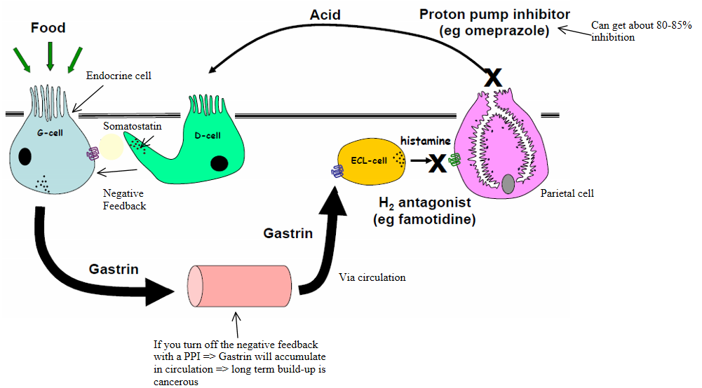
- In gastric pit, have endocrine cell called a G-cell => food enters gastric pit, stimulates release of gastrin (an endocrine hormone) from the G-cell Gastrin is secreted into the circulation and binds to a receptor on ECL cells => this causes the ECL-cells to release histamine Histamine binds to a receptor on parietal cells => parietal cells release HCl
- The acid secreted by the parietal cells will activate the D-cells => they release somatostatin => inhibits the G-cell from releasing Gastrin This is a negative feedback loop to turn off Gastrin secretion => check and balance system
- PPI's are sometimes given (e.g. to patients with gastric ulcers where increased acid is undesirable) => they block the parietal cells from secreting acid (although only about 80-85% => will still be able to digest food; also, as stem cells are continuously renewing cells, will never be able to achieve 100% block) However, if we block the acid secretion, the D-cell will not be activated to secrete Somatostatin to turn off Gastrin secretion from the G-cell => can end up with accumulation of Gastrin in teh circulation => this is undesirable as gastrin also acts in the brain and can cause tumour formation
- Chief cell
- makes pepsinogen
- Endocrine cell
- G-cell
- secretes Gastrin (an endocrine hormone) into the circulation in response to food entering the gastric pit
- Enterochromaffin-Like Cell (ECL-cell)
- A neuroendocrine cell Synthesize and secrete histamine in response to Gastrin
- D-cell
- Somatostatin producing cells
- G-cell
Small Intestine
Large Intestine
Histological Organisation
- The type of cells and tissues that make up the GI tract
- With the exception of the oral cavity, 4 concentric layers of muscle make up the GI tract, from the oesophagus to the large intestine. They are: mucosa, submucosa, muscularis externa and adventitia or serosa
- Vascular and lymphatic vessels aid nutrient absorption => epithelial cells require a constant supply of nutrients for cell maintenance and repair
- Mucosa
- Innermost layer of the GI tract => comes into contact with the digested food (chyme)
- Is itself made up of three layers: *epithelium => with loose connective tissue that is highly vascularized => responsible for degestive, secretory and absorptive processes *lamina propria =>layer of connective tissue with immunocompetent cells *muscularis mucosae => a thin layer of smooth muscle => when it contracts, it throws the mucosa into folds and ridges)
- The mucosa is highly specialized for each different part of the GI tract, according to function
- Oesophagus
- Here the epithelium is squamous stratified (i.e. flattened epithelial cells in layers) as the upper layer gets sloughed off during friction and abrasion with food that is being swallowed => this is for protective purposes It is non-keratinised as it is kept moist by mucus and other fluids
- Stomach
- Epithelium is simple columnar organised into gastric pits, glands and goblet cells, to deal with secretion of gastric juices etc
- Small Intestine
- Epithelium is simple columnar and organised into villi => the enterocytes have their own microvilli => this forms a brush border and massively increases the surface area for absorption
- in the ileum, there are occasionally Peyer's patches in the lamina propria => aggregates of lymphoid tissue to act as immune defence against pathogens being consumed
- Large Intestine
- Epithelium is simple columnar with no villi Goblet cells throughout
- Submucosa
- Consists of large blood vessels and lymphatic vessels. Nerve fibres branch into the mucosa and the muscularis externa. Contains an enteric nervous plexus (collection of ganglia of enteric nervous system) known as meissner's plexus
- Muscularis Externa
- Consists of two layers of muscle (the smooth type, except in the upper oesophagus and anal sphincter, where striated muscle exists): *one inner, circular layer => contraction constricts width of lumen *one outer, longitudinal layer => contraction shortens the digestive tube
- At sphincter points, the thickness of this circular layer increases. It is also thicker in the colon as the faeces are large and heavy, requiring more force to push along
- Serosa
- Also known as adventitia
- consists of several layers of loose, connective tissue => alternately collagenous and elastic Covered by a thin layer of mesothelial cells
- Mucosa Circulation
- The arteries supply an irregular capillary plexus around glands, sending terminal branches into the mucosal villi (lumen of the intestine)
- Generally, in the intestinal and exocrine epithelium, the capillaries are faenestrated to allow passage of small molecules and limited amount of protein.
- Veins arising in the mucosa anastamose (reconnect) in the submucosa and pass out of the intestine alongside arteries
- Mucosa Lymphatic Vessels
- These arise in the mucosa as blind-ended tubes also known as lacteal vessels. Similar to blood vessels, they contain pinocytotic vesicles (vesicles that form around particles from the extracellular fluid) that shuttle marcromolecules and chylomicrons (lipoprotein particles) from the interstitial fluid in the lymphatic circulation.
- In the submucosa, large lymphatic vessels branch freely, crossing the muscle layers and into the serosa
- Innervation of the GI tract
- The autonomic nervous system of the CNS innervates the GI tract, comprising both the intrinsic and extrinsic nervous system
- Extrinsic
- Efferent fibres
- Carry information from the brain stem and spinal cord into the GI tract
- Afferent fibres
- Carry sensory information from chemoreceptors and mechanoreceptors to the brain stem and spinal cord
- Parasympathetic Nervous System
- Usually exciting of the GI tract functions
- Sympathetic Nervous System
- Usually inhibitive of the GI tract's functions. These fibres terminate in the submucosa and cause constriction of blood vessels, inhibit contraction of muscularis externae, but allows constrictions of muscularis mucosae
- Efferent fibres
- Intrinsic (The Enteric Nervous System)
- Coordinates and relays information from the parasympathetic and sympathetic nervous systems to the GI tract, even in the absence of extrinsic innervation, using local reflexes
- These plexus' can cross-talk to one another without the need for central input known as local nerve nets
- Myenteric Plexus (Auerbach's Plexus)
- Primarily controls motility of the GI tract
- Submucosal Plexus (Meissner's Plexus)
- Primarily controls secretion and blood flow
- Endocrine Control of the GI tract
- The GI tract is also under neurocrine, endocrine and paracrine influence => these include gastrin, cholecystokinin. Endocrine cells are widely distributed throughout the stomach, small intestine, appendix and pancreatic epithelium
Oral cavity
- Saliva
- Salivary glands are compound organs that secrete electrolytes and proteins (e.g. amylase) as a fluid called saliva into the oral cavity. Some functions of saliva include: *lubrication of digested food *initial starch digestion by amylase *initial triglyceride digestion by lingual lipase *protection of mouth and oesophagus by buffering and diluting digested foods
- Stimulation of Saliva
- Set off by taste, touch, smell receptors and by chewing (mastication) => conditioned reflexes may also be involved. Its production is controlled by parasympathetic and sympathetic nervous systems (not by hormones)
- It is secreted by 3 major glands: parotid gland, submandibular gland and sublingual gland
- At the maximum flow rate (~1 mL/min) the composition of saliva closely resembles that of plasma
- Salivary Gland Cells
- Salivary glands are divided into lobules. There are two different cell types: *Acini: Acinar cells are secretory cells that form a group called an acinus (plural: acini). They are arranged at the terminal part of the gland, connected to the ductal system, as a single layer of cuboidal epithelium surrounding a small central lumen where saliva is deposited. These open into intercalated ducts that converge into larger ducts tha open into the main excretory ducts draining into the mouth *Ductal cells form lumina. Firstly, intercalated ducts lead directly from the acinus to a striated duct. The striated duct then leads to an interlobular duct.
- Microvasculature
- Highly fenestrated capillaries to allow passage of ions and limited amounts of protein from the blood into the saliva
- During neural activation of salivation, there is a 5-10 fold increase of blood flow through these capillaries, guaranteeing sufficient supply of water, electrolytes and nutrients for saliva production (NOTE! however, that salivation is not due to increased hydrostatic pressure gradient from blood to saliva)
- Parallel capillary networks supply both ductal and acinar cells
- Secretory Mechanism of Saliva
- Saliva is secreted in a two-stage mechanism: *The acinar cells secrete the initial saliva. This is isotonic (with a plasma-like electrolyte composition). This is confirmed by micropuncture techniques *The ductal cells then modify this initial saliva in the striated duct system. The ductal cells: => Reabsorb Na+ and Cl- => Secrete K+ and HCO3- The saliva produced is hypotonic (it has less solute and more water than the previous solution)
- Effects of Parasympathetic and Sympathetic stimulation on Salivary Secretion
- Salivary production is unique, in that it is increased by BOTH sympathetic and parasympathetic nervous systems. However, the effect of the parasympathetic nervous system is greater.
- Saliva
What can go wrong?
- Cholera toxin => instead of being in absorption mode => entire GI tract switches into secretory mode => you become dehydrated
- can have malabsorption => e.g. in the intestine because you don't have the right pancreatic enzyme or liver product => don't absorb food => start losing muscle
- Cancer => e.g. in pancreas => lost ability of pancreas to secrete insulin => glucose homeostasis will be affected => main metabolite of brain is absent
Images and files in the map




Information
- Description
- Graduate Gastro-Intestinal System and Skin Mind Map on Gastro-Intestinal Tract: Physiology, created by katpotter71 on 14/07/2015.
- Created by
- katpotter71
- Views
- 15
- Rating