
Mind map
Pulmonary Apparatus and Chest Wall
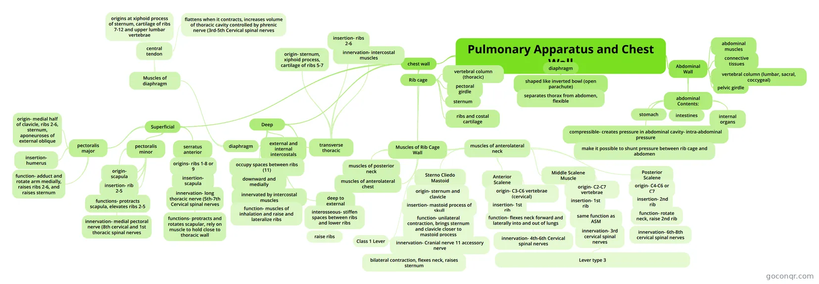
Mind map: Pulmonary apparatus and chest wall
Map content
Pulmonary Apparatus and Chest Wall
chest wall
- Rib cage
- ribs and costal cartilage
- sternum
- pectoral girdle
- vertebral column (thoracic)
- Muscles of Rib Cage Wall
- muscles of anterolateral neck
- Sterno Cliedo Mastoid
- origin- sternum and clavicle
- insertion- mastoid process of skull
- function- unilateral contraction, brings sternum and clavicle closer to mastoid process
- innervation- Cranial nerve 11 accessory nerve
- Class 1 Lever
- bilateral contraction, flexes neck, raises sternum
- Anterior Scalene
- origin- C3-C6 vertebrae (cervical)
- insertion- 1st rib
- function- flexes neck forward and laterally into and out of lungs
- innervation- 4th-6th Cervical spinal nerves
- origin- C3-C6 vertebrae (cervical)
- Middle Scalene Muscle
- origin- C2-C7 vertebrae
- insertion- 1st rib
- same function as ASM
- innervation- 3rd cervical spinal nerves
- Lever type 3
- Posterior Scalene
- origin- C4-C6 or C7
- insertion- 2nd rib
- function- rotate neck, raise 2nd rib
- innervation- 6th-8th cervical spinal nerves
- Sterno Cliedo Mastoid
- muscles of anterolateral chest
- muscles of posterior neck
- muscles of anterolateral neck
- Superficial
- pectoralis major
- origin- medial half of clavicle, ribs 2-6, sternum, aponeuroses of external oblique
- insertion- humerus
- function- adduct and rotate arm medially, raises ribs 2-6, and raises sternum
- pectoralis minor
- origin- scapula
- insertion- rib 2-5
- functions- protracts scapula, elevates ribs 2-5
- innervation- medial pectoral nerve (8th cervical and 1st thoracic spinal nerves
- serratus anterior
- origins- ribs 1-8 or 9
- insertion- scapula
- innervation- long thoracic nerve (5th-7th Cervical spinal nerves
- functions- protracts and rotates scapular, rely on muscle to hold close to thoracic wall
- pectoralis major
- Deep
- diaphragm
- Muscles of diaphragm
- central tendon
- origins at xiphoid process of sternum, cartilage of ribs 7-12 and upper lumbar vertebrae
- flattens when it contracts, increases volume of thoracic cavity controlled by phrenic nerve (3rd-5th Cervical spinal nerves
- central tendon
- Muscles of diaphragm
- external and internal intercostals
- occupy spaces between ribs (11)
- downward and medially
- innervated by intercostal muscles
- function- muscles of inhalation and raise and lateralize ribs
- deep to external
- interosseous- stiffen spaces between ribs and lower ribs
- raise ribs
- interosseous- stiffen spaces between ribs and lower ribs
- transverse thoracic
- origin- sternum, xiphoid process, cartilage of ribs 5-7
- insertion- ribs 2-6
- innervation- intercostal muscles
- diaphragm
- Rib cage
diaphragm
- shaped like inverted bowl (open parachute)
- separates thorax from abdomen, flexible
- shaped like inverted bowl (open parachute)
Abdominal Wall
- abdominal muscles
- connective tissues
- vertebral column (lumbar, sacral, coccygeal)
- pelvic girdle
- abdominal Contents:
- stomach
- intestines
- internal organs
- compressible- creates pressure in abdominal cavity- intra-abdominal pressure
- make it possible to shunt pressure between rib cage and abdomen
Information
- Description
- Mind Map on Pulmonary Apparatus and Chest Wall, created by juliebolton9 on 11/30/2013.
- Created by
- juliebolton9
- Views
- 221
- Rating
More on Lung and Pleura Anatomy