
Mind map
staining mechanisms(8/12)
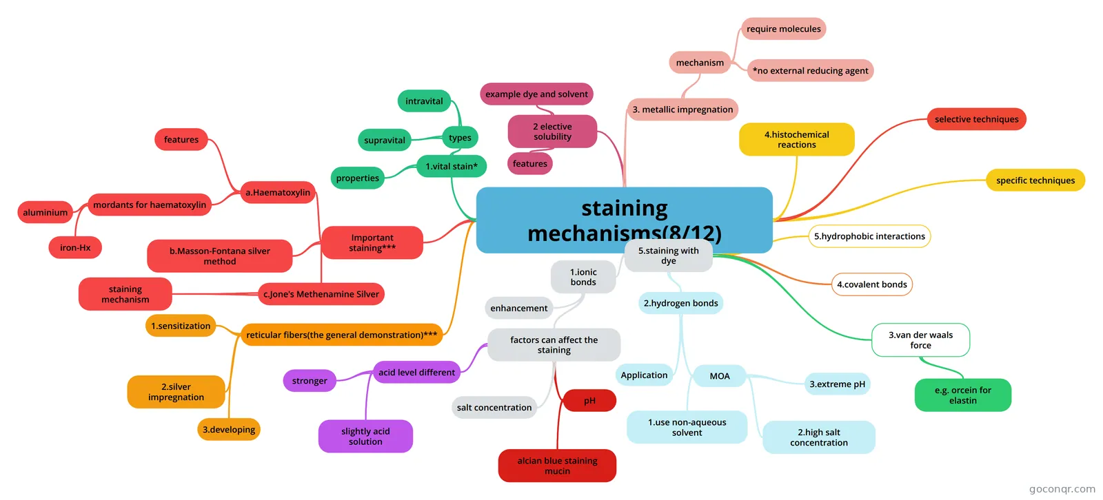
Mind map: staining mechanisms
Map content
staining mechanisms(8/12)
- staining purposes: 1. differentiation between the composite structure with contrasting coloured dyes 2.demonstration for various cell products
3. metallic impregnation
- 1.deposition of metallic silver onto tissue elements
- mechanism
- argyrophil reaction
- e.g.capable of impregnation with silver salt with the use of an external reducing agent(e.g. stain reticulin)
- require molecules
- formaldehyde or hydroquinone
- *no external reducing agent
- argentaffin:tissue contains reducing groups to reduce silver ion, without the need for external reducing agent
- example of argentaffin 1.enterochromaffin cells(is a type of enteroendocrine cells-know as kulchitsky cells) 2.melanin
2 elective solubility
- criteria of a lysochrome(a soluble dye used for histochemical staining of lipid)
- features
- 1.strong coloured 2.highly soluble in tissue constitutes than in solvents(e.g. soluble in tissue lipid) 3.solvent will not dissolve specimen
- example dye and solvent
- 1.oil red O 2.sudan (III,IV and black) 3. nile blue sulphate(NBS)
- Solvents: 1.70% alcohol 2.60% triethyl phosphate 3.propylene glycol 4.60% isopropanol
1.vital stain*
- properties
- 1. non toxin, harmless stain: colored tissues or cells of living organisms. 2. often used to determine live/dead cell ratio in a cell population
- types
- supravital
- it is processed by removing living tissues from body and cells and treated with a non toxic dye solution
- intravital
- a stain absorbed by living cells after injection into the body
- supravital
- properties
4.histochemical reactions
- 1.It may utilize a dye or in itself to produce coloured reaction e.g.. a. Schiff reagent uses basic fuchsin: coloured aldehyde site magenta
- 2.enzyme histochemistry
5.staining with dye
- based on different kind of chemical bondings
- 1.ionic bonds
- it is electrostatic interaction between oppositely charged ions
- enhancement
- 1.binding of dyes depend on tissue ionization 2.pH(can get highly specific staining): -acid pH favours staining with anionic dyes -alkaline pH favours staining with basic dyes
- factors can affect the staining
- how salt/acid concentration affect tissue ionization and staining??
- acid level different
- concerning tissue components
- stronger
- phosphate groups in nucleic acids and sulphate groups found in mucin:can still ioniz and less easily inhibited
- slightly acid solution
- e.g. methylene blue will act as a differential staining:stain nuclear but unstain proteins
- salt concentration
- in high salt concentration:competition between dyes and the salt ions, so staining will be inhibited -weak salt can increase the staining.
- pH
- alcian blue staining mucin
- apparent staining is produced in ideal pH environment: alcian blue can bind to both weak carboxylic-acid containing mucin and strong sulphated mucin.
- alcian blue staining mucin
- 2.hydrogen bonds
- an intermolecular force:between a hydrogen atom in one molecule and a small atom of high electronegativity in other molecules.-play a role in the selectivity of the dye -not affected by pH and salt concentration ,but stronger of the hydrogen-bonding agents.
- Application
- 1.Congo red for amyloid 2.Best carmine's for glycogen
- MOA
- used to inhibit ionic bonds, increase the selectivity of the dye
- 1.use non-aqueous solvent
- water inhibit hydrogen bonding and favours ionization
- 3.extreme pH
- chosen to inhibit the ionization of tissue groups
- 2.high salt concentration
- competition with the dye
- 3.van der waals force
- attraction between non-polar molecules: weak, short range forces
- e.g. orcein for elastin
- 4.covalent bonds
- very strong bonds and are not easily broken: important in PAS(periodic Schiff reagent: histochemical technique)
- 5.hydrophobic interactions
- -not chemical bond -important in selectivity and play a role in lipid staining
selective techniques
- stain several structure: differentiate by other features
specific techniques
- only a particular tissue structure or chemical moiety is demonstrated
Important staining***
- a.Haematoxylin
- -natural product,no obvious chromophore -for staining: first oxidized to heamatein
- features
- 1.oxidation is slower in acid conditions: many solutions kept acidic: (i)ripening-oxidation of heamatoxylin to haematein in atmospheric oxygen. (ii)using oxidizing agents-sodium iodate, potassium permanganate or mercuric oxide
- mordants for haematoxylin
- Aluminium: usually potash alum and ammonium alum
- aluminium
- solubility different in pH: 1.in acid solution: the alum dye lakes are quite soluble and have a strong red color 2.in alkaline solution: dye lakes are less soluble and have a strong blue colour.
- iron-Hx
- -salts used are ferric chloride or ferric ammonium sulphate(ferric salt)-blacker and intense .will resist acidic counter stain -ferric salts are oxidizing agents and will accelerate oxidation of haematoxylin to haematein
- -mordant is therefore either used separately or -mix mordant and haematoxylin before use
- b.Masson-Fontana silver method
- 1. required argentaffin: substances that reduce silver diamine e.g. for melanin demonstration/5 hydroxytrypatmine in enterochromaffin cells
- process for the demonstration: 1.blenching of melanin on one slide MOA:Ag impregnation reducing groups convert silver diamine to colloidal metallic silver
- c.Jone's Methenamine Silver
- lt is used to stain glomerular basement membrane MOA:argentaffin metallic impregnantion
- The structure can be demonstrated: 1. spikes of GBMs due to immune deposits in membranous glomerulonephritis 2.spicules in amyloidosis:due to amyloid infiltration of peripheral capillary loops 3.double contours: mesangial interposition in membranoproliferative glomerulonephritis.
- staining mechanism
- 1. mild oxidation: expose reactive aldehyde groups 2.Ag impregnation: Ag+ bind to aldehyde groups and reduced to Ag deposit
- a.Haematoxylin
reticular fibers(the general demonstration)***
- -agyrophilic -common in the liver,kidney and spleen(perform a support function) -demonstration by silver stains
- 1.sensitization
- metallic complexes is developed at the aldehyde groups
- 2.silver impregnation
- 1.silver salt and ferric complex exchange ions 2.ammoniical silver solution is reduced by the exposed aldehyde groups to metallic silver
- 3.developing
- 1.further reduction of the diamine silver by transfer the section to formaldehyde
Information
- Description
- - histology Mind Map on staining mechanisms(8/12), created by Clare Yu on 29/11/2015.
- Created by
- Clare Yu
- Views
- 1
- Rating