4191414
Description
Mind Map by Sarah Emslie, updated more than 1 year ago
|
|
Created by Sarah Emslie
about 10 years ago
|
|
Amino Acids and Proteins
- Proteins
- Structure
Annotations:
- Favorable interactions: Hydrophobic effect Hydrogen bonds London dispersion forces Electrostatic interactions
- Primary Structure
Annotations:
- All the information required for polypeptides to form structure is encoded in primary structure
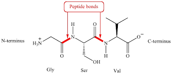
- Peptide bond
Annotations:
- Rigid and almost planar, caused by resonance structures, less reactive than ester linkages, each bond has small dipole moment
- Bonds go C-alpha, C, N starting at N terminus
- Usually trans conformation
- Shorter than a
single bond but
longer than a typical
double bond
Annotations:
- About 0.133 nm long
- Planar
- Phi is N-Calpha bond, Psi is Calpha-C bond
Annotations:
- Unfavorable when both are 0 degrees or one is 180 and one is 0
- Ramachandran plot shows interactions in a backbone
- Native Fold
Annotations:
- Creates biological function, made up of favorable interactions.
- Chaperones and
Chaperonins
Annotations:
- Chaperones: proteins that interact with partially folded or improperly folded proteins to help them fold right. Chaperonins: elaborate protein complexes required for some cellular proteins that don't fold spontaneously
- Secondary Structure
- Alpha helices
Annotations:
- First proposed by Pauling and Corey, identified by Perutz
- Favorable H-bonds between
peptide carbonyl and peptide NH
group four residues up the chain
- Angles allowed (phi -57, psi -47)
- 3.6 residues per turn,
5.41 angstrom pitch
- R groups are perpendicular
to the helix axis and
stabilize favorable
interactions 3-4 residues
apart
- Destabilized by
stretches of
similarly charged
residues
- Has a large dipole
moment since all
peptide bonds have
the same orientation
- Factors determining stability
- Interactions of R groups
3-4 helices apart
- Occurence of Gly and Pro residues
- Interaction between AA
residues at ends of helical
segment
- Electrostatic
interatctions
between
successive AA w/R
groups
- Interactions of R groups
3-4 helices apart
- Beta pleated sheets
Annotations:
- Also first postulated by Corey and Pauling
- Strands can be parallel
or antiparallel
- Rise/residue is 3.47 A
for antiparallel and
3.25 A for parallel
- H bonds between
neighboring chains, alpha
carbons in folds, side chains
protrude up and down
- Not flexible
- Beta Turn
- Allows peptide chain to
reverse direction
- Stabilized by H
bond from
carbonyl to
amide proton 3
residues down
- Often contains Pro or Gly
- Allows peptide chain to
reverse direction
- Alpha helices
- Tertiary Structure
- Fibrous proteins
Annotations:
- Typically insoluble, made from a single secondary structure
- Alpha keratin (alpha
helices), silk fibrin (beta
pleated sheets), collagen
(triple helix)
- Globular proteins
Annotations:
- Water soluble globular proteins, lipid soluble membrane proteins
- Hydrophobic
residues tend to face
inside, polar residues
tend to face out
- Domains are sections
of the protein that can
fold independently
- Secondary structures
form wherever
possible
- Helices and sheets
pack together
- Fibrous proteins
- Quaternary Structure
- Typical Kd for two
subunits is 10^-8-10^-16
M
- Entropy loss due to
association but entropy
gain due to burying of
hydrophobic groups
makes up for it
- Provides stability, genomic
efficiency, brings catalytic sites
together, provides
cooperativity
- Typical Kd for two
subunits is 10^-8-10^-16
M
- Structure
- Amino Acids
- Display acid-base properties
- Able to polymerize into proteins
- All except Gly are chiral
- Metabolites, essential to
human nutrition,
hormones
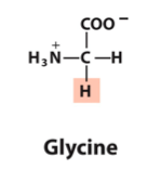
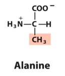
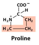
- Nonpolar AAs
- Glycine, Gly, G
- Alanine, Ala, A
- Proline, Pro, P
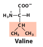
- Valine, Val, V
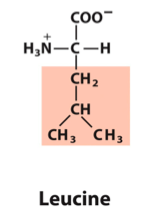
- Leucine, Leu, L
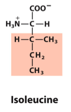
- Isoleucine, Ile, I
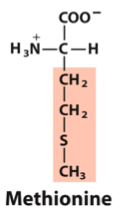
- Methionine, Met, M
- Glycine, Gly, G
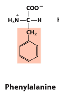
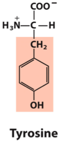
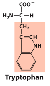
- Aromatic AAs
- Phenylalanine, Phe, F
- Tyrosine, Tyr, Y
- Tryptophan, Trp, W
- Phenylalanine, Phe, F
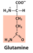
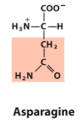
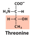
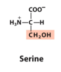
- Polar AAs
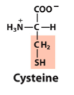
- Serine, Ser, S
- Threonine, Thr, T
- Cysteine, Cys, C
- Asparagine, Asn, N
- Glutamine, Gln, Q
- Serine, Ser, S
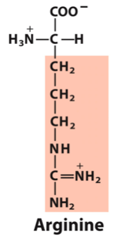
- Positive AAs
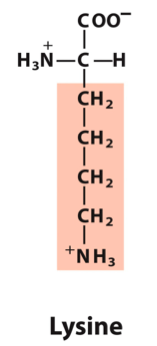
- Lysine, Lys, K
- Arginine, Arg, R
- Histidine, His, H
- Lysine, Lys, K
- Negative AAs
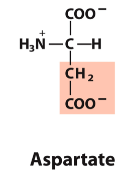
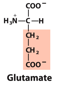
- Aspartate, Asp, D
- Glutamate, Glu, E
- Aspartate, Asp, D
- Display acid-base properties
- Protein Purification & Primary Sequence
Annotations:
- General Method: 1. Obtain/grow cells 2. Lyse cells 3. Fractionation based on protein solubility differences 4. Dialysis 5. Chromatography
- Types of Chromatography
- Column chromatography
- Ion exchange chromatography
Annotations:
- Cation exchange elutes negatively charged proteins first, anion exchange elutes positively charged proteins first.
- Gel filtration chromatography
Annotations:
- Separate by size, smaller particles go through smaller channels and take longer to elute.
- Affinity chromatography
Annotations:
- Binds protein to a ligand, protein of interest elutes last.
- Column chromatography
- Specific Activity
Annotations:
- One unit of enzyme activity = the amount of enzyme that can transform one um of substrate to product/min at 25C
- Total activity or total units is the total number of enzyme units in a sample
- Specific activity is the number of enzyme units per mg of protein
- Yield is the total number of units at each step divided by the initial total in the crude extract x100
- Purification Techniques
- SDS-Page
Annotations:
- PAGE = polyacrylamide gel electrophoresis, SDS = sodium dodecyl sulfate
- SDS binds and unfolds proteins and gives a uniformly negative charge, native shape doesn't matter so they will elute by size (smaller faster)
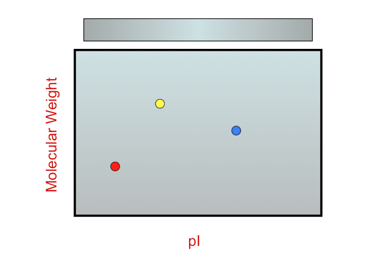
- Isoelectric Focusing
Annotations:
- Protein is applied to gel with immobilized pH gradient, protein will move along strip based on pI values (left side is more alkaline, right side is more acidic)
- Running SDS-PAGE and
isoelectric focusing at the
same time can give
indication of weight and pI
of proteins at same time
- SDS-Page
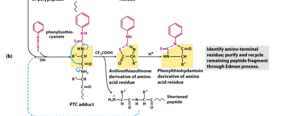
- Primary Sequence Determination
- Small Proteins (<~50 residues)
Annotations:
- Steps: 1. Reduce disulfide bonds and alkylate Cys residues 2. Determine total AA composition via hydrolysis with 6M HCl 3. Determine identity of N-terminal residue by: a. Treating with Sanger's reagent, FDNB, then acid workup (peptide destroyed) or b. Do first step of Edman degradation (liberates one AA at a time, peptide intact) 4. Sequence by Edman degradation
- Large Proteins (>~50 residues)
Annotations:
- Complete steps 1-4 from small protein 5. Digest protein into smaller peptides suitable for Edman degradation via proteases a. Trypsin cleaves on C side of Arg and Lys (+) b. Chymotrypsin cleaves on C side of Phe, Trp, and Tyr (aromatic) c. Cyanogen bromide cleaves on C side of Met 6. Sequence by Edman degradation on each fragment (deduce AA sequence from overlap)
- Reagent used for
N-terminal determination
is in pink, rest of molecule
is AA
- Small Proteins (<~50 residues)
- Protein Function
- Ligand
Annotations:
- Molecule that binds reversibly to a protein (can be another protein)
- Binding site
Annotations:
- Site on the protein where the ligand binds, complimentary to the ligand in size, shape, charge, and hydrophilic/phobic character
- Induced fit
Annotations:
- Binding of ligand to protein results in conformational change that results in increased binding
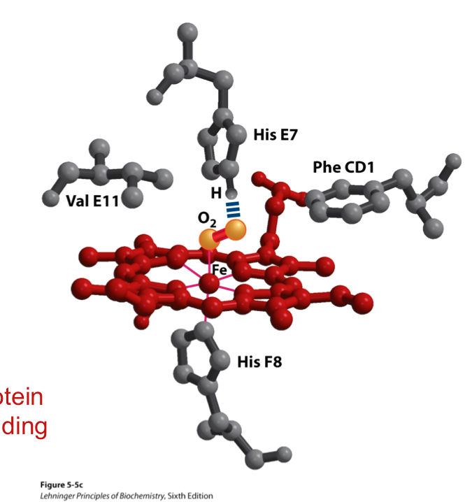
- Myoglobin
- Oxygen storage protein
- Ineffective for
transport b/c it
has a very high
affinity for O2
Annotations:
- Has a hyperbolic curve
- Good
transporters must
vary affinity with
pO2
- Ineffective for
transport b/c it
has a very high
affinity for O2
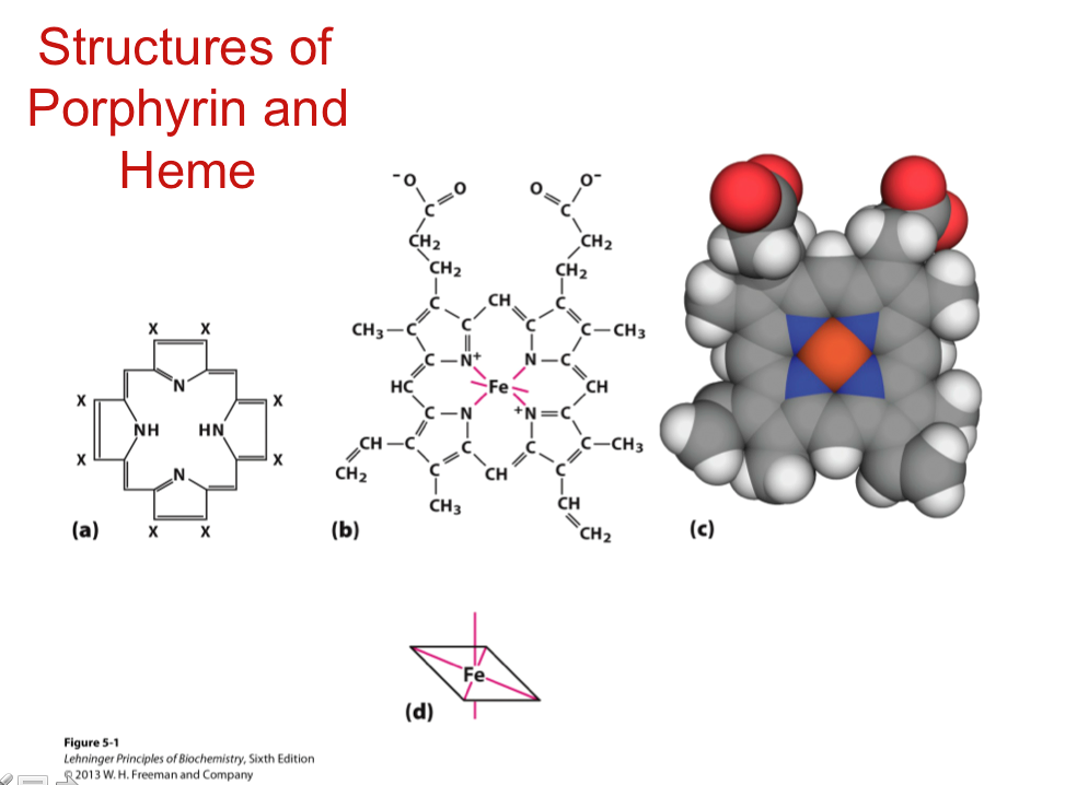
- Contains a heme group so
Fe2+ can be oxidized to Fe3+
without creating free radicals
- Structural Features
Annotations:
- -First protein structure determined by x-ray crystallography -Peptide bond in trans config -All alpha domain -3/4 of Pro were at bends -All hydrophobic residues interior -All but 2 polar R groups exterior -Dense, only room for 4 H20 -8 alpha helices (A-H) -Heme group is in hydrophobic pocket between E and F -Fe(II) at center of heme has *4 porphyrin N atoms *His F8 on proximal side *O2 on distal side (E7 binds to this) -Val E11 and Phe CD1 on O2 side of heme helps keep it in place
- Porphyrin and Heme
- Affinity for CO
- Similar size and shape
to O2 but has lone e-
pair that can be donated
- CO binds over 20k
times better than O2
- Similar size and shape
to O2 but has lone e-
pair that can be donated
- Oxygen storage protein
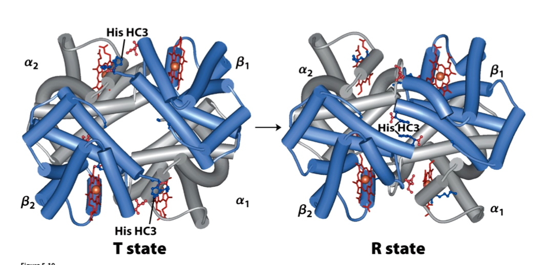
- Hemoglobin
- Structure
- Tetramic,
contains 4
alpha2beta2
subunits
- Each subunit has 1
heme and binds 1 O2
(Mr=16000)
- Most interactions between
alpha and beta subunits (not
alpha-alpha or beta-beta)
- Each subunit has 1
heme and binds 1 O2
(Mr=16000)
- Tetramic,
contains 4
alpha2beta2
subunits
- Very sensitive to
changes in [O2],
[CO2], [H+], and
[BPG]
- Uses cooperativity
to have affinity
changes
- Positive cooperativity: first
binding event increases
affinity at remaining sites
- Negative cooperitivity:
first binding event
decreases affinity at
remaining sites
- Generally has
sigmoidal binding
curves
- Positive cooperativity: first
binding event increases
affinity at remaining sites
- Conformations
- Oxyhemoglobin (HbO2):
relaxed (R) state, greater
affinity for O2
- Deoxyhemoglobin (Hb): tense
(T) state, lower affinity for O2,
stabilized by more ion pairs
than R state
- Movement from T-->R state
triggered by movement of
heme iron
Annotations:
- When O2 binds Fe is pulled into the heme plane pulling the His F8 and F helix with it. This binding changes Fe's spin state from high to low so it can fit in the porphyrin ring
- Solvent filled channel
narrows as ab dimers
slide 15 degrees past
each other, switch from
T-->R is simultaneous
- Oxyhemoglobin (HbO2):
relaxed (R) state, greater
affinity for O2
- Allosteric Protein
Annotations:
- All or none model: two conformations are in equilibrium and the ligand can bind to either conformation. Molecular symmetry is conserved
- Sequential model: ligand binding to a subunit causes a conformational change that may cause changes in adjacent subunits. The more ligand is bound the more likely more conformational changes will occur
- T--->R conversion more closely follows concerted model
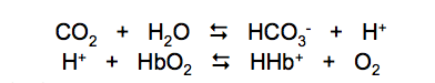
- Bohr Effect
- Affinity of Hb for O2
decreases with decreasing
pH (increasing [H+])
- Hb releases more O2 at
more acidic pHs because
increased [H+] favors ion pair
formation driving R-->T
transition, releasing O2
- Affinity of Hb for O2
decreases with decreasing
pH (increasing [H+])
- Effect of CO2
- CO2 produces HCO3- and
H+ in water, H+ then
decreases Hb affinity
- In tissues this drives R-->T releasing O2
- In lungs O2 binds driving T-->R
which releases H+ and creates CO2
from the HCO3- present
- In tissues this drives R-->T releasing O2
- CO2 produces HCO3- and
H+ in water, H+ then
decreases Hb affinity
- BPG
- Binds to deoxyhemoglobin (T)
- Helps stabilize T state by
binding in the channel in
the middle
- Binds to deoxyhemoglobin (T)
- Fetal hemoglobin has
a higher affinity for
O2 than adult Hb
- In sickle cell anemia, the
Hb interacts and
crystallizes
- Structure
- Ligand
Media attachments
Want to create your own Mind Maps for free with GoConqr? Learn more.