4202004
Description
Mind Map by Lauren Jane Speed, updated more than 1 year ago
|
|
Created by Lauren Jane Speed
about 10 years ago
|
|
Topic 1A - Biological Molecules
- Carbohydrates
- Most cabrohydrates are
POLYMERS (Large complex
molecules made by long
chains of monomers
joined together)
- MONOMERS are small basic units
- MONOMERS are small basic units
- The monomer they are
made from are
MONOSACCHARIDES
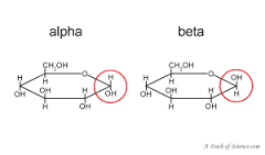
- Glucose is a hexose sugar
with 6 monosaccaride units.
There are 2 times of glucose
alpha glucose and beta glucose
- There are ISOMERS (molecules
with the same molecular formula
as eachother but connected in
different ways)
- There are ISOMERS (molecules
with the same molecular formula
as eachother but connected in
different ways)
- Glucose is a hexose sugar
with 6 monosaccaride units.
There are 2 times of glucose
alpha glucose and beta glucose
- CONDENSATION REACTIONS join
monosaccharides together to
form a new chemical bond and a
water molecule is RELEASED
- Monosaccharides are joined together by
condensation reactions forming a
GLYCOSIDIC BOND between them when
water is released - A DISSACHARDIE if
formed when 2 monosacchardies join
together
- Polymers can be broken into
monomers by a HYDROLYSIS
REACTION which breaks chemical
bonds by ADDING WATER
- Polymers can be broken into
monomers by a HYDROLYSIS
REACTION which breaks chemical
bonds by ADDING WATER
- Monosaccharides are joined together by
condensation reactions forming a
GLYCOSIDIC BOND between them when
water is released - A DISSACHARDIE if
formed when 2 monosacchardies join
together
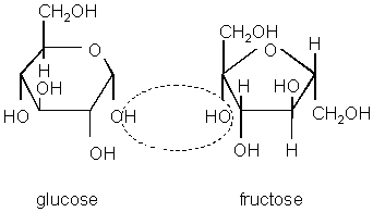
- a glucose + a glucose = Maltose
- glucose + fructose = sucrose
- glucose + galactose = lactose
- Benedict's test for sugar
- REDUCING SUGARS include all
monosacchardies & maltose & lactose
- Add benedics regent (blue) to a
sample and boil it in a hot bath
- If the tests positive it wll form a coloured
precipitate going from BLUE - GREEN -
YELLOW - ORANGE - RED the higher the
concentration, the further the colour change
- If the tests positive it wll form a coloured
precipitate going from BLUE - GREEN -
YELLOW - ORANGE - RED the higher the
concentration, the further the colour change
- Add benedics regent (blue) to a
sample and boil it in a hot bath
- If the result is negative you use a NON
REDUCING SUGARS TEST to test sucrose
- You must get them into their
monosaccharides, you do this buy adding
HCL and heat it in a water bath. You
then dilute it using sodium hydrogen
carbonate
- You then carry out the previous test, if it
chanes colour, a reducing sugar is present.
If it remains blue it doesn't include a sugar
- You then carry out the previous test, if it
chanes colour, a reducing sugar is present.
If it remains blue it doesn't include a sugar
- You must get them into their
monosaccharides, you do this buy adding
HCL and heat it in a water bath. You
then dilute it using sodium hydrogen
carbonate
- REDUCING SUGARS include all
monosacchardies & maltose & lactose
- POLYSACCHARDIES
are lots of
MONOSACCHARIDES
joined together by
glycosidic bonds
- STARCH (Main energy
storage in plants)
- cells get energy from glucose, plants
store excess as glucose - when a
plant needs more energy it breaks
don starch to produce glucose
- Starch is a mixture of two
pollysacchardides of alpha glucose -
AMYLOSE & AMYLOPECTIN
- AMYLOSE - LONG, UNBRANCHED
chain of a glucose. COILED structure
means its COMPACTS and GOOD
FOR STORAGE a you can fit more in
a small space
- AMYLOPECTIN - LONG,
BRANCHED chain of a glucose.
Side branches allow enzymes to
break down the molecules to
release glucose quickly & break
glycocidic bonds
- Starch is INSOLUBLE in water &
so DOESN'T effect WATER
POTENTIAL, so DOESN'T CAUSE
OSMOSIS and so if GOOD FOR
STORAGE
- IODINE TEST for
STARCH - add
iodine and if the
solution turns
bluey/purple
STARCH IS
PRESENT
- IODINE TEST for
STARCH - add
iodine and if the
solution turns
bluey/purple
STARCH IS
PRESENT
- Starch is INSOLUBLE in water &
so DOESN'T effect WATER
POTENTIAL, so DOESN'T CAUSE
OSMOSIS and so if GOOD FOR
STORAGE
- AMYLOPECTIN - LONG,
BRANCHED chain of a glucose.
Side branches allow enzymes to
break down the molecules to
release glucose quickly & break
glycocidic bonds
- AMYLOSE - LONG, UNBRANCHED
chain of a glucose. COILED structure
means its COMPACTS and GOOD
FOR STORAGE a you can fit more in
a small space
- Starch is a mixture of two
pollysacchardides of alpha glucose -
AMYLOSE & AMYLOPECTIN
- cells get energy from glucose, plants
store excess as glucose - when a
plant needs more energy it breaks
don starch to produce glucose
- GLYCOGEN (main
energy storage in
animals)
- Animals get energy from
glucose, they store glucose as
GLYCOGEN (another a glucose
polysaccharides)
- similar to amylopectin but
MORE SIDE BRANCHES this
means glucose can be
RELEASED QUICKLY
- Its also COMPACT so good for STORAGE
- Its also COMPACT so good for STORAGE
- similar to amylopectin but
MORE SIDE BRANCHES this
means glucose can be
RELEASED QUICKLY
- Animals get energy from
glucose, they store glucose as
GLYCOGEN (another a glucose
polysaccharides)
- CELLULOSE (major
component to plants
cell walls)
- LONG, UNBRANCED b glucose
- When b glucose molecules bond
they form straight cellulose chains
- These are joined by HYDROGEN BONDS
to form strong fibres called microfibrils.
This provides the cell with STRON
STRUCTUAL SUPPORT
- These are joined by HYDROGEN BONDS
to form strong fibres called microfibrils.
This provides the cell with STRON
STRUCTUAL SUPPORT
- When b glucose molecules bond
they form straight cellulose chains
- LONG, UNBRANCED b glucose
- Most cabrohydrates are
POLYMERS (Large complex
molecules made by long
chains of monomers
joined together)
- Lipids
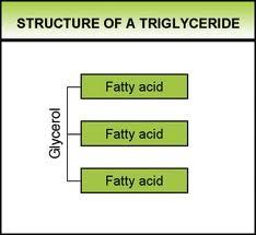
- TRYGLICERIDES (used as
ENERGY STORAGE molecules)
- 1 GLYCEROL & 3 FATTY ACIDS
- Fatty acids have long
tails made of
hydrocarbons. The
TAILS are
HYDROPHOBIC (repel
water) this makes tails
insoluble in water. The
HEADS are
HYDROPHILIC
- Tryglycerides are formed by
CONDENSATION REACTIONS -
when the water is released an
ESTER BOND IS PRODUCED
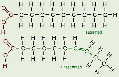
- There are 2 different types of fatty
acids, UNSATURATED & SATURATED -
this depends on the 'tails'
- SATURATED fatty acids DONT
have DOUBLE BONDS between
carbon atoms
- UNSATURATED fatty acids have
atlleast one DOUBLE BONDS
between carbon atoms
- SATURATED fatty acids DONT
have DOUBLE BONDS between
carbon atoms
- Tryglycerides are formed by
CONDENSATION REACTIONS -
when the water is released an
ESTER BOND IS PRODUCED
- Fatty acids have long
tails made of
hydrocarbons. The
TAILS are
HYDROPHOBIC (repel
water) this makes tails
insoluble in water. The
HEADS are
HYDROPHILIC
- The long chain of
hydrocarbon tails contain
lots of chemical energy. -
lots of energy is released
when they break down
because of this lipids
contain twice the amount of
energy than carbohydrates
- theyre INSOLUBLE so DONT
effect WATER POTENTIAL & cause
water to enter by osmosis.
- theyre INSOLUBLE so DONT
effect WATER POTENTIAL & cause
water to enter by osmosis.
- 1 GLYCEROL & 3 FATTY ACIDS
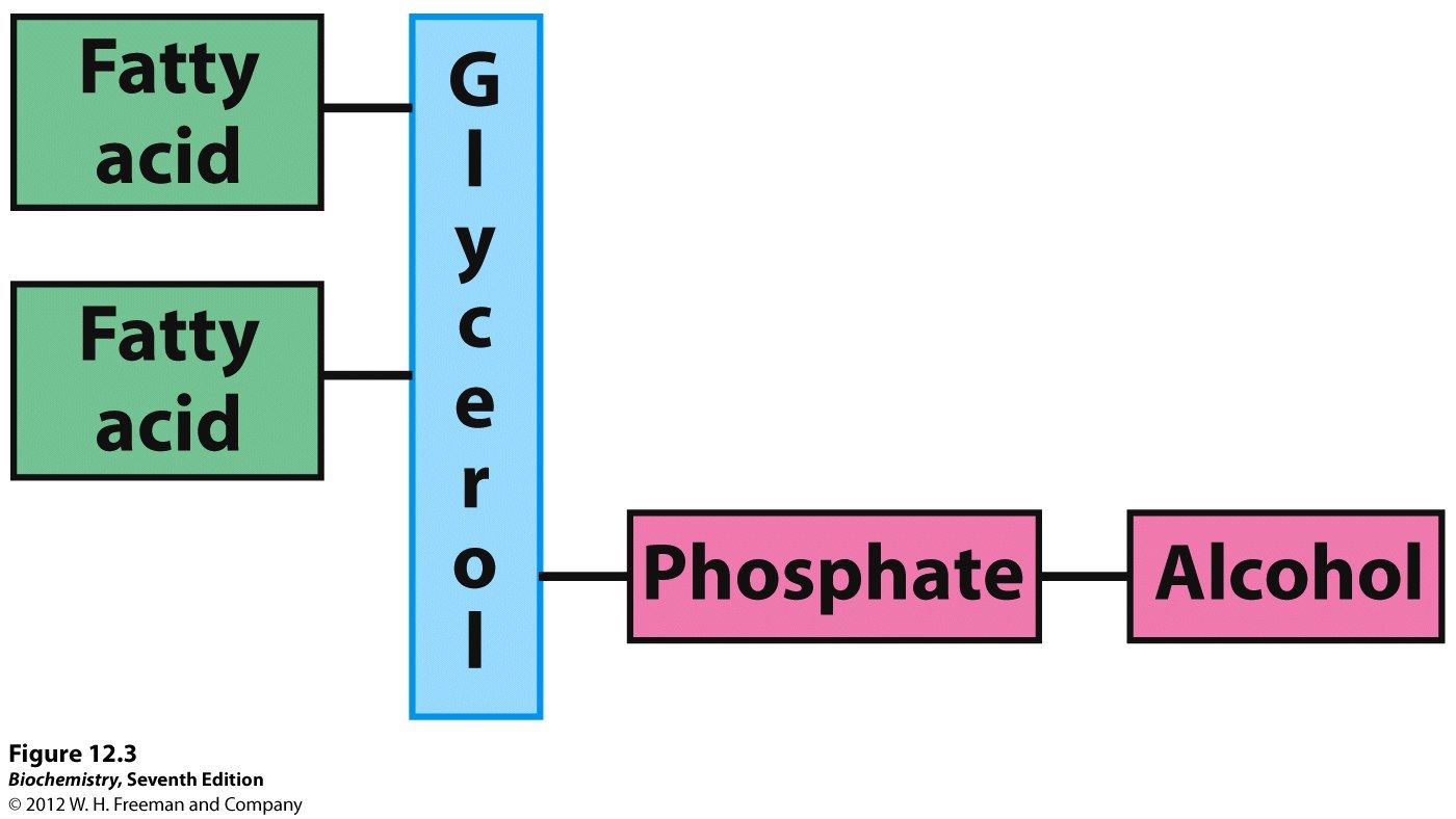
- PHOSPHOLIPIDS - make up the
bilayer of the cell membrane
(Controls what goes in & out)
- Found in the cell membrane
- 1 GLYCEROL,, 2 FATTY ACIDS
& 1 PHOSPHATE GROUP
- The phosphate group is HYDROPHILIC and
the fatty acids tails are HYDROPHOBIC
- The phosphate group is HYDROPHILIC and
the fatty acids tails are HYDROPHOBIC
- Form a double layer because
the hydrophilic heads face out
and the hydrophobic tails in
- Water soluble substances can't
easily pass through, so the
membrane acts as a barrier
- Water soluble substances can't
easily pass through, so the
membrane acts as a barrier
- Found in the cell membrane
- TRYGLICERIDES (used as
ENERGY STORAGE molecules)
- Proteins
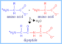
- The monomer of
protein is amino acids
- A dipeptide is formed when
two amino acids join together
- A polypeptide is formed when two
or more amino acids join together
- Proteins are made up of
one or more polypeptides
- Proteins are made up of
one or more polypeptides
- A polypeptide is formed when two
or more amino acids join together
- A dipeptide is formed when
two amino acids join together
- Amino acids have the same
general strucure. A
CARBOXYLL GROUP -COOH,
AMINO GROUP NH2 & a
Carbon containing R GROUP
- All living things share a
bank of only 20 amino
acids, the only difference
between them is whats
in the R group
- All living things share a
bank of only 20 amino
acids, the only difference
between them is whats
in the R group
- Amino acids are linked
together by condensation
reactions to form
polypeptides. A molecule of
water is released. The
bonds between are called
PEPTIDE BONDS
- 4 structural levels
- PRIMARY STRUCTURE -
sequence of AMINO ACIDS
in a POLYPEPTIDE CHAIN
- SECONDARY STRUCTURE -
Hydrogen bonds are formed
between the amino acid in
the chain. This makes the
structure COIL into an
ALPHA HELIX or FOLD into a
BETA PLEATED SHEET
- TERTIARY STRUCTURE - coiled of
folder chain is often COILED OR
FOLDED FURTHER. more bonds are
formed including HYDROGEN BONDS
AND IONIC BONDS. DISULFINE BONDS
are also formed where two amino
acids CYSTIENE come cllose. Proteins
with a single polypeptide chain, this is
often THE FINAL 3D STRUCTURE
- QUARTANERY STRUCTURE -
some proteins are made of
many polypeptide chains held
together., the quartenary
structure is the way these
polypeptide chains are arranged.
- PRIMARY STRUCTURE -
sequence of AMINO ACIDS
in a POLYPEPTIDE CHAIN
- Functions
- ENZYMES - roughly sherical due to the tight folding of
polypeptide chains. Theyre soluble and often have a role
in metabolism (e.g some enzymes break down large
food molecules, others help to make large molecules)
- ANTIBODIES - involved in immune response. made of two
short and two long polypeptide bonds joined together.
- TRANSPORT PROTEINS - (eg channel proteins in the cell
membrane) channel proteins contain hydrophobic and
hydrophilic amino acids which cause proteins to fold up and
cause a channel
- STRUCTUAL PROTEINS - Physically strong. Consists
of long polypeptide chains lying parallel to eachother.
these include kollagen (found in connective tisue) and
keratin found in hair and nails
- ENZYMES - roughly sherical due to the tight folding of
polypeptide chains. Theyre soluble and often have a role
in metabolism (e.g some enzymes break down large
food molecules, others help to make large molecules)
- Biuiret test for
proteins
- 1) The test solution needs to be
alkaline & so first need to add
sodium hydroxide solution
- 2) You then add a few drops of copper sulfate
solution, if protein IS PRESENT it will turn PURPLE. If
protein ISN'T PRESENT the solution will STAY BLUE
- 1) The test solution needs to be
alkaline & so first need to add
sodium hydroxide solution
- The monomer of
protein is amino acids
- Enzyme Action
- BIOLOGICAL
CATALYSTS THAT
SPEEDS UP A
REACTION WITHOUT
BEING USED UP
- Enzymes catalyse
metabolic reactions, at
cellular level (respiration)
and for the organism at
a whole level (eg
digestion)
- Enzymes can effect
STRUCTURE of an organism
(eg production of collagen) and
FUNCTION (eg respiration)
- Enzyme action can be
INTERCELLULAR (inside the
cell) or EXTRACELLULAR
(outside the cell) ENZYMES
ARE PROTEINS
- HIGHLY SPECIFIC DUE TO
TERTIARY STRUCTURE
- Enxymes have an ACTIVE
SITE which has a specific
shape. The active site is part
of the enzyme where the
substrate binds to
- ACTIVATION ENERGY - the
amount f energy that
needs to be suplied for the
reaction to start - often
provided as heat
- ENZYMES LOWER ACTIVATION
ENERG, this means reactions
can take place at a lower
temperature, this speeding up
the rate of reaction
- when a substrate fits an active
site it forms an
enzyme-substate complex. If
two substartes need to be
JOINED the enzyme holds them
together so they can bond more
easily
- If the enzyme is catalysing
a breakdown, fittin into the
active site puts a strain on
the bonds so break up
more easily
- If the enzyme is catalysing
a breakdown, fittin into the
active site puts a strain on
the bonds so break up
more easily
- when a substrate fits an active
site it forms an
enzyme-substate complex. If
two substartes need to be
JOINED the enzyme holds them
together so they can bond more
easily
- ENZYMES LOWER ACTIVATION
ENERG, this means reactions
can take place at a lower
temperature, this speeding up
the rate of reaction
- Lock & Key
method
- Enzymes only work with
substrates that fit their active
sites, scietists came up with
the lock & key model. where
the substrate fits into the
enzyme the same way as a
key
- New evidence shows that the
enzyme-substrate complex
changes shape slightly to
complete the fit., scientists then
came up with the induce fit
model
- The INDUCED FIT MODEL explains
why enzymes are so specific. to
substrates. Substrates don't only
have to be the right shape to fit
into the active site, the active site
also has to change shape aswell
- The INDUCED FIT MODEL explains
why enzymes are so specific. to
substrates. Substrates don't only
have to be the right shape to fit
into the active site, the active site
also has to change shape aswell
- Enzymes only work with
substrates that fit their active
sites, scietists came up with
the lock & key model. where
the substrate fits into the
enzyme the same way as a
key
- Enzymes have
TERTIARY
STRUCTURES
- 1) Enzymes are very specific, they often only
catalyse one reaction eg sucrase only breas down
sucrase this is because only one complementory
substrate will fit
- 2) The active sites shape is determined by its tertiary
structure which is dtermined by its primary structure. Each
enzyme has a different tertiary stucture, so a different active
site.. If a substrate doesnt match the active site, the substance
wont be formed and reacton wont be cataylsed
- 3) If the tertiary stucture is changed the
shape of the active site will change, This
means substrates wont fit and the
enzyme will no longer be able to carry out
its function
- 4) Tertiary structure can be changed to to pH
or temperature. The primary structure is
determined by a gene, if a mutation occurs it
could change the tertiary stucture of the
protein produced
- 4) Tertiary structure can be changed to to pH
or temperature. The primary structure is
determined by a gene, if a mutation occurs it
could change the tertiary stucture of the
protein produced
- 3) If the tertiary stucture is changed the
shape of the active site will change, This
means substrates wont fit and the
enzyme will no longer be able to carry out
its function
- 2) The active sites shape is determined by its tertiary
structure which is dtermined by its primary structure. Each
enzyme has a different tertiary stucture, so a different active
site.. If a substrate doesnt match the active site, the substance
wont be formed and reacton wont be cataylsed
- 1) Enzymes are very specific, they often only
catalyse one reaction eg sucrase only breas down
sucrase this is because only one complementory
substrate will fit
- BIOLOGICAL
CATALYSTS THAT
SPEEDS UP A
REACTION WITHOUT
BEING USED UP
- Factors effecting
enzyme activity
- Temperature
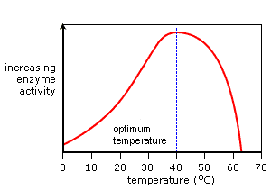
- The ROR INCREASES when TEMP INCREASES.
MORE HEAT = MORE KINETIC ENERGY, so
molecules move faster. this makes enzymes
MORE LIKELY to collide with molecules.
- If the TEMP gets TOO HIGH the reaction STOPS. This is because the rise in temp
makes enzymes VIBRATE MORE.. If the temperature gets too high the vibrations
BREAK BONDS that hold the enzyme together. The ACTIVE SITE SHAPE CHANES
and substrates can no longer fit. The enzyme is then DENATURED
- The ROR INCREASES when TEMP INCREASES.
MORE HEAT = MORE KINETIC ENERGY, so
molecules move faster. this makes enzymes
MORE LIKELY to collide with molecules.
- pH
- All enzymes have an optimum pH,
most work best at 7 (neutral) nut
some such as pepsin found in the
stomach prefer acidic conditions -
pH 2.. Above & below the optimum
pH the hydrogen and hydroxide
found in acids and alkalies break
the ionic & hydrogen bonds that
hold the tertiary structure in place.
This denatures the enzyme
- Enzyme
concentration
- The MORE ENZYMES there is
in a solution, the MORE LIKELY
A SUBSTRATE WILL COLLIDE,
so and INCREASED
CONCENTRATION INCREASES
ROR
- But, if the SUBSTARTE IS
LIMITED, there becomes a
point that no matter the
amount of enzymes there are
no reactions avaliable and so
no further effects
- But, if the SUBSTARTE IS
LIMITED, there becomes a
point that no matter the
amount of enzymes there are
no reactions avaliable and so
no further effects
- The MORE ENZYMES there is
in a solution, the MORE LIKELY
A SUBSTRATE WILL COLLIDE,
so and INCREASED
CONCENTRATION INCREASES
ROR
- Substrate
concentration
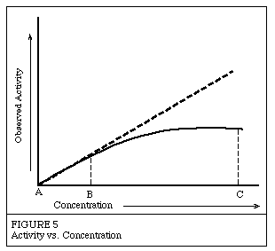
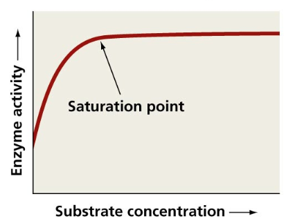
- The HIGHER the SUBSTRATE
CONC the HIGHER the ROR. This
means more substartes will
collide with enzymes active sites
more often. This is only true up
to the point of SATURATION
where by the active sites of
enzymes are all used up sp
increasing concentration wont
make a difference
- substrate concentration
decreases within time
unless more substrates are
added. so if no variables are
changed the ROR will
decrease.
- substrate concentration
decreases within time
unless more substrates are
added. so if no variables are
changed the ROR will
decrease.
- The HIGHER the SUBSTRATE
CONC the HIGHER the ROR. This
means more substartes will
collide with enzymes active sites
more often. This is only true up
to the point of SATURATION
where by the active sites of
enzymes are all used up sp
increasing concentration wont
make a difference
- Competitive
Inhibitors
- 1) Competitive inhibitors have
similar shapes to that of the
substrate, they compete with the
substrate molecules to bind to the
active site but NO REACTION
- 2) They BLOCK the active site so NO substrate
molecule can fit. If theres A HIGH CONC of INHIBITORS
it'll take up nearly all the active sites & hardly any of
the substrates will get to the enzyme. But if theres a
HIGHER CONC of SUBSTRATE then the chnaces of
getting to an active site before inhibitors increase
- 1) Competitive inhibitors have
similar shapes to that of the
substrate, they compete with the
substrate molecules to bind to the
active site but NO REACTION
- Non-Competitive
Inhibitors
- 1) Non competitive inhibitors BIND TO
ENZYMES AWAY FROM THEIR ACTIVE
SITES. This causes the ACTIVE SITE TO
CHANGE SHAPE so molecules can no
longer bind to it
- 2) They don't COMPETE with the
substate molecule, to bind to the
active site because they are a
different shape. INCREASING
SUBSTRATES WONT INCREASE ROR
as enzymes will still be inhibited
- 2) They don't COMPETE with the
substate molecule, to bind to the
active site because they are a
different shape. INCREASING
SUBSTRATES WONT INCREASE ROR
as enzymes will still be inhibited
- 1) Non competitive inhibitors BIND TO
ENZYMES AWAY FROM THEIR ACTIVE
SITES. This causes the ACTIVE SITE TO
CHANGE SHAPE so molecules can no
longer bind to it
- Temperature
Media attachments
Want to create your own Mind Maps for free with GoConqr? Learn more.