4442505
Description
Mind Map by Michelle Lamb, updated more than 1 year ago
|
|
Created by Michelle Lamb
over 8 years ago
|
|
Chapter 3: The
Biological Bases of
Behaviour (map 1 of 2)
- Communication in the Nervous System
- Nervous Tissue: The Basic
Hardware
- Neurons
Annotations:
- -Neurons are individual cells in the nervous system that receive, integrate, and transmit information. -Vast majority of them communicate only with other neurons. -A small minority receive signals from outside the nervous system(from sensory organs) or carry messages from the nervous system to muscles that move the body.
- Come in tremendous
variety of types and
shapes
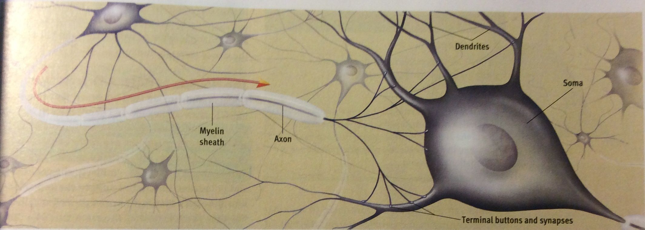
- Parts of a Neuron
- Dendritic Tree(Dendrites)
Annotations:
- -Dendrite is a Greek word for tree. -Each individual branch is a dendrite -dendrites are the parts of a neuron that are specialized to receive information. -most neurons receive information from many (thousands)of others.
- Soma (cell body)
Annotations:
- Contains the cell nucleus and much of the chemical machinery common to most cells.
- Axon
Annotations:
- - From Greek for axle. -The axon is a long, thin fibre that transmits signals away from the soma to other neurons or muscles or glands. -Axons can be over a meter long.
- Myelin Sheath
Annotations:
- -The myelin sheath is insulating material, derived from glial cells, that encases some axons. - myelin is a high concentration of a white fatty substance -Myelin sheath speeds up the transmission of signals that move along the axon. if an axon's myelin sheath deteriorates, its signal may not be transmitted effectively, ie Multiple Sclerosis.
- Terminal Buttons
Annotations:
- -The axon ends in a cluster of terminal buttons, which are small knobs that secrete chemicals called neurotransmitters. these chemicals serve as messengers that may activate neighbouring neurons.
- Synapse
Annotations:
- - A synapse is a junction where information is transmitted from one neuron to another. -synapse is Greek for junction.
- Picture below is from pg 81 of psychology
themes and variations(4th edition) showing the
structure of a neuron
- Dendritic Tree(Dendrites)
- Parts of a Neuron
- Glia
- Found throughout the nervous
system
- Provide support for neurons
- Glia literally means glue
- glia cells supply
nourishment to, remove
waste products from
neurons
- Glia cells play a role inthe
development of the nervous
system in the human embryo
- New research suggests that
glia may also send & receive
chemical signals
- New research suggests that
glia may also send & receive
chemical signals
- Glia cells play a role inthe
development of the nervous
system in the human embryo
- glia cells supply
nourishment to, remove
waste products from
neurons
- Glia literally means glue
- Provide support for neurons
- Found throughout the nervous
system
- The Neural Impulse
- Neuron at rest
- The cell membrane is
semipermeable, permitting
movement of some ions.
- The resting potential of a neuron is its
stable, negative charge when the cell
is inactive. About 270 millivolts.
- The resting potential of a neuron is its
stable, negative charge when the cell
is inactive. About 270 millivolts.
- The cell membrane is
semipermeable, permitting
movement of some ions.
- Action Potential
Annotations:
- -Action Potential is a very brief shift in a neuron's electrical charge that travels along an axon.
- When the neuron is stimulated,
channels in its cell membrane open,
briefly allowing postively charged
sodium ions to rush in, creating an
action potential.
- Voltage change races down the axon
- After firing the channels in the membrane close
up.Some time is needed before they are ready to
open again, until that time the neuron cannot fire
again. (absolute refractory period)
Annotations:
- -Absolute refractory period is the minimum length of time after an action potential during which another action potential cannot begin. -Only 1 or 2 milliseconds.
- During the Relative Refractory Period. the neuron can
fire, but its threshold for firing is elevated, so more
intense stimulation is required to initiate an action
potential.
- All or None Law
Annotations:
- -Neurons fire or they don't, all action potentials are all the same size, that is weaker stimuli do not produce smaller action potentials -neurons can convey information about the strength of a stimulus. -In general stronger stimulus will cause a cell to fire a more rapid volley of neural impulses than a weaker stimulus will.
- All or None Law
- After firing the channels in the membrane close
up.Some time is needed before they are ready to
open again, until that time the neuron cannot fire
again. (absolute refractory period)
- Voltage change races down the axon
- Neuron at rest
- The Synapse
- Depends on chemical messengers
- Sending Signals
- Neurons don't actually touch.
They are seperated by the
Synaptic Cleft.
Annotations:
- -The synaptic Cleft is, a microscopic gap between the terminal button of one neuron and the cell membrane of another neuron. -signals have to cross this gap to permit neurons to communicate.
- Presynaptic neuron sends
the signal across the gap
- Postsynaptic neuron receives
the signal
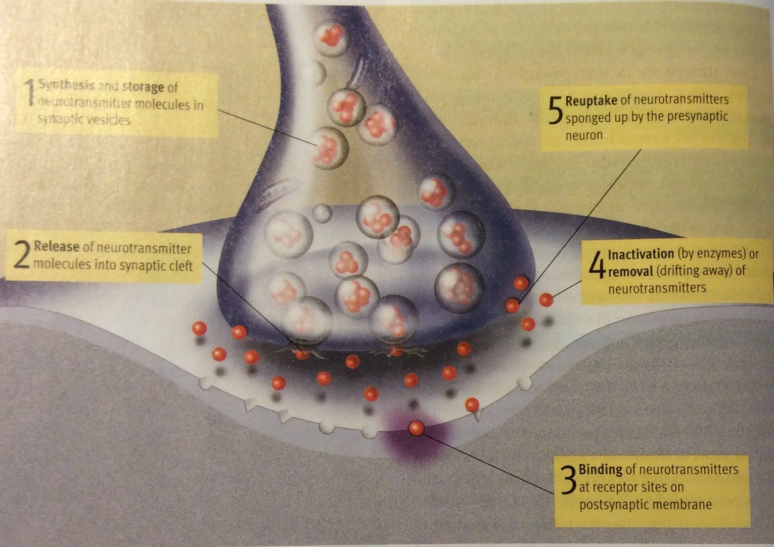
- Arrival of the action potential at the terminal
buttons triggers the release of Neurotransmitters.
Annotations:
- -Neurotransmitters are chemicals that transmit information from one neuron to another. -With in the buttons most of these chemicals are stored in small sacs called the Synaptic vesicles. -Neurotransmitters are released when a vesicle fuses with the membrane of the presynaptic cell and the contents spill into the synaptic cleft.
- Neurotransmitters may bind with special molecules in
the postsynaptic cell membrane at various receptor
sites.These sites are specifically tuned to respond to
some , but not other neurotransmitters.
- Picture below is from pg 83 of psychology themes and
variations(4th edition) showing the synapse
- Picture below is from pg 83 of psychology themes and
variations(4th edition) showing the synapse
- Neurons don't actually touch.
They are seperated by the
Synaptic Cleft.
- Receiving Signals
- When a neurotransmitter and a
receptor molecule combine,reactions
in the cell membrane cause a
postsynaptic potential (PSP)
Annotations:
- -Postsynaptic potential (PSP), a voltage change at a receptor site on a postsynaptic cell membrane. -PSP do NOT follow the all or none law.
- PSP are graded
Annotations:
- -They vary in size and they increase or decrease the probability of a neural impluse in the receiving cell in proportion to the amount of voltage change.
- 2 types of messages can be sent
from cell to cell
- Excitatory PSP
- increases the likelihood
that the postsynaptic
neuron will fire action
potentials
- Reuptake
Annotations:
- - Reuptake is a process in which neurotransmitters are sponged up from the synaptic clef by the presynaptic membrane. - this allows for synapses to recycle their materials.
- picture below is taken from pg84 od psychology themes and
variations (4th edition) showingsynaptic transmission
- Reuptake
- increases the likelihood
that the postsynaptic
neuron will fire action
potentials
- Inhibitory PSP
- decreases the
likelihood that the
postsynaptic
neuron will fire
action potentials
- Reuptake
- Reuptake
- decreases the
likelihood that the
postsynaptic
neuron will fire
action potentials
- Excitatory PSP
- When a neurotransmitter and a
receptor molecule combine,reactions
in the cell membrane cause a
postsynaptic potential (PSP)
- Integrating Signals
- Neurons must integrate signals
arriving at many synapses before
it "decides" to fire a neural
impulse.
- Elimination of old synapses appears to
play a larger role in the sculpting of
neural networks than the creation of
new synapses.
- Synaptic pruning is a key process in the
formation of the neural networks that
are crucial to communication in the
nervous system.
- Repeated synaptic activity
leads to a stregthening of the
synapse
- Repeated synaptic activity
leads to a stregthening of the
synapse
- Synaptic pruning is a key process in the
formation of the neural networks that
are crucial to communication in the
nervous system.
- Elimination of old synapses appears to
play a larger role in the sculpting of
neural networks than the creation of
new synapses.
- Neurons must integrate signals
arriving at many synapses before
it "decides" to fire a neural
impulse.
- Sending Signals
- Neurotransmitters & Behaviour
- Neurotransmitters are fundamental to
behaviour, playing a key role in everything from
muscle movements to moods and mental health.
- Agonist is a chemical that MIMICS the
action of a neurotransmitter
- Antagonist is a chemical that OPPOSES
the action of a neurotransmitter
Annotations:
- -Key slides in the lock, but it doesn't work -temporarily block the action of the natural transmitter ny occupying its receptor sites rendering them unusable
- Agonist is a chemical that MIMICS the
action of a neurotransmitter
- Monoamines
- Dopamine (DA)
- used by neurons that control
voluntary movements
Annotations:
- -degeneration of such neurons in a specific area of the brain causes PARKINSON's
- over activity at DA synapses associated with
schizophrenia
- Cocaine and amphetamines elevate activity at DA
synapses
- Cocaine and amphetamines elevate activity at DA
synapses
- used by neurons that control
voluntary movements
- Norepinephrine (NE)
- Contributes to modulation of
mood and arousal
- Cocaine and amphetamines elevate activity at NE
synapses
- Cocaine and amphetamines elevate activity at NE
synapses
- Contributes to modulation of
mood and arousal
- Serotonin
- Play a prominent role in the regulation of sleep
& wakefulness, eating, and aggression
- Abnormal levels may contribute to
depression and OCD
- Antidepressant drugs affect serotonin
circuits
- Antidepressant drugs affect serotonin
circuits
- Abnormal levels may contribute to
depression and OCD
- Play a prominent role in the regulation of sleep
& wakefulness, eating, and aggression
- Dopamine (DA)
- Acetylcholine (ACh)
- Only transmitter
between motor neurons
and voluntary muscles
- Appears to contribute to
attention,arousal and memory
- Some ACh receptors stimulated by
nicotine
- Some ACh receptors stimulated by
nicotine
- Appears to contribute to
attention,arousal and memory
- Only transmitter
between motor neurons
and voluntary muscles
- Amino Acids
- Glutamate
- Has both excitatory and inhibitory
effects
- Implicated in learning and memory
- Implicated in learning and memory
- Has both excitatory and inhibitory
effects
- Gamma-aminobutyric Acid (GABA)
- Serves as widely distributed inhibitory
transmitter
- Antianxiety drugs work at GABA synapses
- Antianxiety drugs work at GABA synapses
- Serves as widely distributed inhibitory
transmitter
- Glutamate
- Endorphins
- Contribute to pain relief & perhaps to some
pleasurable emotions
- Resemble opiate drugs in structure and effects
- Resemble opiate drugs in structure and effects
- Contribute to pain relief & perhaps to some
pleasurable emotions
- Neurotransmitters are fundamental to
behaviour, playing a key role in everything from
muscle movements to moods and mental health.
- Depends on chemical messengers
- Neurons
- Nervous Tissue: The Basic
Hardware
- Organization of the Nervous System
- Central Nervous System (CNS)
- Consists of the brain and the spinal cord
- Brain
- Protected by enclosing sheaths called meninges. Also bathed in
cerebrospinal fluid (CSF)
- Protected by enclosing sheaths called meninges. Also bathed in
cerebrospinal fluid (CSF)
- Spinal Cord
- Connects the brain to the rest of the
body through the peripheral nervous
system
- Connects the brain to the rest of the
body through the peripheral nervous
system
- Brain
- Consists of the brain and the spinal cord
- Peripheral Nervous System
Annotations:
- The peripheral nervous system ia made up of all those nerves that lie outside the the brain and spinal cord. Nerves are bundles of neuron fibres (axons) that are routed together in the peripheral nervous system.
- Somatic Nervous System
- made up of nerves that connect to
voluntary skeletal muscles and to
sensory receptors
- Has afferent nerve fibres that carry information
inward to the CNS.Efferent nerve fibres carry
information outward from the CNS
- Has afferent nerve fibres that carry information
inward to the CNS.Efferent nerve fibres carry
information outward from the CNS
- made up of nerves that connect to
voluntary skeletal muscles and to
sensory receptors
- Autonomic Nervous System (ANS)
- made up of nerves that connect to the heart, blood
vessels, smooth muscles and glands.
- is a separate autonomous system that controls things
like heart rate, digestion etc.
- Fight or Flight response
- ANS has two branches
- Sympathetic division
mobilizes the body's
resources for emergencies (go!!!)
- Parasympathetic
division generally
conserves bodily
resources (stop)
- Sympathetic division
mobilizes the body's
resources for emergencies (go!!!)
- ANS has two branches
- Fight or Flight response
- is a separate autonomous system that controls things
like heart rate, digestion etc.
- made up of nerves that connect to the heart, blood
vessels, smooth muscles and glands.
- Central Nervous System (CNS)
- Looking Inside the Brain; Research Methods
- geography or structure of the brain can
be mapped by examining brains that
have been removed
- mapping brain function requires a
working brain
- Neuroscientists conduct research on
the brain and other parts of the
nervous system.
- Neuroscientists conduct research on
the brain and other parts of the
nervous system.
- mapping brain function requires a
working brain
- Electrical Recordings
- Electroncephalograph (EEG)
- Is a device that monitors the electrical activity of the brain over
time by means of recording electrodes attached to the surface of
the scalp.
- EEG recordings are translated into line tracings
commonly called brain waves.
- EEG is often used in the clinical diagnosis of brain
damage and neurological disorders.
- EEG is often used in the clinical diagnosis of brain
damage and neurological disorders.
- EEG recordings are translated into line tracings
commonly called brain waves.
- Is a device that monitors the electrical activity of the brain over
time by means of recording electrodes attached to the surface of
the scalp.
- Electroncephalograph (EEG)
- Lesioning
- Used to study the relationship between brain and
behaviour more precisely, scientists sometimes observe
what happens when specific brain structures in animals
are purposely disabled.
- Lesioning involves destroying a piece of
the brain.
- Typically done by inserting an electrode into a brain
structure and passing a high frequency electric current
through it to burn the tissue and disable the structure.
- Typically done by inserting an electrode into a brain
structure and passing a high frequency electric current
through it to burn the tissue and disable the structure.
- Lesioning involves destroying a piece of
the brain.
- Used to study the relationship between brain and
behaviour more precisely, scientists sometimes observe
what happens when specific brain structures in animals
are purposely disabled.
- Electrical Stimulation
of the Brain (ESB)
- Involves sending a weak electric
current into a brain structure to
stimulate (activate) it.
- Has lead to advances in the
understanding of many aspects of
brain-behaviour relationships.
- Has lead to advances in the
understanding of many aspects of
brain-behaviour relationships.
- Involves sending a weak electric
current into a brain structure to
stimulate (activate) it.
- Transcranial Magnetic Stimulation (TMS)
- Noninvasive and permits scientists to temporarily
enhance or depress activity in a specific area of the
brain.
- Chief limitation is that it cannot be used to
study areas deep within the brain.
- By varying the timing and duration of the magnetic pulses, a
researcher can either increase or decrease the excitability of
neurons in the local tissue
- By varying the timing and duration of the magnetic pulses, a
researcher can either increase or decrease the excitability of
neurons in the local tissue
- Chief limitation is that it cannot be used to
study areas deep within the brain.
- Noninvasive and permits scientists to temporarily
enhance or depress activity in a specific area of the
brain.
- Brain Imaging Procedures
- Computerized tomography (CT) scan
- Is a computer enhanced X-ray of brain
structure
- Is a vivid image of a horizontal slice of the brain.
- Is a vivid image of a horizontal slice of the brain.
- Is a computer enhanced X-ray of brain
structure
- Positron Emission Tomography (PET)
- Can examine brain function, mapping
actual activity in the brain over time.
- Uses radioactively tagged chemicals introduced into the brain.
which can be monitored with X-rays.
- Uses radioactively tagged chemicals introduced into the brain.
which can be monitored with X-rays.
- Can examine brain function, mapping
actual activity in the brain over time.
- Magnetic Resonance Imaging (MRI) Scan
- uses magnetic fields, radio waves and computerized
enhancement to map brain structure
- Provides better images of brain structure than CT scan
- insightful about depressive disorders
- Functional Magnetic Resonance Imaging (fMRI)
- New variation on the MRI that monitors blood flow and oxygen in the brain
to identify areas of high activity. It can map actual activity in the brain over
time with greater precision
- New variation on the MRI that monitors blood flow and oxygen in the brain
to identify areas of high activity. It can map actual activity in the brain over
time with greater precision
- Functional Magnetic Resonance Imaging (fMRI)
- insightful about depressive disorders
- Provides better images of brain structure than CT scan
- uses magnetic fields, radio waves and computerized
enhancement to map brain structure
- Computerized tomography (CT) scan
- geography or structure of the brain can
be mapped by examining brains that
have been removed
Media attachments
Want to create your own Mind Maps for free with GoConqr? Learn more.