
Mind map
Salivary Gland Pathology
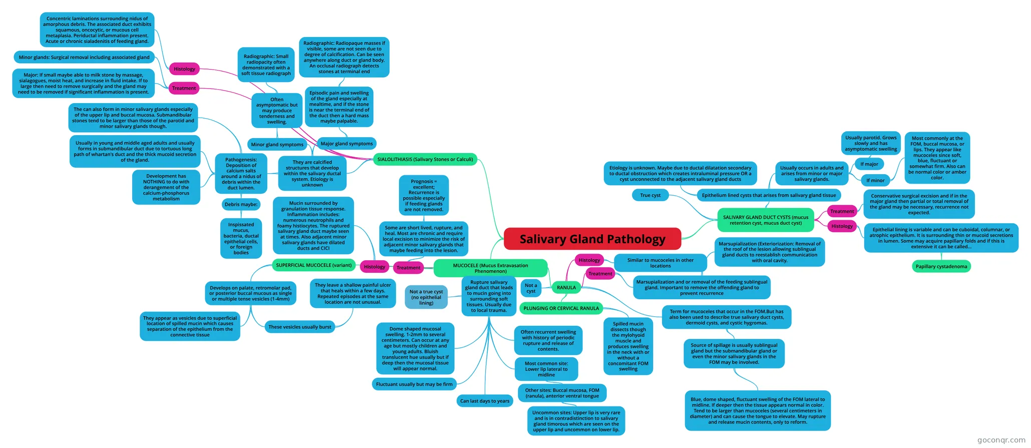
Mind map: Salivary gland pathology
Map content
Salivary Gland Pathology
MUCOCELE (Mucus Extravasation Phenomenon)
- Rupture salivary gland duct that leads to mucin going into surrounding soft tissues. Usually due to local trauma.
- Not a true cyst (no epithelial lining)
- Dome shaped mucosal swelling. 1-2mm to several centimeters. Can occur at any age but mostly children and young adults. Bluish translucent hue usually but if deep then the mucosal tissue will appear normal.
- Fluctuant usually but may be firm
- Can last days to years
- Often recurrent swelling with history of periodic rupture and release of contents.
- Most common site: Lower lip lateral to midline
- Other sites: Buccal mucosa, FOM (ranula), anterior ventral tongue
- Uncommon sites: Upper lip is very rare and is in contradistinction to salivary gland timorous which are seen on the upper lip and uncommon on lower lip.
- Other sites: Buccal mucosa, FOM (ranula), anterior ventral tongue
- SUPERFICIAL MUCOCELE (variant)
- Develops on palate, retromolar pad, or posterior buccal mucous as single or multiple tense vesicles (1-4mm)
- These vesicles usually burst
- They leave a shallow painful ulcer that heals within a few days. Repeated episodes at the same location are not unusual.
- They appear as vesicles due to superficial location of spilled mucin which causes separation of the epithelium from the connective tissue
- These vesicles usually burst
- Develops on palate, retromolar pad, or posterior buccal mucous as single or multiple tense vesicles (1-4mm)
- Histology
- Mucin surrounded by granulation tissue response. Inflammation includes: numerous neutrophils and foamy histiocytes. The ruptured salivary gland duct maybe seen at times. Also adjacent minor salivary glands have dilated ducts and CICI
- Treatment
- Some are short lived, rupture, and heal. Most are chronic and require local excision to minimize the risk of adjacent minor salivary glands that maybe feeding into the lesion.
- Prognosis = excellent; Recurrence is possible especially if feeding glands are not removed.
- Some are short lived, rupture, and heal. Most are chronic and require local excision to minimize the risk of adjacent minor salivary glands that maybe feeding into the lesion.
- RANULA
- Term for mucoceles that occur in the FOM.But has also been used to describe true salivary duct cysts, dermoid cysts, and cystic hygromas.
- Source of spillage is usually sublingual gland but the submandibular gland or even the minor salivary glands in the FOM may be involved.
- Blue, dome shaped, fluctuant swelling of the FOM lateral to midline. If deeper then the tissue appears normal in color. Tend to be larger than mucoceles (several centimeters in diameter) and can cause the tongue to elevate. May rupture and release mucin contents, only to reform.
- Source of spillage is usually sublingual gland but the submandibular gland or even the minor salivary glands in the FOM may be involved.
- Histology
- Similar to mucoceles in other locations
- Treatment
- Marsupialization and or removal of the feeding sublingual gland. Important to remove the offending gland to prevent recurrence
- Marsupialization (Exteriorization: Removal of the roof of the lesion allowing sublingual gland ducts to reestablish communication with oral cavity.
- Marsupialization and or removal of the feeding sublingual gland. Important to remove the offending gland to prevent recurrence
- PLUNGING OR CERVICAL RANULA
- Spilled mucin dissects though the mylohyoid muscle and produces swelling in the neck with or without a concomitant FOM swelling
- Not a cyst
- Term for mucoceles that occur in the FOM.But has also been used to describe true salivary duct cysts, dermoid cysts, and cystic hygromas.
- Rupture salivary gland duct that leads to mucin going into surrounding soft tissues. Usually due to local trauma.
SALIVARY GLAND DUCT CYSTS (mucus retention cyst, mucus duct cyst)
- True cyst
- Epithelium lined cysts that arises from salivary gland tissue
- Etiology is unknown. Maybe due to ductal dilatation secondary to ductal obstruction which creates intraluminal pressure OR a cyst unconnected to the adjacent salivary gland ducts
- Usually occurs in adults and arises from minor or major salivary glands.
- If minor
- Most commonly at the FOM, buccal mucosa, or lips. They appear like mucoceles since soft, blue, fluctuant or somewhat firm. Also can be normal color or amber color.
- If major
- Usually parotid. Grows slowly and has asymptomatic swelling
- If minor
- Treatment
- Conservative surgical excision and if in the major gland then partial or total removal of the gland may be necessary, recurrence not expected.
- Histology
- Epithelial lining is variable and can be cuboidal, columnar, or atrophic epithelium. It is surrounding thin or mucoid secretions in lumen. Some may acquire papillary folds and if this is extensive it can be called...
- Papillary cystadenoma
- Epithelial lining is variable and can be cuboidal, columnar, or atrophic epithelium. It is surrounding thin or mucoid secretions in lumen. Some may acquire papillary folds and if this is extensive it can be called...
SIALOLITHIASIS (Salivary Stones or Calculi)
- They are calcified structures that develop within the salivary ductal system. Etiology is unknown
- Pathogenesis: Deposition of calcium salts around a nidus of debris within the duct lumen.
- Debris maybe:
- Inspissated mucus, bacteria, ductal epithelial cells, or foreign bodies
- Development has NOTHING to do with derangement of the calcium-phosphorus metabolism
- Usually in young and middle aged adults and usually forms in submandibular duct due to tortuous long path of whartan's duct and the thick mucoid secretion of the gland.
- The can also form in minor salivary glands especially of the upper lip and buccal mucosa. Submandibular stones tend to be larger than those of the parotid and minor salivary glands though.
- Debris maybe:
- Minor gland symptoms
- Often asymptomatic but may produce tenderness and swelling.
- Radiographic: Small radiopacity often demonstrated with a soft tissue radiograph
- Often asymptomatic but may produce tenderness and swelling.
- Major gland symptoms
- Episodic pain and swelling of the gland especially at mealtime, and if the stone is near the terminal end of the duct then a hard mass maybe palpable.
- Radiographic: Radiopaque masses if visible, some are not seen due to degree of calcification. Can be seen anywhere along duct or gland body. An occlusal radiograph detects stones at terminal end
- Episodic pain and swelling of the gland especially at mealtime, and if the stone is near the terminal end of the duct then a hard mass maybe palpable.
- Pathogenesis: Deposition of calcium salts around a nidus of debris within the duct lumen.
- Treatment
- Major: If small maybe able to milk stone by massage, sialagogues, moist heat, and increase in fluid intake. If to large then need to remove surgically and the gland may need to be removed if significant inflammation is present.
- Minor glands: Surgical removal including associated gland
- Histology
- Concentric laminations surrounding nidus of amorphous debris. The associated duct exhibits squamous, oncocytic, or mucous cell metaplasia. Periductal inflammation present. Acute or chronic sialadenitis of feeding gland.
- They are calcified structures that develop within the salivary ductal system. Etiology is unknown
Information
- Description
- Mind Map on Salivary Gland Pathology, created by cbaker43 on 06/02/2016.
- Created by
- cbaker43
- Views
- 0
- Rating