
Mind map
1_Neck and Head
Map content
1_Neck and Head
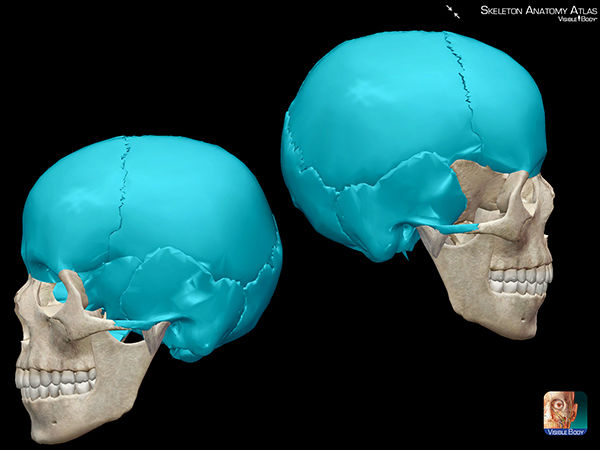
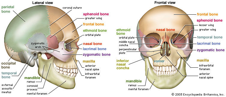
Bones of head= cranium
- Between which is sutures which are fibrous joints
- Flat bones
- Formed by intramembranous ossification from head mesenchyme from neural crest
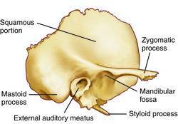
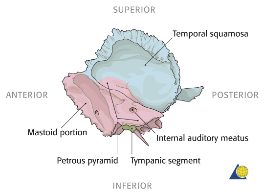
- Temporal bone
- Petrous part
- on the inside
- dense part, surrounds inner ear
- on the inside
- carotid canal
- continuation of foramen lacerum
- jugular fossa is right next to it
- articular tubercle
- bump on zygomatic process
- Groove for sigmoid sinus
- near internal acoustic meatus.
- Stylomastoid foramen
- where facial nerve exits
- Bell's palsy
- where facial nerve exits
- Mastoid process
- mastoid air cells
- Petrous part
- Calvaria= skullcap= mainly flat bones
- Occipital (posterior)
- frontal
- Parietal (on the sides and top back)
- Cranial base made mainly of irregular bones
- Formed by endochondral ossification
- Zygoma
- cheek bone
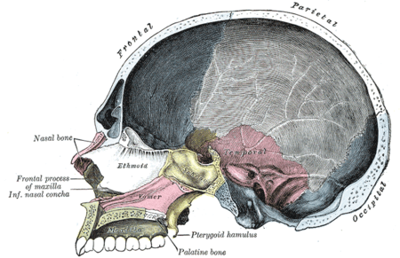
- Nasal bone
- Sphenoid bone
- Just behind zygoma
- and in eye
- inside
- lesser and greater wing
- Has sinuses
- Holes is skull
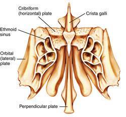
- Ethmoid bone
- Just in front of the zygoma
- More medial than sphenoid
- Ethmoid perpendicular plater
- inside skull is crista galli in cribriform
- olfactory nerves
- inside skull is crista galli in cribriform
- lacrimal bone
- Lacrimal bone
- medial part of eye socket
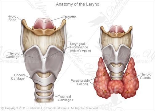
Larynx
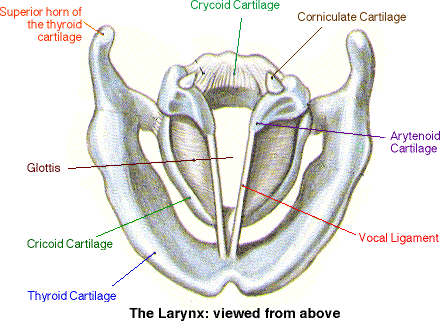
- Thyroid cartilage
- Cricoid cartilage- complete ring of cartilage
- Thyroid
- Cricothyroidotomy
- Insert tube in the gap between cricoid and thyroid cartilage
- Move thyroid out the way
- Tracheostomy
- Between 2nd and 3rd tracheal rings
- Insert tube in the gap between cricoid and thyroid cartilage
- Cricoid cartilage- complete ring of cartilage
- Nerves
- Superior laryngeal nerve
- From vagus nerve
- External branch
- Cricothyroid muscle
- Increases pitch
- Cricothyroid muscle
- Internal branch
- Sensation of larynx
- Cough reflex
- Recurrent laryngeal nerve
- Lateral cricoarytenoid
- Adducts vocal cords- closes to air
- Posterior cricoarytenoid
- abducts vocal cords
- Comes apart to allow air in lungs
- abducts vocal cords
- Interarytenoid
- Adducts vocal cords- air trapped in lungs
- Thyroarytenoid
- Decreases pitch
- Comes from vagus nerve, passes under loop of aorta (left) or brachiocephalic on right
- Lateral cricoarytenoid
- Superior laryngeal nerve
- Arytenoid cartilage
- Hyoid bone
- Pathology
- Cervical spondylosis
- Neck pain caused by age-related wear and tear
- Headaches
- Causes
- spinal discs get worn out
- body compensates by making more bone spurs
- this can cause spine to become too rigid
- Risk factors
- obesity
- athritis
- Treatment
- relieving pain
- NSAIDs
- diclofenac
- codeine
- NSAIDs
- Sit properly
- exercise
- relieving pain
- Pancoasts tumour
- Apical lung tumour
- Invades cervical plexus
- Some sympathetic to pulmonary plexus
- Leads to Horner's syndrome
- Ptosis
- Upper eyelid droops
- Anhydrosis
- No sweating
- Leads to dry skin on face
- No sweating
- Miosis
- Pupil constricted
- Ptosis
- Invades brachial plexus
- Shoulder and arm pain
- Invades recurrent larygeal nerve
- Hoarse voise
- Bovine cough
- Like pushing air out fast
- Not proper cough
- Like pushing air out fast
- Respiratory stridor
- Treat
- Chemo
- Surgery
- Prognosis
- 5 years
- Cervical spondylosis
- Epilottis
- Vallecula
- Thyroid cartilage
Ear
- Semi-circular canals (labyrinth)
- Up
- Down
- Side to side
- Ear drum (tympanic membrane)
- Hammer (malleus)
- Incus (anvil)
- Stirrup (stapes)
- Cochlea- hair cells
- Ossicles connected by synovial joints
- Semi-circular canals (labyrinth)
Triangles of the neck
- Anterior
- Body of mandible
- Midline of neck
- Mastoid process
- Anterior borderSternocleidomastoid
- Suprasternal notch
- Glands: thyroid and submandibular
- Carotid sheath
- vagus
- common carotid
- internal jugular
- In triangle made between the two sternocleidomastoid heads
- deep cervical
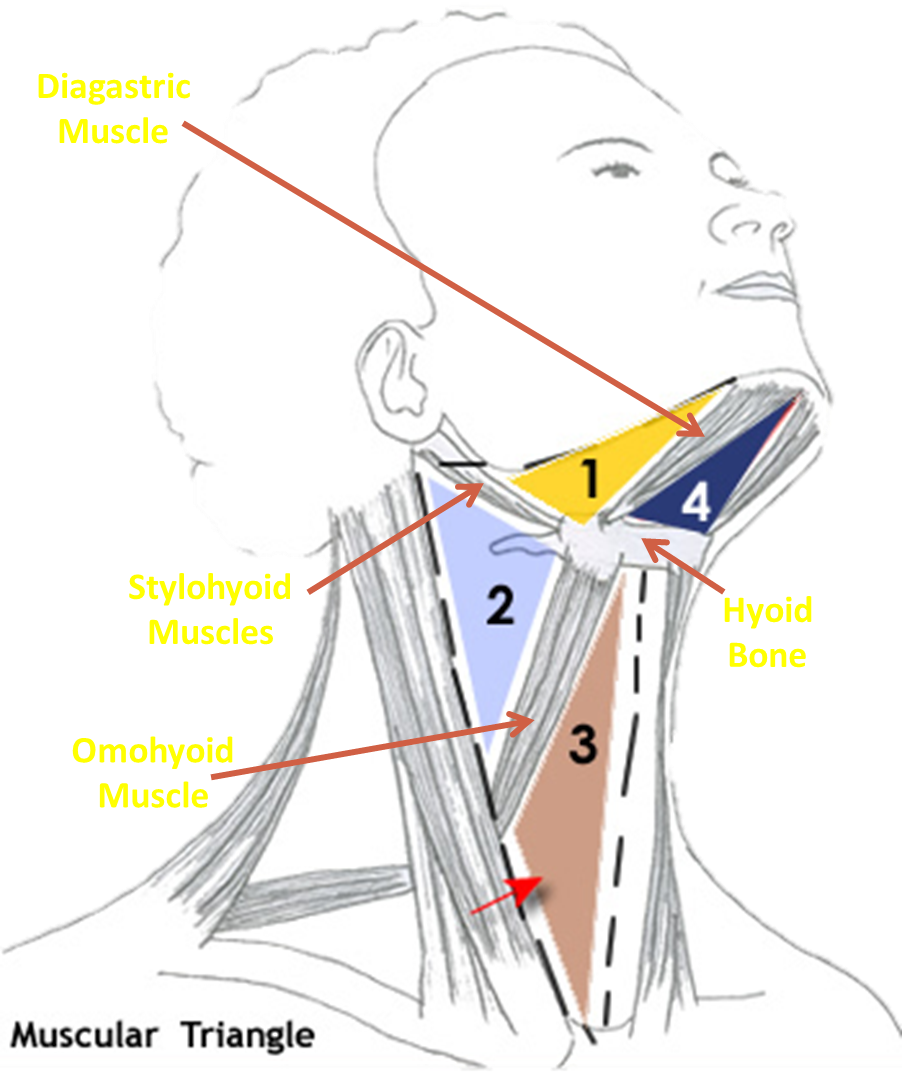
- Split into 4 further triangles
- Diagastric/ submandibular
- Submental
- Carotid
- Muscular
- Posterior
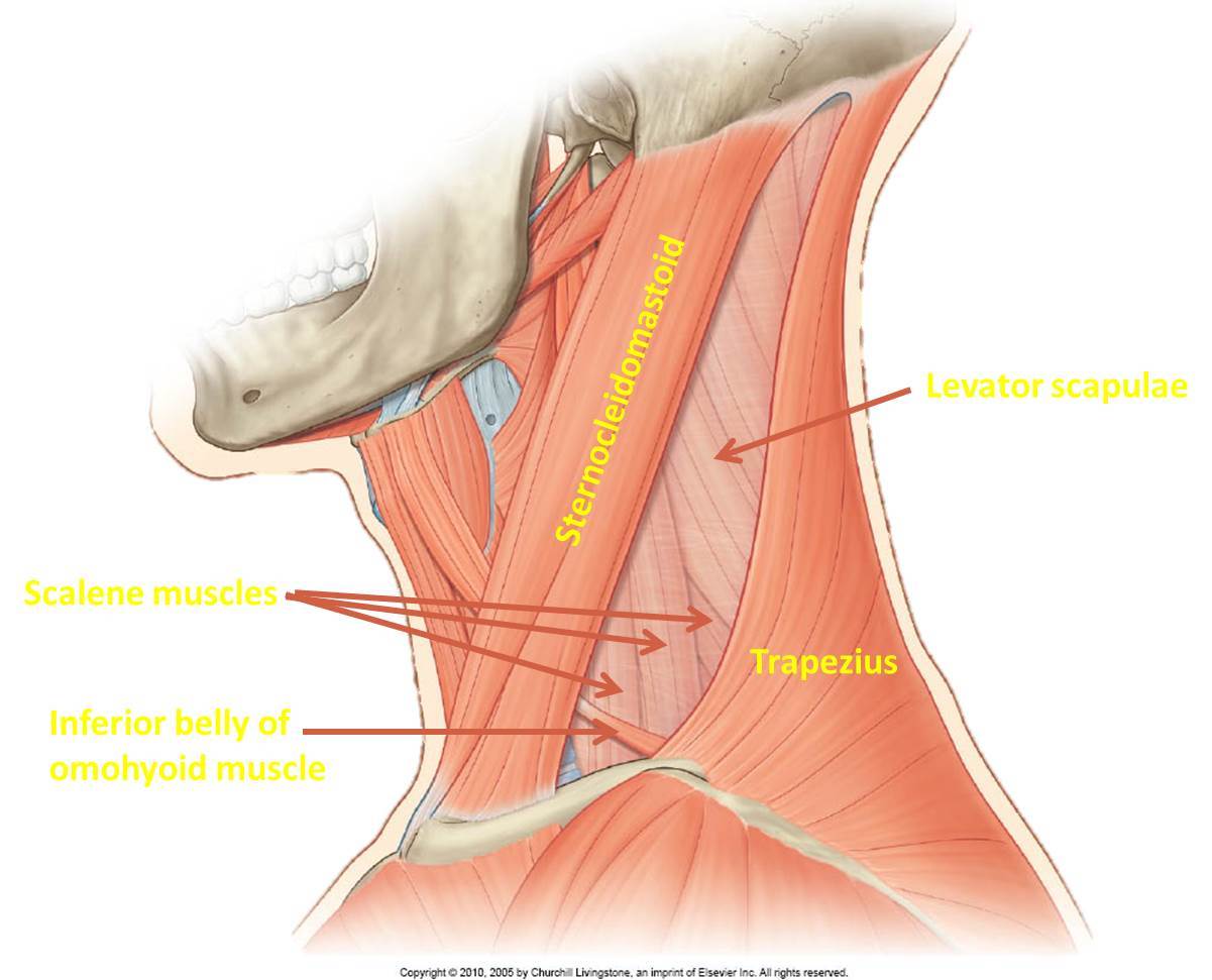
- Posterior sternocleidomastoid
- Clavicle
- Divide into thirds
- Middle third has subclavian vein
- Divide into thirds
- Trapezius muscle
- Mastoid process
- External jugular
- Subclavian artery
- Accessory nerve
- Trunks of brachial plexus
- Posterior cervical and supraclavicular
- Surface Anatomy
- Suprasternal notch
- aka jugular notch
- Mastoid process
- behind ear
- trapezius muscle
- when make a face
- sternocleidomastoid
- when look to the side
- Suprasternal notch
- Lymph nodes
- Submandibular
- Lateral tongue
- Submental
- floor of mouth, tip of tongue, skin of cheek
- Cervical
- mastoid
- occipital
- superficial and deep cervical nodes
- Posterior tongue, oropharynx, parotid gland, anterior pinna
- Deep cervical drains posterior tongue
- Submandibular
- Anterior
Fascial layers of the neck
- Carotid sheath
- Vagus nerve
- Common carotid artery
- Carotid sinus
- Bulge in internal artery
- Detect BP
- Carotid body
- Chemoreceptors
- Detects oxygen and CO2
- at the intersection between internal and external
- Chemoreceptors
- Bulge in internal artery
- Carotid sinus
- Jugular vein
- Pre-tracheal
- Trachea
- Oesophagus
- Thyroid
- Pre-vertebral
- Investing layer
- sternocleidomastoid
- trapezius
- Carotid sheath
Meninges
- Dura mater (outer)
- Meningioma is tumour of meninges, usually dura mater and benign.
- Subdural hematoma is quite common
- same thing as subdural haemorrhage
- Headache, confusion, muddled speech
- Arachnoid mater (middle)
- Subarachnoid haemorrhage
- Thunderclap headache
- comes on suddenly
- Usually result of brain aneurysm bursting
- Vomiting
- Photophobia
- Thunderclap headache
- Subarachnoid haemorrhage
- Pia mater (inner)
- Intracranial haemorrhage= anywhere in skull
- Dura mater (outer)
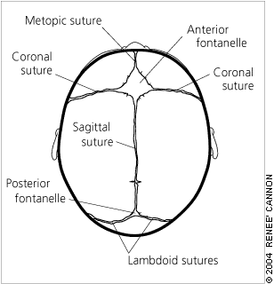
Sutures
- frontal
- only in children, separate frontal bones
- coronal
- frontal- pareital
- Sagittal
- Between parietal bones
- Lambdoid suture
- sagittal and occipital
- Squamous suture
- Temporal and parietal
- Fontanelles
- Anterior
- becomes Bregma
- Posterior
- becomes lambda
- sphenoid fontanelle becomes pterion
- Mastoid
- between temporal, parietal and occipital
- Anterior
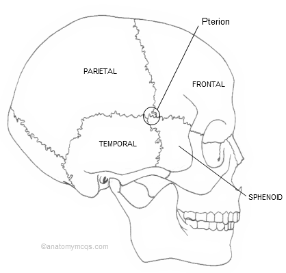
- Pterion
- where temporal, parietal, frontal and sphenoid bones fuse
- Shaped like a H
- Behind it is middle meningeal artery
- if damaged can cause extra dural haemorrhage
- frontal
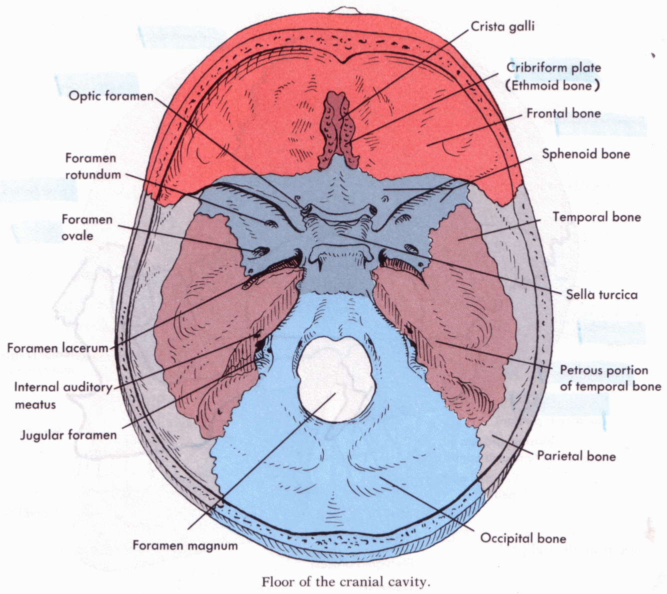
Holes in skull
- cribriform plate
- olfactory nerve (I)
- optic canal
- optic nerve II
- opthalamic artery
- Foramen rotundum
- CN V2
- Superior orbital fissure
- Oculomotor nerve III
- Trochlear IV
- First branch off trigeminal CN V1
- Abducens VI
- Foramen ovale
- CN V3
- Foramen spinosum
- middle meningeal artery
- Foramen lacerum
- internal carotid artery
- Doesn't actually go through here, goes pass here
- goes through carotid canal
- Internal acoustic meatus
- CN VII (facial) and VIII (vestibulocochlear)
- Jugular foraman
- Internal jugular vein
- CN IX (glossopharyngeal) X(vagus), XI (spinal accessory
- Hypoglossal canal
- CN XII hypoglossal nerve
- Foramen magnum
- spinal cord
- cribriform plate
GCS
- Glasgow Coma Scale
- Eyes
- Spontaneous
- Verbal
- Pressure
- use blunt object on distal fingernail
- No response
- <8 may want to incubate
- Motor
- Obey commands
- With pain: localising
- Moves hands up to push away trapezius pinch
- use thumb and fingers to pinch trapezius muscle or supraorbital ridge pressure DO NOT USE sternal rub, proximal nail bed, interdigit pressure, skin pinch/twist, nipple twist
- Moves hands up to push away trapezius pinch
- Withdraws from pain
- Abnormal flexion
- DECORTICATE POSITION
- Extension
- DECEREBRATE POSITION
- No response
- Verbal
- Orentiated
- Confused
- Words
- Sounds
- No response
- 15= good
Nasopharynx
- Turbinates
- narrow curved bony shelves in nasal cavity
- Turbinates
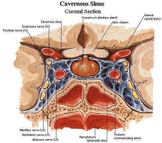
Cavernous sinus
- Between sphenoid and temporal bone. Has many veins. Has many nerves around it and the ICA (circle of Willis). Pituitary gland in the middle also near optic chiasm, and mamillary bodies (to do with memory)
Imaging
- MRI
- T2
- CSF, fluid and fat is bright white
- T1
- CSF is black
- T2
- MRI
Muscle in the neck
- Sternocleidomastoid
Images and files in the map
Information
- Description
- revision
- Created by
- alshuaibiabrar
- Views
- 0
- Tags
- History
-
- Created by Danielle Richardson · Feb 25, 2016
- Copied by alshuaibiabrar · Feb 25, 2016
- Copied by alshuaibiabrar · Feb 25, 2016
- Rating