
Mind map
Viles L2 - Protein Folding Pathway and Stability
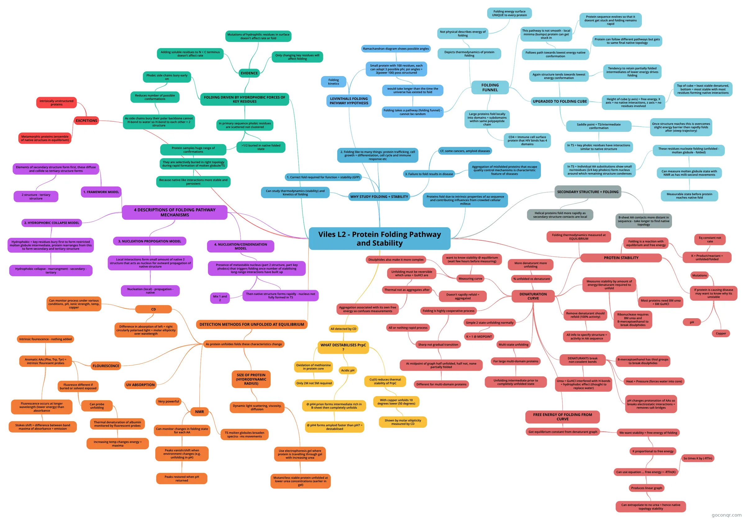
Mind map: Protein folding pathway and stability
Map content
Viles L2 - Protein Folding Pathway and Stability
WHY STUDY FOLDING + STABILITY
- 1. Correct fold required for function + stability (GFP)
- 2. Folding like to many things: protein trafficking, cell growth + differentiation, cell cycle and immune response etc
- 3. Failure to fold results in disease
- CF, some cancers, amyloid diseases
- Aggregation of misfolded proteins that escape quality control mechanisms is characteristic feature of diseases
- Can study thermodynamics (stability) and kinetics of folding
Proteins fold due to intrinsic properties of aa sequence and contributing influences from crowded cellular milleux
LEVINTHALS FOLDING PATHWAY HYPOTHESIS
- Folding kinetics
- Small protein with 100 residues, each can adopt 3 possible phi, psi angles = 3(power 100) poss structured
- Ramachandran diagram shows possible angles
- would take longer than the time the universe has existed to fold
- Folding takes a pathway (folding funnel) - cannot be random
- FOLDING FUNNEL
- Depicts thermodynamics of protein folding
- Not physical describes energy of folding
- Folding energy surface UNIQUE to every protein
- Not physical describes energy of folding
- This pathway is not smooth - local minima (bumps) protein can get stuck in
- Follows path towards lowest energy native conformation
- Protein can follow different pathways but gets to same final native topology
- Protein sequence evolves so that it doesnt get stuck and folding remains rapid
- Follows path towards lowest energy native conformation
- Large proteins fold locally into domains + subdomains within same polypeptide chain
- CD4 = Immune cell surface protein that HIV binds has 4 domains
- UPGRADED TO FOLDING CUBE
- Again structure tends towards lowest energy conformation
- Tendency to retain partially folded intermediates of lower energy drives folding
- Height of cube (y axis) = free energy, X axis = no native interactions, z axis = no residues involved
- Top of cube = least stable denatured, bottom = most stable with most residues forming native interactions
- Saddle point = TS/intermediate conformation
- Once structure reaches this is overcomes slight energy barrier then rapidly folds after (steep trajectory)
- In TS = key phobic residues have interactions similar to native structure
- In TS = Individual AA substitutions show small no/residues (3/4 key phobics) form nucleus around which remaining structure condenses
- These residues nucleate folding (unfolded - molten globule - folded)
- Can measure molten globule state with NMR as has milli-second movements
- Measurable state before protein reaches native fold
- Can measure molten globule state with NMR as has milli-second movements
- These residues nucleate folding (unfolded - molten globule - folded)
- In TS = Individual AA substitutions show small no/residues (3/4 key phobics) form nucleus around which remaining structure condenses
- Again structure tends towards lowest energy conformation
- Depicts thermodynamics of protein folding
- FOLDING FUNNEL
- Folding takes a pathway (folding funnel) - cannot be random
FOLDING DRIVEN BY HYDROPHOBIC FORCES OF KEY RESIDUES
- EVIDENCE
- Adding soluble residues to N + C terminus doesn't affect rate
- Mutations of hydrophilic residues in surface doesn't affect rate or fold
- Only changing key residues will affect folding
- Phobic side chains bury early on
- Reduces number of possible conformations
- As side chains bury their polar backbone cannot H-bond to water so H-bond to each other = 2 structure
- Reduces number of possible conformations
- In primary sequence phobic residues are scattered not clustered
- >1/2 buried in native folded state
- Protein samples huge range of confirmations
- They are selectively buried in right topology during rapid formation of molten globule/TS
- Because native like interactions more stable and persistent
- They are selectively buried in right topology during rapid formation of molten globule/TS
- EVIDENCE
SECONDARY STRUCTURE + FOLDING
- Helical proteins fold more rapidly as secondary structure contacts are local
- B-sheet AA contacts more distant in sequence - take longer to find native topology
4 DESCRIPTIONS OF FOLDING PATHWAY MECHANISMS
- 1. FRAMEWORK MODEL
- Elements of secondary structure form first, these diffuse and collide so tertiary structure forms
- 2 structure - tertiary structure
- Elements of secondary structure form first, these diffuse and collide so tertiary structure forms
- 2. HYDROPHOBIC COLLAPSE MODEL
- Hydrophobic + key residues bury first to form restricted molten globule intermediate, protein rearranges from this to form secondary and tertiary structure
- Hydrophobic collapse - rearrangment - secondary - tertiary
- Hydrophobic + key residues bury first to form restricted molten globule intermediate, protein rearranges from this to form secondary and tertiary structure
- 3. NUCLEATION PROPOGATION MODEL
- Local interactions form small amount of native 2 structure that acts as nucleus for outward propagation of native structure
- Nucleation (local) - propagation - native
- Local interactions form small amount of native 2 structure that acts as nucleus for outward propagation of native structure
- 4. NUCLEATION/CONDENSATION MODEL
- Presence of metastable nucleus (part 2 structure, part key phobics) that triggers folding once number of stabilising long-range interactions have built up
- Then native structure forms rapidly - nucleus not fully formed in TS
- Mix 1 and 2
- Presence of metastable nucleus (part 2 structure, part key phobics) that triggers folding once number of stabilising long-range interactions have built up
- 1. FRAMEWORK MODEL
PROTEIN STABILITY
- Folding thermodynamics measured at EQUILIBRIUM
- Folding is a reaction with equilibrium and free energy
- K = Product/reactant = unfolded/folded
- Eq constant not rate
- K = Product/reactant = unfolded/folded
- If protein is causing disease may want to know why its unstable
- Mutations
- Copper
- pH
- DENATURATION CURVE
- Simple 2 state unfolding normally
- K = 1 @ MIDPOINT
- % unfolded vs denaturant
- More denaturant more unfolding
- Folding is highly cooperative process
- Sharp not gradual transition
- At midpoint of graph half unfolded, half not, none partially folded
- Different for multi-domain proteins
- All or nothing rapid process
- At midpoint of graph half unfolded, half not, none partially folded
- Sharp not gradual transition
- Multi-state unfolding
- For large multi-domain proteins
- Unfolding intermediate prior to completely unfolded state
- For large multi-domain proteins
- Measures stability by amount of energy/denaturant required to unfold
- Ribonuclease requires 8M urea and B-merceptoethanol to break disulphides
- Most proteins need 8M urea + 6M GuHCl
- Remove denaturant should refold (100% activity)
- All info to specify structure + activity in AA sequence
- DENATURANTS break non-covalent bonds
- Urea + GuHCl interfered with H-bonds + hydrophobic effect (thought to replace water)
- B-merceptoethanol has thiol groups to break disulphides
- Heat + Pressure (forces water into core)
- pH changes protonation of AAs so breaks electrostatic interactions + removes salt bridges
- Measuring curve
- want to know stability @ equilibrium (wait few hours before measuring)
- Unfolding must be reversible which urea + GuHCl are
- Thermal not as aggregates after
- Aggregation associated with its own free energy so confuses measurements
- Doesn't rapidly refold = aggregated
- Thermal not as aggregates after
- Disulphides also make it more complex
- FREE ENERGY OF FOLDING FROM CURVE
- Got equilibrium constant from denaturant graph
- We want stability = free energy of folding
- K proportional to free energy
- Can use equation ... Free energy = -RTln(K)
- So times K by (-RTln)
- Produces linear graph
- Can extrapolate to no urea = hence native topology stability
- Can use equation ... Free energy = -RTln(K)
- K proportional to free energy
- We want stability = free energy of folding
- Got equilibrium constant from denaturant graph
- Simple 2 state unfolding normally
WHAT DESTABILISES PrpC ?
- Cu(II) reduces thermal stability of PrpC
- With copper unfolds 10 degrees lower (50 degrees)
- Shown by molar eliipticity measured by CD
- With copper unfolds 10 degrees lower (50 degrees)
- Oxidation of methionine in protein core
- Only 2M not 5M required
- Acidic pH
- @ pH4 prion forms intermediate rich in B-sheet then completely unfolds
- @ pH4 forms amyloid faster than pH7 = destabilised
- @ pH4 prion forms intermediate rich in B-sheet then completely unfolds
- All detected by CD
- Cu(II) reduces thermal stability of PrpC
DETECTION METHODS FOR UNFOLDED AT EQUILIBRIUM
- As protein unfoldes folds these characteristics change
- CD
- Difference in absorption of left + right circularly polarised light = molar ellipticity over wavelength
- Can monitor process under various conditions, pH, ionic strength, temp, copper
- FLOURESCENCE
- Aromatic AAs (Phe, Trp, Tyr) = intrinsic flouresent probes
- Fluorescence occurs at longer wavelength (lower energy) than absorbance
- Stokes shift = difference between band maxima of absorbance + emission
- Intrinsic fourescence - nothing added
- Fluoresce different if buried or solvent exposed
- Can probe unfolding
- Thermal denaturation of albumin monitored by fluorescent probes
- Increasing temp changes energy + maxima
- Thermal denaturation of albumin monitored by fluorescent probes
- Can probe unfolding
- Fluorescence occurs at longer wavelength (lower energy) than absorbance
- Aromatic AAs (Phe, Trp, Tyr) = intrinsic flouresent probes
- UV ABSORPTION
- NMR
- Very powerful
- Can monitor changes in folding state for each AA
- Peaks vanish/shift when environment changes (e.g. unfolding in pH)
- Peaks restored when pH returned
- Peaks vanish/shift when environment changes (e.g. unfolding in pH)
- TS molten globules broaden spectra - ms movements
- SIZE OF PROTEIN (HYDRODYNAMIC RADIUS)
- Dynamic light scattering, viscosity, diffusion
- Use electrophoresis gel where protein is travelling through gel with increasing urea
- Mutant/less stable protein unfolded at lower urea concentrations (earlier in gel)
- Use electrophoresis gel where protein is travelling through gel with increasing urea
- Dynamic light scattering, viscosity, diffusion
- CD
- As protein unfoldes folds these characteristics change
EXCPETIONS
- intrisically unstructured proteins
- Metamorphic proteins (ensemble of native structues in equilibrium)
Information
- Description
- Mind Map on Viles L2 - Protein Folding Pathway and Stability, created by George West on 20/05/2016.
- Created by
- George West
- Views
- 0
- Rating