
Notes
Breast anatomy
Content of the notes (3)
Page 1
BREAST ANATOMY
- Modified sweat glands
- Specifically function as modified apocrine glands
- Secondary sex characteristic / Gender identifying structures ==> Female phenotype
- Roughly hemispherical, occupy part of the anterior chest wall
- Extend from the 2nd-6th ribs craniocaudally and from the margin of the sternum to the midaxillary line from the centre laterally
- Part of the breast tissue extend into the axilla: Tail of Spence
- Lies: between the SUPERFICIAL and DEEP layers of the superficial pectoral fascia,which is contiguous with the Scarpa fascia of the anterior abdominal wall. The breast is enclosed within the superficial pectoral fascia, and lies on top of the deep pectoral fascia
- There are fibrous bands that run between the deep and superficial layers of the superficial pectoral fascia, these are dense, and called Cooper ligaments or suspensory ligaments of the breast. They are more prominent at the lower part of the breast (inframammary fold) and are important for the shape and structure of the breast
- The functional unit of the breast is the lobular unit: Cuboidal and myoepithelial cells within a vascular stroma form the glands (modified sweat glands)--> intralobular ducts --> extralobular ducts--> collecting ducts--> lactiferous duct (drain a lobule) --> dilated part of the lactiferous duct AMPULLA--> nipple
- Montgomery glands (modified sebaceous glands)--> around the areola, ducts termini
- The anatomical/structural unit of the breast is the lobe. Each breast has 15-20 segments LOBES --> each lobe has 20-40 LOBULES --> each lobule has 10-100 ALVEOLI. The breast has 5-10 large collecting ducts
Page 2
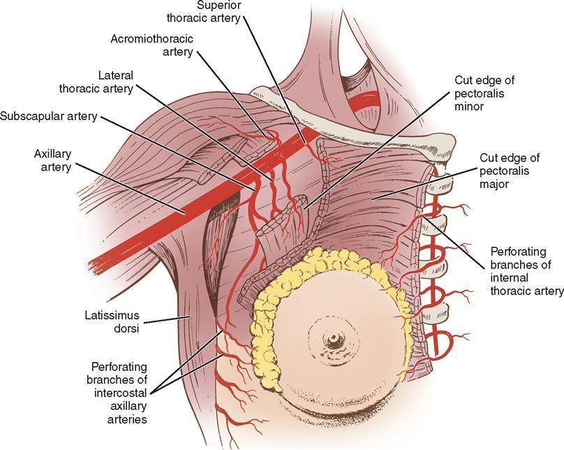
Blood supply:
- Internal thoracic (previously known as internal mammary) artery: Perforating branches, medially, from 1-4th intercostal spaces
- Axillary artery: From central to peripheral
(1) Superior thoracic artery
(2) Pectoral branch of acromiothoracic artery
(3) Lateral thoracic artery
(4) Subscapular artery, and
- Intercostal artery, perforating branches (laterally)
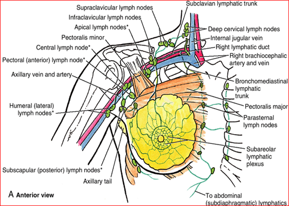
Lymphatic drainage
- Axillary lymph nodes 20-30 LN,
(1) Lateral group: 4-6 LN, posterior and medial to the axillary vein
(2) Anterior group: 4-5LN, along the inferior border of pectoralis minor, adjacent to the lateral thoracic vessels
(3) Posterior or subscapular group: 6-7 LN, along the subscapular vein
(4) Central group: Drain the above 3 groups, 3-4 LN, large nodes, in the axillary fat pad
(5) Apical group: Posterior to and above the pectoralis minor muscle, along the medial aspect of the axillary vein. Drain the above, and into the subclavian trunk. The latter drains, on the left side usually directly into the thoracic duct, on the right side into the jugulosubclavian junction or into a common right lymphatic trunk
LEVEL I: Nodes inferior to pectoralis minor muscle, (lateral, anterior and posterior groups of axillary LN)
LEVEL II: Nodes behind the pectoralis minor muscle, (apical group)
LEVEL III: Nodes above the pectoralis minor muscle (apical group).
- Internal thoracic LN
small, along the internal thoracic vessels, 2-3 cm from the sternal edge
3-5 LN are found on each side of the sternum.
Drain (1) the anterior chest wall, (2) the anterior part of the diaphragm, (3) the upper part of the rectus sheath and muscle, (5) the superior part of the liver, and (5) the inner aspect of the mammary gland.
- Intercostal LN.
Receive deep lymph vessels from the posteromedial aspect of the chest and
Receive some drainage from the lateral extremity of the mammary gland.
~ 75% of all lymphatic drainage of the breast passes to the
axillary nodes. The remainder principally drains to the internal
thoracic nodes.
* Any part of the breast may drain to either group, though there is a greater tendency for tumours situated in the
medial part of the breast to disseminate to the internal thoracic nodes than for tumours in the lateral part of the breast to do so.
* Involvement of the supraclavicular nodes in breast cancer usually represents retrograde spread along blocked lymphatic
channels when the apical axillary nodes are heavily involved. Efferent channels pass directly from these nodes to the inferior
deep cervical chain so that involvement of cervical nodes may occur via this route.
* Lymphatics do not normally drain to lymphatics across the opposite side of the body; early lymphatic spread of a tumour
from one breast to another does not occur. Such bilateral cases represent synchronous or early metachronous double primary tumours. In very advanced cases, extensive blockage of lymphatic channels allows subcutaneous lymphatic permeation to occur to the opposite side.
Innervation:
Sensory innervation is dermatomal in nature.
Mainly from anterolateral and anteromedial branches of T3-T5 thoracic intercostal nerves.
Supraclavicular nerves from the lower fibers of the cervical plexus provide innervation to the upper and lateral portions of the breast.
Researchers believe sensation to the nipple derives largely from the lateral cutaneous branch of T4.
Page 3
https://reference.medscape.com/article/1273133-overview#a2
Information
- Description
- ABR Breast imaging (Breast anatomy) Note on Breast anatomy, created by Eleni Kariki on 23/09/2018.
- Created by
- Eleni Kariki
- Views
- 2
- Rating