
Notes
Veterinary Virology
Content of the notes (15)
Page 1
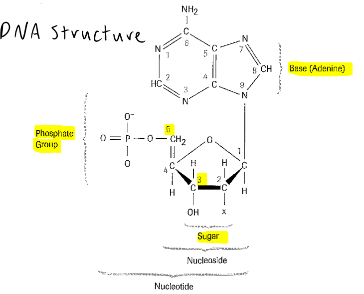
DNA structures
Page 2
RNA:
The extra -O- in the ribose backbone prevents formation of stable double-helices.
ssRNA very unstable and can degraded easily - RNases
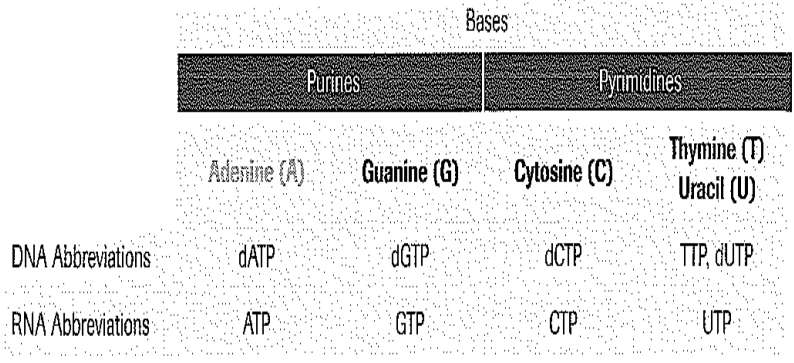
RNA cannot be directly PCR – Taq polymerase does not ‘read’ dUTPs in RNA
Page 3
Eukaryotes vs Prokaryotes vs Viruses:
Eukaryotes: Linear genomes plus mitochondrial DNA
Prokaryotes: Genomes ss/ds/ circular with extra genetic material -plasmids
Viruses: RNA or DNA – Never both - Polarity of genome
Exons and Introns in Eukaryotes - Variable number of tandem repeats (VNTR) (also in bacteria):
Only 10% exons – coding for proteins
Tandem repeats: short lengths of DNA that are repeated multiple times within a gene
Genome organization – use of mitochondrial DNA for phylogeny:
Own genome outside the nucleus – coding for proteins
High copy number
Size of genomes:
Different sizes in microorganisms
Page 4
Prokaryotes
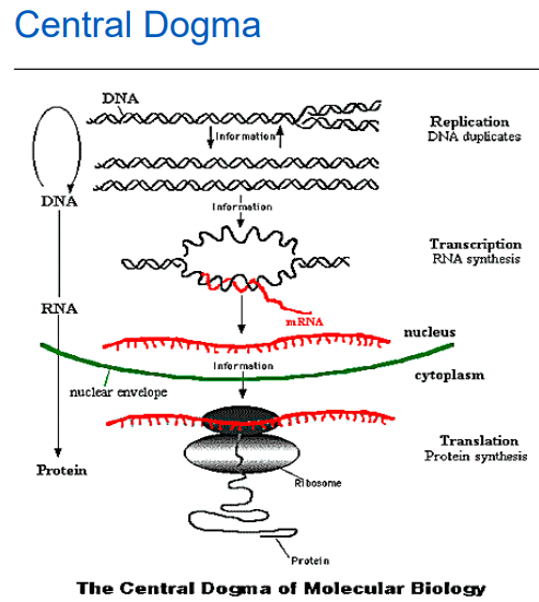
Replication: making a copy for reproduction (binary fission) – DNA polymerase
Transcription: making a single strand mRNA copy for production of protein – RNA polymerase
Translation: translating the transcribed mRNA into a protein chain – ribosomes
DNA is a single circle, packaged by DNA binding proteins
DNA forms loops or domains (50-100) constrained by a protein core attached to the cell wall
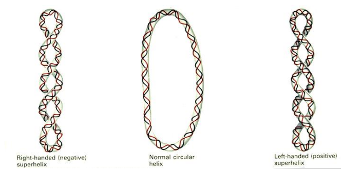
Bacterial DNA is further compacted by twisting the DNA in each domain around itself, called supercoiling
Bacterial DNA is negatively supercoiled
Topoisomerase
Supercoiling is controlled by a group of enzymes called topoisomerases.
During replication, the unwinding of DNA may cause the formation of tangling structures, such as supercoils.
Major role is to prevent DNA tangling.
Topoisomerase I causes relaxation of the supercoil.
Topoisomerase II (DNA gyrase) induces negative supercoils in newly replicated DNA.
Page 5
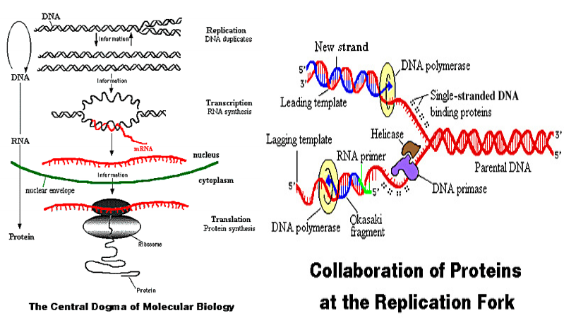
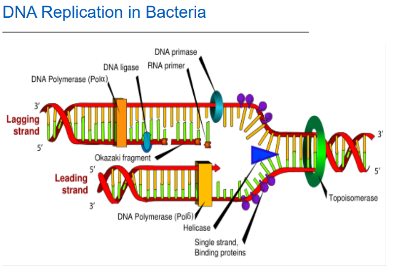
DNA Replication:
DNA helicase breaks a strand and unzips the DNA - Replication fork
Stabilising proteins hold the DNA open and protect it
Topoisomerase 1: relaxes supercoil
Primase – synthesises an RNA primer
DNA polymerase: catalyses addition of bases to 3’ end of DNA molecule - Continuous/discontinuous
RNAse removes RNA primer on lagging strand – DNA polymerase fills gap
DNA ligase repairs the nick (joins backbone molecules together)
DNA gyrase (Topoisomerase 2) supercoils new DNA
Page 6
Replication Fidelity:
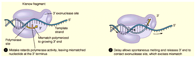
Some DNA polymerases have proof reading ability: they can detect mistakes in nucleotide pairing.
If mistake detected, polymerization stops: Uses 3’ to 5’ exonuclease activity to remove incorrect nucleotide, allows another attempt.
DNA polymerase proof reading ability varies and effects rates of spontaneous mutation.
Page 7
Transcription
Carried out by RNA polymerase.
Newly synthesised mRNA reflects the template DNA strand.
Only one strand of the DNA is transcribed. • mRNA is comparatively short molecule (often just a few kilobases)
Start signals on DNA called promoters, also convey info on which direction and which strand to transcribe.
The one DNA strand can be the template for multiple RNA polymerases to make multiple copies of mRNA’s at once.
Terminator sequence responsible for stopping transcription at end of gene.
5' binds to 3'
Page 8
Protein Synthesis:
Expression of genes occurs predominantly through production of proteins.
Conversion of nucleic acid (DNA) info to proteins involves transcription and translation.
Transcription – Conversion of DNA information into messenger RNA (mRNA) – RNA differs from DNA in structure of bond between bases and in use of uracil rather than thymine – RNA typically single stranded
Translation – Conversion of RNA information into a chain of amino acids
Page 9
Translation:
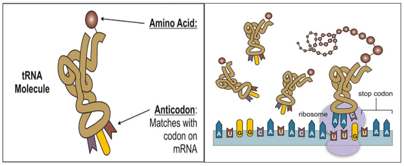
Initiation – Attachment of ribosomes (rRNA) to mRNA
Elongation – Movement of ribosome along mRNA, reading codons (triplet of mRNA bases) – Recognition of codon by transfer RNA (tRNA) and binding – Attachment of amino acid bound to tRNA to string of amino acids (polypeptide chain)
Termination – Termination of reading of mRNA sequence, release of rRNA and polypeptide chain
Reading Frame:
mRNA read in consecutive groups of 3 bases
Could code for 3 different proteins, depending on where it starts, i.e., there are 3 potential reading frames.
Protein synthesis starts at a specific triplet. – usually AUG, initiation codon
Position of the ribosome binding site and the initiation codon determines the reading frame.

Page 10
Post Translational Modification
Protein has to fold correctly. - Complicated and difficult to predict
Multiple polypeptide chains often associate to form a complete protein.
Sections of protein may be cleaved to produce mature protein.
Proteins may have non-protein components added. - E.g. glycoproteins
Page 11
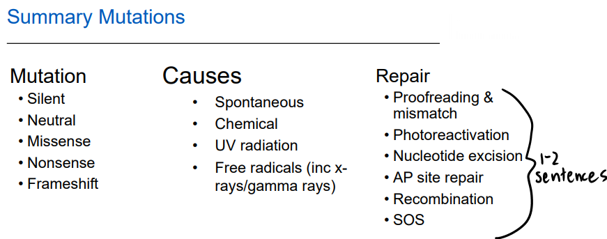
Mutations:
Population of bacteria and viruses are not homogenous. - Some variation from cell to cell.
Mutagenesis – the production of genetic mutations
Mutation: permanent, heritable alterations in the base sequence of DNA. - Can result from spontaneous errors in replication, or as a consequence of the damaging effects of physical or chemical agents (mutagens).
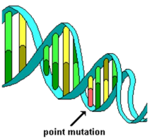
Point Mutation: simplest form - Sequence of DNA altered at a single position - Base substitution: one nucleotide is replaced by another
Consequences of Mutations:
Consequence of the change can vary dramatically.
Depends on the nature of the change and its location.
If the change is in the coding region of a gene (region which is ultimately translated into protein) it may give rise to an alteration in the amino acid sequence at that point, which may affect function of the protein.
The alteration may have little or no effect, either because the changed codon gives rise to the same amino acid, or because the new amino acid is sufficiently similar to the original that the function of the protein remains unaffected.
Types of Mutations:
Silent Mutation: A change in a base pair does not result in a change in the amino acid sequence in a protein
Neutral Mutation: A change in a base pair results in an amino acid change but the new amino acid has the same chemical properties as the old amino acid (i.e. hydrophobic to hydrophobic; acidic to acidic)
Missense Mutation: A mutation results in a change in an amino acid where the new amino acid has a different property than the old amino acid. The protein with the new primary structure may have reduced or no activity
Nonsense Mutation A mutation results in a new stop translation codon formed before the naturally occurring one. Translation is stopped prematurely and a shortened protein is made.
Frameshift mutation: Deletion or insertion of one base results in a change in the translational reading frame.
Protein synthesis is likely to be terminated soon after the insertion / deletion.
For most genes, the alternative reading frames are blocked by termination codons. - prevents synthesis of aberrant proteins.
Non-translated regions also often contain frequent stop codons.
Mechanisms of mutation:
1. Spontaneous: Thermodynamically unfavoured alternative forms
2. Chemical:
Nucleoside analogues: Can be incorporated instead of normal base, E.g. 5-bromouracil instead of thymine
Nucleotide altering agents: Make chemical changes to base, changing H bonding, Changes - which base will bind when replicated. E.g. nitrous acid
Intercalating agents: Insert between 2 double strands – causing bulge, Replication can get inserts and deletes. E.g. ethidium bromide
3. Ultraviolet radiation:
Causes formation of new chemical bonds. In particular causes adjacent thymine residues on the same strand to link covalently (thymine dimer).
Cell cannot replicate, therefore lethal to cell if not repaired. Attempts to repair can lead to incorrect base incorporation.
4. Free radicals:
Generated in cells (e.g. oxygen free radicals, intermediates of toxins such as aflatoxin)
X-rays and gamma rays - Causes molecules to lose electrons, can cause strand breaks or base/sugar destruction.
Page 12
DNA Repair
Enzymes are used e.g.: Exonucleases remove bases to past the mismatch site, Helicase, DNA polymerase and DNA ligase use the parent strand as a template and fill in the gaps
UV-light damage - Photoreactivation: Defense against UV damage, Uses enzyme called photolyase, In the presence of visible light; breaks the covalent bonds linking the two pyrimidine residues.
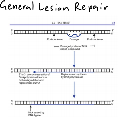
General Lesion Repair:
Endonuclease cuts DNA on both sides of the lesion.
DNA helicase unravels the cut strand for removal.
DNA polymerase replaces nucleotides in that region.
DNA ligase repairs the nick.
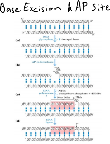
Base Excision Repair and AP Site Repair:
Base damaged (or lost).
Damaged - removed by DNA glycosylase.
Yields an AP site (i.e. apurinic or apyrimidinic site) that must be correctly filled.
This is done in a similar manner to nucleotide excision repair - Backbone nicked by endonuclease, Polymerase 1 and ligase do repairs
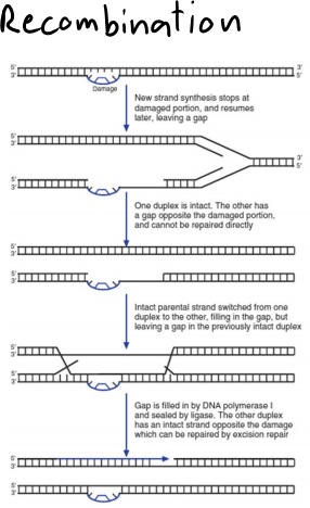
Recombination (know in general - multiple choice or summary):
Lesion in DNA – DNA polymerase can’t move through it and stops.
DNA polymerase starts again further downstream of the lesion – results in a section of single stranded DNA.
This portion of the DNA cannot be repaired by excision repair (which requires one intact strand).
The gap can be filled using a portion of the DNA from the other pair of strands in a recombination process (cutting and rejoining the DNA).
The original damage can now be repaired using general lesion repair, while the gap in the other DNA molecule can be filled by DNA polymerase and DNA ligase.
SOS Repair:
Used when faced with overwhelming levels of DNA damage preventing normal replication
Can temporarily modify or abolish specificity of the DNA polymerase.
Enables DNA polymerase to continue making new DNA strand, but may contain many mistakes.
Page 13
Page 14
Mutation Applications:
Mutation:
permanent, heritable alterations in the base sequence of DNA
Consequences:
All offspring carry altered base
Consequences in eukaryotes – some offspring may carry alteration (depending on location of alteration)
Heritable diseases
Tracking changes over time and space – determining origins of outbreaks
Identification of related organism – e.g. forensics, paternal testing
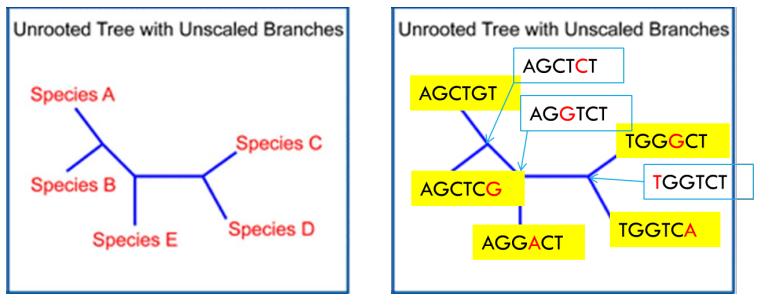
SNPS (Single nucleotide polymorphisms):
A single base substitution
Commonly in the population: DNA fingerprinting, Susceptibility to disease, How drugs affect individuals, Virulence factors of microorganisms
Presence or absence of SNPs may also provide information on importance of genes to survival
Page 15
Phylogenetic Trees:
Information
- Description
- TV1002 Note on Veterinary Virology, created by Erika Cobb on 01/09/2019.
- Created by
- Erika Cobb · Sep 2019
- Updated
- Sep 2, 2019
- Views
- 0
- Rating