10992620
Descrição
FlashCards por Anna Hogarth, atualizado more than 1 year ago
|
|
Criado por Anna Hogarth
aproximadamente 8 anos atrás
|
|
| Questão | Responda |
| Define connective/supportive tissue | A tissue that lies between two other tissues, consists of cells and ECM |
| What are the four kinds of connective tissue? | 1) Loose connective tissue - e.g. serous membranes, blood and adipose. 2) Dense connective tissue - dermis of skin 3) Bone 4) Cartilage |
| What are the five different cells which synthesis ECM? | 1) Fibroblast 2) Adipocyte 3) Chondrocytes 4) Osteocytes 5) Haemopoeitc |
| What does the ECM determine? | Physical properties of tissue |
| List the five roles of ECM? | 1) Cell migration 2) Shape 3) Proliferation 4) Survival 5) Tissue development |
| Describe the secretion of ECM | Constitutive secretion - not tightly controlled |
| How is the nature of a tissue changed? | ECM is altered with development. |
| What are the qualities of the dermis and why is this important? | Elastic and strong so that shearing forces don't cause damage. |
| Describe the ECM of: 1) Bone 2) Neural tissue 3) Tendons 4) Dermis | 1) Abundant calcified ECM 2) Almost no ECM 3) Tough, rope-like ECM 4) Pliable, acts as a shock absorber |
| LIst the components you would expect to find in the dermis (6) | 1) Collagen fibres 2) Macrophages 3) Capillaries 4) Mast cells 5) Elastic fibres 6) Fibroblast |
| What separates the dermis from the epithelial layer (epidermis)? | Basement membrane |
| What is a proteoglycan? Where are they assembled and how are they secreted? | 1) A proteoglycan monomer consists of a protein core with many glycosaminoglycans attached 2) Proteoglycans are assembled in the ER and Golgi and are delivered to the ECM via secretory bodies (constitutive secretion) |
| What is the primary function of GAGs and proteoglycans? | They can bind water and form hydrated gels, filling the space between cells. This can function to lubricate joint cavities as well as resisting compressive forces. |
| Other than resisting compressive forces and lubricating joint cavities, what do GAGs ad proteoglycan gels facilitate? | Permit the rapid diffusion of nutrients, hormones and metabolites. |
| What is a glycosaminoglycan (GAG)? | A linear chain of 20-100 dissaccharides |
| What modification has usually been made to disachharides? | Usually sulfated |
| Give three examples of GAGs | 1) Chondroitin sulfate 2) Heparin sulfate 3) Dermatan Sulfate |
| Give two examples disaccharides which are found in GAGs | 1) Iduronic acid 2) N-acetylgalactosamino-4-sulfate |
| What is hyaluronic acid? | A glycosaminoglycan which consists of 25,000 disaccharides |
| Why is it important that cells can migrate through hyaluronic acid? How can this be exploited clinically? | 1) Important for wound healing 2) Can use a synthetic dermis consisting of HA to encourage cell migration and dermal healing |
| Give three examples of proteoglycans | 1) Biglycan 2) Syndecan 3) Aggrecan |
| Of the three examples listed previously, which is the longest? Which is attached to the plasma membrane? | 1) Aggrecan is much longer than biglycan and syndecan 2) Syndecan |
| Which GAGs make up: 1) Aggrecan 2) Syndecan 3) Biglycan | 1) Chondroitin sulfate chains and keratin sulfate chains. Can attach through a linker protein to hyaluronan 2) Heperin sulfate chains 3) Dermatan sulfate chains |
| Which is the most abundant protein in the human body? What is its primary function? | Collagen - makes up 1/3 to 1/4 of protein in the body. Provides tensile strength |
| How many different types of collagen are there? Which is the most common? | 1) 20 2) Type 1 |
| What makes up the basement membrane? | Type III collagen (reticulin), type IV collagen, laminin, fibrin and ground substance. |
| What is orthogonal collagen? | Collagen which runs in perpendicular bundles to increase tensile strength. |
| What is elastin? What does it form? Where is it commonly found? | 1) A fibrous protein: polymer of tropoelastin associated with fibrillin (glycoprotein) 2) Elastic fibres 3) Arteries |
| What is Marfin syndrome? | 1) Fibrillin gene mutation which results in ruptured arteries. |
| What is Ehlers-Danlos syndrome? | Can be inherited (autosomal dominant or recessive) or occur due a mutation during the early years. Can affect more than a dozen of genes - usually affects the structure, productions, or processing of collagen or proteins associate with collagen. Results in elasticated skin, loose joints, abnormal scar production, chronic pain, or early osteoarthritis. |
| What causes wrinkles? | Breakdown of collagen and elastin, often as a result of sun damage. |
| Where are fibronectin fibres found in the body? | In the ECM: circulating soluble fibronectin fibres in plasma and body fluid |
| What are the functions of fibronectin? (3) | 1) Binds cells (integrins) and ECM (collagen) 2) Cell attachment and matrix organisation 3) Guides cell migration - important for development and wound healing |
| What does the basement membrane underlie? | 1) Epithelial and endothelial cells |
| What is the function of the basement membrane? | Separates epithelial/endothelial cells from underlying connective tissue. |
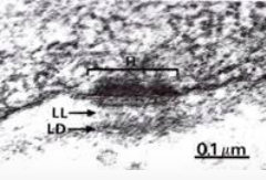
| What two components make up the basement membrane? How does the basement membrane connect to epithelial cells? | 1) Lamina lucida and lamina densa 2) Hemidesmosome |
| What roles does the basement membrane have? (6) | Supportive, anchoring, protective role, selective cell movement, molecular filtering, signalling, and helps to prevent oncogenesis (Prevents contact with the underlying connective tissue). |
| What is the underlying connective tissue under the basal lamina? | Reticular tissue |
| What makes up the basal lamina/basement membrane? | Type IV collagen, heparan sulfate proteoglycan, laminin, and nidogen (entactin). |
| Which laminin in the basal lamina connects to integrin in the epithelial cells? | Laminin 10 |
| What is Junctional Epidermolysis Bullosa? | A genetic disease which effects laminin in the basement membrane. Causes the skin to blister as the epidermis and the dermis separate. N.B. this is similar to epidermolysis bullosa where the mutation is within keratin. |
| How does a carcinoma in situ turn to malignant cancer? | Cancerous cells pass through the basement membrane and enter the bloodstream - metastases |
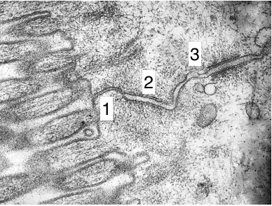
| What are the different types of junctions (and their functions)? (superior to inferior) | Between adjacent epithelial cells 1) Tight/separate - prevents the passage of molecules across the epithelium 2) Adherens - Tether adjacent cells together 3) Desmosomal - Resist mechanical stress (strongest junction, abundant in the skin) 4) Gap - allow passage of small molecules between adjacent cells Between epithelial cells and basement membrane 1) Hemidesmosome/focal adhesion - Anchor epithelium to basal lamina |
| In histology exams which tissue will usually be used to demonstrate junctions? | Gut epithelia - easiest to see |
| Why is it important that there is a seal between epithelial cells? | Transfer from lumen into organ is ideally through a cell as this is a controlled process |
| 1) Tight junction by villi 2) Adherens - gap and reasonably dense borders 3) Desmosome - very dense border | |
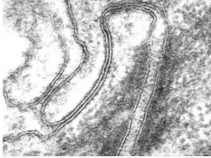
| Gap junctions - note that it looks like 3 lines | |
| Hemidesmosome | |
| What does desmosome related disease result in? | Compromised cardiac and/or cardiac function |
| Give an example of an infectious disease targeted at desmosomes | Staphylococcus - scalded skin syndrome, bacterial proteases directed as desmosomal cadherins causing epidermal blisters. |
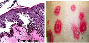
| Give an example of an autoimmune disease directed at desmosomes | Pemphigus vulgaris - antibodies directed against desmosomal cadherins causing epidermal blisters. |
| What is desmoplakin? Which genetic disease can it contribute to? | Protein which contributes to desmosome structures in cardiac muscle and epidermal cells. Mutation in Naxos disease - causes skin blisters, 'wooly' hair and cardiomyopathy. |
| Where are primary (non-motile) cilia found? What is there function? | 1) Nearly all cells (usually one long cilia). 2) Function as sensory antennae - chemosensors, photosensors, mechanosensors; signalling - Hedgehog and wnt signalling are involved in cancer developement |
| Where are motile cilia found? | Trachea and fallopian tubes - help move mucus and germinal cells respectively |
| How do flagella compare to cillia? | Flagella - much longer than cilia, propel sperm cells |
| Describe the cytoskeleton of cilia and flagella | Microtubule based cytoskeleton - axoneme (circular structure, for primary cilia this a ring of 9 microtubule doublets; for motile cilia this consists of 9 outer microtubule doublets with two central microtubules - 9+2 axoneme) |
| Give three examples of genetic ciliopathies | 1) Bardet-Biedl Syndrome - characterised principally by obesity, retinitis pigmentosa, polydactyly, hypogonadism and renal failure in some cases 2) Polycystic kidney disesae 3) Lack of cilia in fallopian tubes - ectopic pregnancy |
| What are psuedopodia | Cell protrusions - actin polymerisation for movement. Found on neutrophils. |
| What affect do amoeboid cancerous cells have on cancer prognosis? | These are highly migratory and greatly worsen the prognosis |
| What are lamellipodia and filopodia important for? | Cell movement, contact and environmental sensing |
| What are the structures that lamellipodia generally referred to as? What type of cells use them? | 1) Ruffles 2) Fibroblast cell migration (wound healing) and epithelial cells |
| What are the structures that filopodia form commonly referred to? Which cells use them? What are they predominantly for? When are they abundant? | 1) Spikes 2) Migrating neural growth cones (if the filopodia don't make contact the cells tend to die) and fibroblasts 3) Environmental sensing 4) Abundant filopodia are found in invasive cancer cells |
| What are microvilli? Where are they commonly found? | 1) Cell surface extension of secretory and absorptive cells 2) Kidney and intestinal cells |
| How many microvilli are found on the apical surface of a single small intestinal cell? What affect does this have on surface area? | 1) Several thousand 2) ~600 fold |
| Describe the structure of microvilli | Amorphous densely staining region-digestive enzymes, outer plasma membrane and polymerised actin filament bundles within. Terminal web at the base. Much more of an internal structure than cilia. |
| What causes persistent osmotic diarrhea | Infection by enteropathogenic Escherichia coli, leads to malabsorption and consequently persistent osmotic diarrhea. |
| What effect do toxins have on intestinal microvilli? | Leads to destruction of intestinal microvilli and intestinal tight junctions resulting in the inhibition of water re-absorption. |
Quer criar seus próprios Flashcards gratuitos com GoConqr? Saiba mais.