
Mapa mental
Ischemic Stroke
Conteúdo do mapa
Ischemic Stroke
Treatment
- Surgical
- MERCI retriever (Lewis et al., 2011).
- Carotid endarterectomy (Lewis et al., 2011).
- Repair of aneurysm (Lewis et al., 2011).
- Removal of tutor (Lewis et al., 2011).
- Drainage of hematoma (Lewis et al., 2011).
- Burr hole (Lewis et al., 2011).
- Angioplasty (Lewis et al., 2011).
- Pharamcological
- Thromolytic; lyse (break down) existing clots i.e. Streptokinase, t-PA, rTpa
- tPA
- Must be administered within 3 to 4.5 hours of the onset of clinical signs of ischemic stroke (Lewis et al., 2011).
- Administered IV to re-establish blood flow through a blocked artery to prevent cell death (Lewis et al., 2011).
- The door-to-needle time for tPA remains less than 60 mins or 1 hour after onset of clinical signs. Thrombolytics given in this time frame reduce disability, but at the expense of an increase in deaths within 7 to 10 days and an increase in intracranial hemorrhage (Lewis et al., 2011).
- Must be administered within 3 to 4.5 hours of the onset of clinical signs of ischemic stroke (Lewis et al., 2011).
- tPA
- Anticoagulants; Inhibit platelet aggregation, prevent platelet plugs e.g. ASA, Plavix
- No anticoagulant or antiplatelet drugs are given for 24 hours after tPA treatment (Lewis et al., 2011).
- Not recommended in the emergency phase because of the risk for intracranial hemorrhage (Lewis et al., 2011).
- Aspirin at a dose of 325 mg may be initiated within 24 to 48 hours after the onset of an ischemic stroke
- Complications of aspirin: GI bleeding. Therefore used cautiously in those who have a history of peptic ulcer disease (Lewis et al., 2011).
- Antiplatelets
- Used only after patient’s condition has been stabilized and to prevent further clot formation (Lewis et al., 2011).
- Vasodilators (Lewis et al., 2011).
- Anti-hyperlipidemics (Lewis et al., 2011).
- Anti-hypertensives e.g. metoprolol
- Hypertension may be a protective response to maintain cerebral perfusion
- Use of drugs to lower BP is recommended only if BP is markedly increased (MAP>130 mm Hg or systolic BP>220 mm Hg) (Lewis et al., 2011).
- Hypertension may be a protective response to maintain cerebral perfusion
- Thromolytic; lyse (break down) existing clots i.e. Streptokinase, t-PA, rTpa
- Surgical
Signs and Symptoms
- Cognitive (Ignatavicius & Workman, 2013).
- Denial of illness
- Spatial and proprioveptive dysfunction
- Impairment of memory, judgment, problem solving, and decision making
- Decreased concentration
- Motor (Ignatavicius & Workman, 2013).
- Hemiplegia (paralysis) or hemiparesis (weakness) contralateral to which part of the brain is affected
- Quadriparesis if the brainstem is involved
- Ataxia (gait) if the cerebellum is affected
- Hypotonia or flaccid paralysis
- Hypertonia or spastic paralysis
- Agnosia: unable to use objects correctly
- Apraxia: unable to carryout purposeful motor activity or speech
- Bladder and bowel incontinence from the loss of neurological control in the cerebral cortex
- Sensory (Ignatavicius & Workman, 2013).
- Decreased sensation of touch and pain contralateral to where the stroke is in the brain
- Unilateral body neglect syndrome
- Amaurosis fugax: brief episode of blindness in one eye
- Hemiaopsia: blindness in half the visual field
- Homonymous hemianopsia blindness in the same side of both eyes
- Nystagmus: or other involuntary eye movements caused from brainstem and cerebellar damage
- Cranial Nerve Dysfunction (Ignatavicius & Workman, 2013).
- CN V: absent gag reflex and ability to chew
- CN IX and X: decreased ability to swallow
- CN XII: impaired tongue movement
- CN VII: facial paralysis
- Cardiovascular (Ignatavicius & Workman, 2013).
- Embolic strokes are associated with heart murmurs, dysrhythmias (atrial fibrillation is the most common, and HTN.
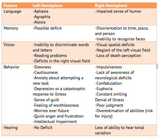
- Left vs. Right Hemisphere (Ignatavicius & Workman, 2013).
- Cognitive (Ignatavicius & Workman, 2013).
What is an ischemic stroke?
- Ischemic stroke is caused by occlusion of cerebral artery by either a thrombus or embolus (Lewis et al., 2010).
- Brain stores no oxygen or glucose and thus needs constant blood flow for normal function and waste removal (Lewis et al., 2010).
- Stroke ranks third for mortality rates in developed countries (Kearney, 2014).
- Ischemic stroke is caused by occlusion of cerebral artery by either a thrombus or embolus (Lewis et al., 2010).
Pathophysiology
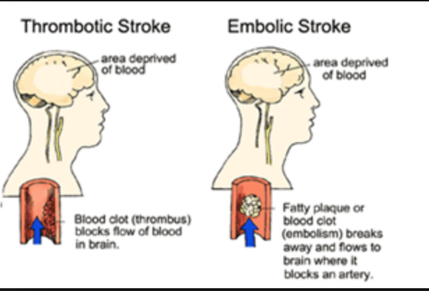
- Thrombic Stokes
- Consists of more than half of all strokes (Ignatavicius & Workman, 2013).
- Caused by the development of atherosclerosis (Ignatavicius & Workman, 2013).
- A rupture of one of these plaques exposes cells to clot promoting elements (Ignatavicius & Workman, 2013).
- End result is a formation of a blood clot in an artery supplying blood to the brain. This blood clot is large enough to interrupt blood flow to cerebral tissue (Ignatavicius & Workman, 2013).
- A rupture of one of these plaques exposes cells to clot promoting elements (Ignatavicius & Workman, 2013).
- Thrombic strokes are characterized by a slow onset, taking minutes to hours (Ignatavicius & Workman, 2013).
- Embolic Strokes
- Caused by thrombus or thrombi breaking off an artery anywhere in the body and traveling to cerebral arteries (Ignatavicius & Workman, 2013).
- Thrombus formation can be caused from heart disease, MI, heart valve prosthetics, non valvular atrial fibrillation, and plaque (Ignatavicius & Workman, 2013).
- Embolic strokes are characterized by a sudden development, rapid neurological deficits (Ignatavicius & Workman, 2013).
- Can lead to hemorrghaic stroke because arterial vessel wall is vulnerable to ischemic damage (Ignatavicius & Workman, 2013).
- Caused by thrombus or thrombi breaking off an artery anywhere in the body and traveling to cerebral arteries (Ignatavicius & Workman, 2013).
- Thrombic Stokes
Diagnostic Tests
- Laboratory Values
- Elevated hematocrit and hemoglobin with severe or major stroke because body is compensating for decreased oxygen to the brain (Ignatavicius & Workman, 2013).
- PT, PTT, and INR: used to establish baseline information for anticoagulation therapy (Ignatavicius & Workman, 2013).
- Elevated WBC: can indicate presence of infection (Ignatavicius & Workman, 2013).
- Lumbar puncture: used to rule out meningitis and subarachnoid hemmorhage (Ignatavicius & Workman, 2013).
- Imaging Studies
- MRI: to determine the extent of brain injury and has greater specificity in determining the location of vascular lesions and blockages than CT (Jauch, 2016).
- CT scan: indicates the size and location of the lesion and helps to differentiate between ischemic and hemorrhagic stroke (Jauch, 2016).
- Ultrasonography and echocardiography: help determine additional cardiovascular risks (Jauch, 2016).
- Laboratory Values
Risk Factors
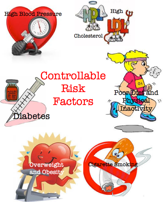
- Modifiable (Choudhury et al., 2015; Ignatavicius & Workman, 2013).
- Hypertension and atherosclerosis
- Cardiovascular disease (i.e. atrial fibrillation)
- Diabetes and glucose metabolism
- High cholesterol levels
- Smoking
- Alcohol consumption
- Illicit drug use (particularly cocaine)
- Lifestyle factors (i.e. obesity, physical activity diet)
- Sleep apnea
- Blood clotting disorders (high risk for thrombotic stroke and require preventive anticoagulants)
- TIA patients (should seek anticoagulation therapy)
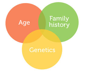
- Non modifiable (Choudhury et al., 2015; Ignatavicius & Workman, 2013).
- Age; increased risk as age increases
- Gender; men 30% higher incidence, post menopausal women
- Race or ethnicity; American Indian, Alaskan Natives, Blacks, Hispanics have a higher chance
- Heredity
- Myocardial Infarction
- Sickle Cell Disease
- Modifiable (Choudhury et al., 2015; Ignatavicius & Workman, 2013).
Collaborative Care
- Nursing
- Managing circulation, airway, breathing → Patients may have difficulty keeping an open and clear airway because of a decreased LOC or decreased or absent gag and swallowing reflexes (Lewis et al., 2011).
- Oxygenation
- Maintaining oxygenation is important - Both hypoxia and hypercarbia are to be prevented because they can contribute to secondary neuronal injury (Lewis et al., 2011).
- Oxygen administration, artificial airway insertion, intubation, and mechanical ventilation may be required (Lewis et al., 2011).
- Neurological Assessment
- Glasgow Coma Scale to address LOC, orientation, motor, pupils, speech.language, vital signs, and blood glucose (RNAO, 2005).
- Fluid and electrolytes
- keep the patient adequately hydrated to promote perfusion and decrease further brain injury (Lewis et al., 2011).
- Adequate fluid intake during acute care via oral, IV, or tube feedings should be 1500 to 2000 mL/day (Lewis et al., 2011).
- IV solutions and water ware avoided because they are hypotonic and may further increase cerebral edema and ICP(Lewis et al., 2011).
- Hyperglycemia may be associated with further brain damage and should be treated (Lewis et al., 2011).
- Management of Increased ICP
- Improve venous drainage (i.e. elevate HOB)
- Maintain head and neck in alignment (Lewis et al., 2011).
- Quiet environments will reduce headaches and reduce aggravation of increased ICP (Ignatavicius & Workman, 2013).
- Avoid extreme hip and neck flexions that increase intrathoracic pressure making ICP more difficult to control. Extreme neck flextion interferes with venous drainage as well (Lewis et al., 2011).
- Avoid clustering nursing procedures all at once because the effect of ICP elevation is more dramatic (Ignatavicius & Workman, 2013).
- CSF drainage (Ignatavicius & Workman, 2013).
- Diuretic drugs (e.g. mannitol, furosemide) (Lewis et al., 2011).
- Removal of bone flap to allow for cerebral edema without increases in ICP (Lewis et al., 2011).
- Management of Constipation
- Constipation can be caused from decreased mobility (Kasaeaneni & Hayes, 2014).
- High fiber food such as fruits or vegetables can be added to the diet to help increase the volume present and cause bowel movement by distension (Kasaeaneni & Hayes, 2014).
- Fiber supplements such as bran, psyllium, or methylcellulose can be added to help increase the fiber consumed by the patient (Kasaeaneni & Hayes, 2014).
- Encourage fluid intakes (Kasaeaneni & Hayes, 2014).
- Avoid Hyperthermia
- Contributes to increased cerebral metabolism. A temperature elevation of even one degree increases brain metabolism by 10% and contribute to further brain damage (Lewis et al., 2011).
- Treatment: Aspirin Acetaminophen Cool blankets (Lewis et al., 2011).
- Education
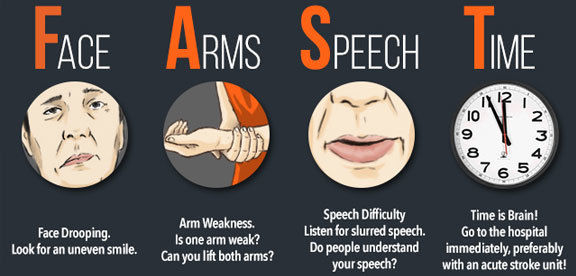
- Teach patients to recognize the symptoms of a stroke by using the acronym FAST
- Return demonstrations assist in evaluating family' ability to perform patient care tasks; provide verbal and oral instruction Ignatavicius & Workman, 2013).
- Teach family about depression that can occur within 3 months of a stroke. Post stroke depression is associated with increased morbidity (Ignatavicius & Workman, 2013).
- Health teaching for the patient includes drug therapy, ambulation skills, nutritional management, and self management skills (Ignatavicius & Workman, 2013).
- Health Care Resources (Ignatavicius & Workman, 2013)
- Available online resources to share with patients and family include American Heart Association, National Stroke Association, Agency of Health care Research and Quality
- SLP (Speech Language Pathologist)
- Appointments for outpatients speech may be arranged to help the patient relearn to talk (Ignatavicius & Workman, 2013).
- Collaborate with the SLP to conduct a bedside swallowing screening and evaluation. Patient may remain NPO until SLP determines the patient can tolerate fluids without aspirating (Ignatavicius & Workman, 2013).
- Home
- Collaborate with case manager to plan patient's discharge; coordinate with rehabilitation therapist to identify need of assistive devices (Ignatavicius & Workman, 2013).
- PT (Physiotherapist)
- To restore movement, balance, and coordination
- Climb stairs, transfer in and out of bed and chair, transfer to and from car, mobility aids (Ignatavicius & Workman, 2013).
- To restore movement, balance, and coordination
- OT (Occupational Therapist)
- To relearn basic skills such as bathing and dressing
- PT (Physiotherapist)
- Collaborate with case manager to plan patient's discharge; coordinate with rehabilitation therapist to identify need of assistive devices (Ignatavicius & Workman, 2013).
- Community Care
- Patients with stroke may be discharged to a rehabilitation centre or skilled nursing fac.ility depending on the extent of their disability and the availability of caregiver/family support (Ignatavicius & Workman, 2013).
- Nursing
Complications
- Increase ICP
- Occurs in response to cerebral edema (American Heart Association, 2015).
- Pneumonia
- Due to decreased mobility after a stroke (American Heart Association, 2015).
- Aspiration pneumonia
- Can be caused by impaired tongue movement, decreased swallowing, or absent gag reflex (American Heart Association, 2015).
- Urinary Tract Infection
- Caused by foley catheter being placed due to bladder incontinence (American Heart Association, 2015).
- Seizure
- Abnormal electrical activity in the brain causing convulsions from cerebral damage (American Heart Association, 2015).
- Clinical depression
- Causes unwanted emotional and physical reactions to changes and losses (American Heart Association, 2015).
- Pressure ulcers
- Results from decreased ability to move and pressure on areas of the body because of immobility (American Heart Association, 2015).
- Limb contractures
- Shortened muscles in an arm or leg from reduced ability to move the affected limb or lack of exercise (American Heart Association, 2015).
- Deep Vein Thrombosis
- Blood clots form in veins of the legs because of immobility from stroke. (American Heart Association, 2015).
- Increase ICP
Imagens e arquivos do mapa

Informações
- Descrição
- Nursing Mapa Mental sobre Ischemic Stroke, criado por Elfete Sadiki em 07-03-2017.
- Criado por
- Elfete Sadiki
- Visualizações
- 172
- Tags
- Avaliação