
Notas
Spinal cord
Conteúdo das notas (5)
Página 1
Surface anatomy
i) General anatomy
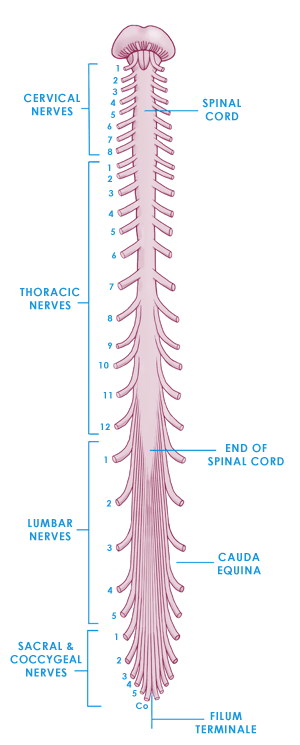
Approx. 45cm long - 1 - 1.5cm in diameter
Begins at Foramen Magnum
Continuous with Medulla Oblongata
Occupies upper 2/3 of vertebral canal - Cervical, Thoracic & some Lumbar
Ends at level L1-L2 in adults - near L-3 in children
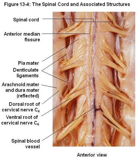
Anterior median fissure
Remember parts of the vertebral column! - 7, 12, 5, 5, 4
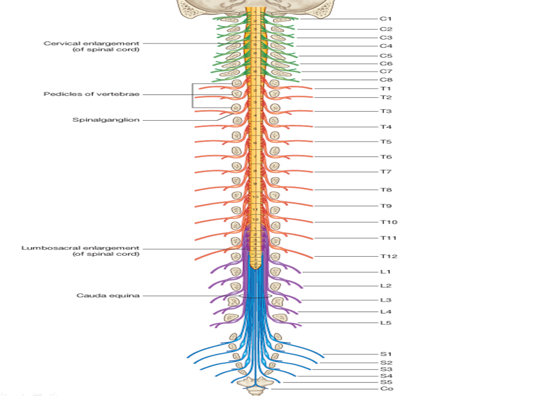
SC enlarged at 2 places
Cervical enlargement - origin of Brachial plexus (C3-T2)
Lumbar enlargement - origin of Lumbosacral plexus (L1-S3)
(Link to 2. Internal anatomy i) Gray matter - gray matter density in SC)
ii) Meninges
3 meningeal layers (like brain!)
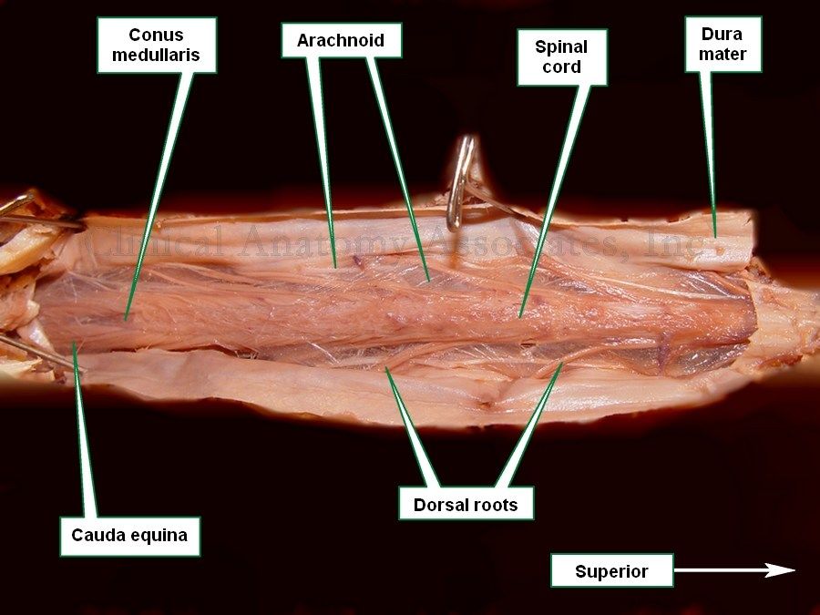
Dura - outermost layer
Arachnoid
Pia - innermost layer
Denticulate ligament = continuation of pia that runs between dorsal & ventral roots
iii) Tail of SC
Conus Medullaris = most inferior point of SC
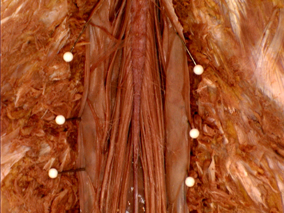
Cauda Equina = extension of bundle of nerve roots beyond SC (horses's tail)
Filum Terminale = prolongation of pia mater - descends from Conus Medullaris & attaches to coccyx - acts as an anchor - thin small strand
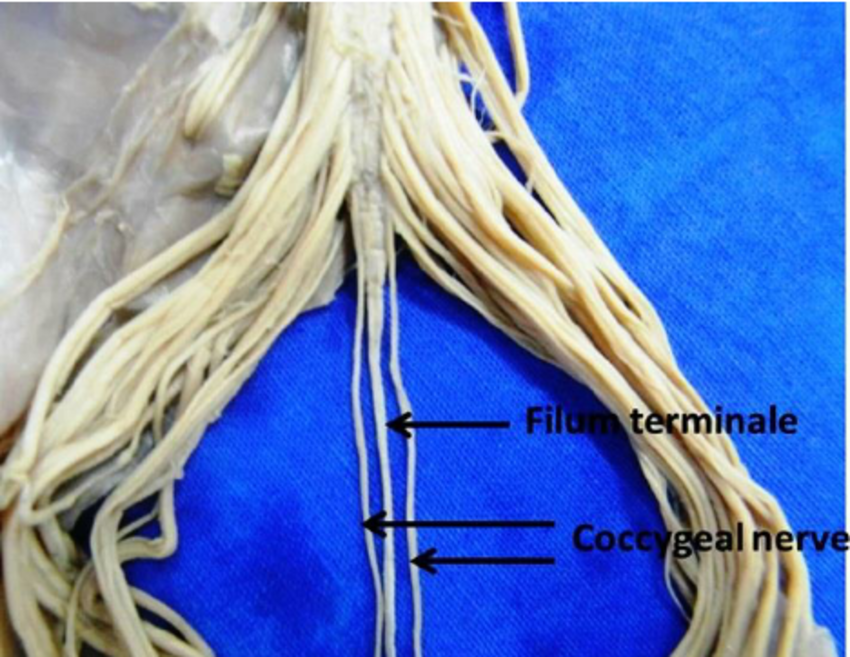
iv) Spinal nerves
31 pairs
8 cervical - pass over their respective vertebrae (except C8)
12 thoracic - pass under
5 lumbar - pass under
5 sacral - pass under
1 coccygeal
Part of PNS
Emerge from vertebral canal via intervertebral foramina
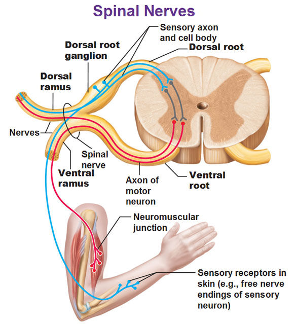
Attach to SC by:
ventral roots at ventrolateral aspect - ** Motor ** - efferent fibres sending info from SC to muscles/organs
dorsal roots at dorsolateral aspect - ** Sensory ** - afferent fibres sending info to SC & up to brain
Dorsal root ganglion
Each root attaches cord with rootlets
Spinal nerve branches into:
Dorsal ramus
Ventral ramus
Spinal nerves contain following fibres:
somatic efferent
visceral efferent
somatic afferent
visceral afferent
Página 2
Internal Anatomy
i) Gray Matter
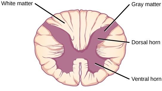
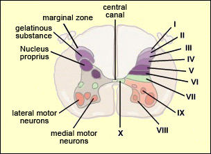
Spinal cord composed inner core of gray matter - nerve cell bodies, glia, blood vessels
Divided into:
Dorsal column/horn
Ventral column/horn
Lateral column (intermediate zone)
Rexed's laminae (10 zones)
Dorsal gray column nerve cell groups
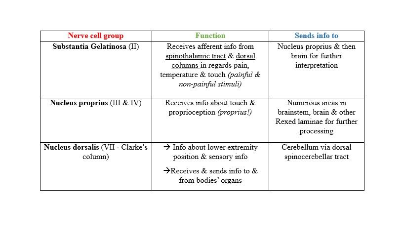
Extra info about dorsal nuclei!
Substantia gelatinosa contains large amount of opiate responsive neurons (morphine & heroin) (Link to Pain lecture)
Nucleus dorsalis most prominent at C8-L2 level
Sympathetic & parasympathetic autonomic NS have pre-ganglionic neurons in Nucleus dorsalis
Ventral gray column nerve cell groups
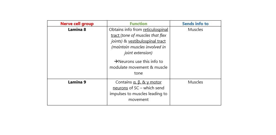
Extra info about ventral nuclei!
Neurons in Lamina 9 topographically organised based on:
Muscle they control (flexor or extensor)
Where in body that muscle located (axial or peripheral)
Medially located neurons control axial muscles
Laterally located neurons control limbs
Anteriorly located neurons control extensors
Posteriorly located neurons control flexors
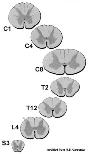
Gray matter density varies at diff. levels of SC
Largest conc. at cervical & lumbosacral enlargements (Link to Surface anatomy i) General anatomy - enlargements)
Página 3
ii) White matter
Divided into 3 regions on each side:
Ventral funiculus - between anterior median fissure & anterolateral sulcus
Lateral funiculus - between anterolateral sulcus & posterolateral sulcus
Dorsal funiculus - between posterolateral sulcus & dorsal median sulcus
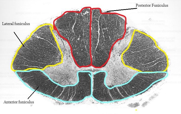
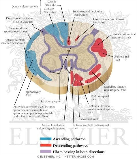
WM contains 3 types of tracts:
Ascending tracts - carry impulses from pain, tactile, thermal & muscle + joint receptors to brain
Intersegmental tracts - carry info from one segment of SC to another - spinal reflexes
Descending tracts - carry info from cerebral cortex & brainstem to muscles - movement, posture, muscle tone
Fibres passing in both directions surround outside of gray matter & project from dorsal horns
Página 4
Blood supply
i) Arteries
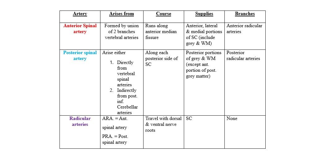
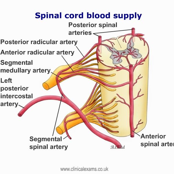
3 arteries
Anterior spinal
Posterior spinal arteries (2!)
Radicular arteries
ii) Veins
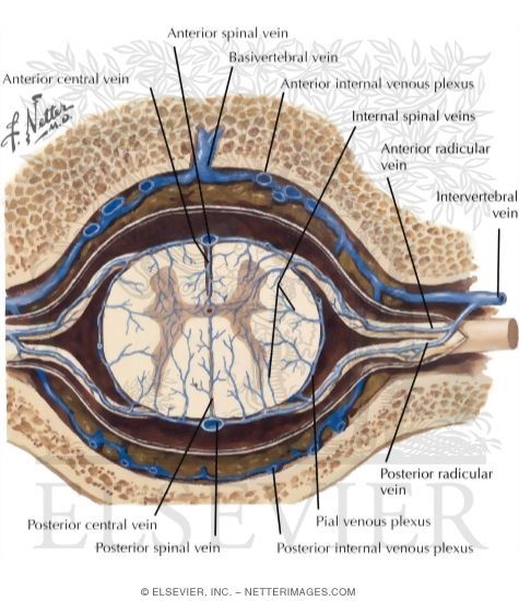
Blood drains from 6 longitudinal channels located around SC (outside dura mater)
Into anterior & posterior radicular veins (inside dura mater)
Which join together & drain into the intervertebral venous plexus
Which drains into the Ant. & Post. spinal veins
Página 5
Spinal Cord injury
V little nerve regeneration in CNS
Loss of sensory & motor control below level of injury
Resp stop when lesion above Phrenic nerve origin (C3,4,5) - intercostal & diaphragmatic paralysis
Possible injuries that can occur to the SC include:
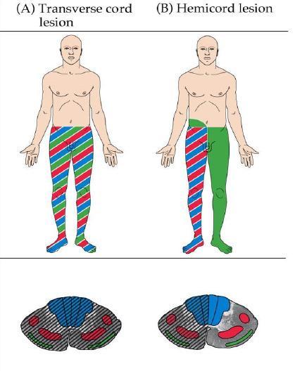
Complete transection - SC is completely torn - usually leads to Lower Motor neuron paralysis @ level of injury & Upper Motor neuron paralysis below LOI
Hemisection - Aka Brown-Sequard syndrome - damage occurs on one side of SC - causes weakness or paralysis on one side of body & loss of sensation on opposite side
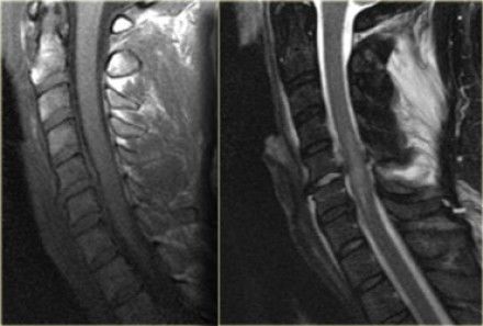
Contusion - 'bruising' of the spinal cord - can result in oedema & compression damage occurring to nerves - common
C4 injury = tetraplegia (including shoulders)
C6 = tetraplegia (not including shoulders)
T6 = paraplegia (above level of pubic symphisis)
L1 = paraplegia (below level of PS)
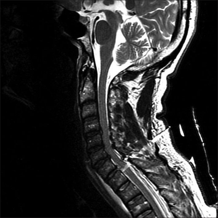
Image 1 = SC T-1 transection due to stab wound - http://www.onlinejets.org/article.asp?issn=0974-2700;year=2012;volume=5;issue=2;spage=204;epage=204;aulast=Yeung
Image 3 = Spinal cord odema due to hyperflexion sprain - http://www.radiologyassistant.nl/en/p49021535146c5/spine-cervical-injury.html
Some treatments for spinal cord injuries include:
Spinal alignment & stabilisation
Decompression
Drugs such as Methylprednisolone (corticosteriod that reduce inflammation & such any 2ndary damage to SC)
Some clinical presentations of SC injury:
Paralysis
Flaccid limbs
No tendon reflexes upon testing
Loss of sensation
Incontinence
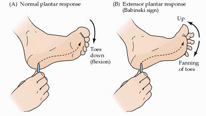
Babinski sign:
Medial aspect sole of foot is 'stroked' from heel towards little toe
Normal rxn = Plantar flexion (toes point down & foot flexes)
Abnormal rxn = Extensor plantar response (toes point up & fan outward) - this is normal rxn in children under age 2
Informações
- Descrição
- Central Nervous System (CNS Anatomy) Notas sobre Spinal cord , criado por Niamh McLoughlin em 18-10-2017.
- Criado por
- Niamh McLoughlin
- Visualizações
- 1
- Avaliação
Mais sobre Spinal Cord Anatomy